Biosafety of Nanoparticles:
|
|
|
- Sheena O’Neal’
- 5 years ago
- Views:
Transcription


1 Biosafety of Nanoparticles: Evaluation of Nanomaterials By a Suite of Cellular Assays Mellisa Theodore 1, Nathan Boggs 1, Bakhtiyor Rasulev PhD 2, Jerzy Leszczynski PhD 2, and Joany Jackman PhD 1 1- JHU APL 2- Jackson State University Science & Technology Business Area


2 Biosafety of Nanoparticles Our Approach Is there a need for concern? Nanoparticles are being used in many industries A variety of shapes and surface chemistry exist at this time- little is known about the effect of nanoparticles on the host Carbon nanotubes have shown asbestos-like health risks Colloidal silver ingestion causes argyria Pharmaceutical, biological research, medical applications Wait and see methodology not acceptable Exposure and fate both very important t considerations What do we want to know? If biomedically-relevant nano-materials can cause damage to cells, cell pathways or genomic DNA If DNA damage correlates to organ specific effects or cytotoxicity How are we investigating the concerns? 2 Building on a suite of in vitro cellular assays for hazard assessment of nanoparticles Prescreening biomarkers in vitro which might translate to in vivo biomarkers of hazard assessment Predicatively modeling cellular effects of nanoparticles


3 Need for Concern? Industrial Problem: Nanoparticles May Not Be Eliminated by HEPA Filters How HEPA filters work: High Efficiency Particulate Air Filter 99.9% 9% efficient at 0.3 micron 3 CRITICAL ISSUE NOTE: Efficiency drops in this size range and
4 Need for Concern? Nanoparticles: Protection? Fate? Elimination? Nanoparticle size and respiratory disposition (Maynard and Kuempel, 2005) this is size where greatest deposition occurs in lungs...and cannot be eliminated by filtration in the kidneys 4


5 Need for Concern? Medical and Industrial Problem: Nanostructures Have Been Linked To Cancer Research Question: What is relationship of chemistry, nanoparticle size and shape to risk? 5

 of the lungs, making breathing")


6 Need for Concern? Hazards of non toxic dust: Effects may not be acute Pulmonary alveolar proteinosis is a rare disease in which a type of protein builds up in the air sacs (alveoli) of the lungs, making breathing difficult In some cases, the cause of pulmonary alveolar proteinosis is unknown In other cases it is associated with infection or an immune problem It also can occur with cancers of the blood system, and after exposure to high levels of dust This rare disorder generally affects people years old and is seen in men more often than in women 6

- apoptosis/ necrosis Flow Cytometry- Cell cycle analysis/")

7 How are we investigating? Suite of Assays and Cells Assays- Cellular Screen Acute measures of toxicity MTT Assay- cellular metabolic activity Live/Dead Assay- cellular membrane integrity Long-term measures DNA Ladders; Flow Cytometry (Annexin-V)- apoptosis/ necrosis Flow Cytometry- Cell cycle analysis/ growth kinetics Cytokine Detection Assay- multiplex immunoassay for human cytokines Cells Lines HL60- promyloblast- peripheral blood THP1- monocytes- peripheral blood HepG2- hepatocyes- liver A549- alveolar epithelial-lung MCF7- breast adenocarcinoma- mammary gland 7

8 Nanoparticle Variations Nanoparticles are significantly different in all aspects Our studies will consider the following distinctions in particles: Composition Size Shape (aspect ratio) Surface charge Initial Studies: Gold nanoparticles characterized by NIST 2nm,10nm, 30nm, 60nm Results shown for HepG2 and A549 cell lines Follow up Experiments: Comparison of Carbon nanotubes and Fullerenes Immune cell responses to incubations i ROS Evaluations 8


9 Results: MTT ASSAY 24H-72H Assay results shown up to 72H of incubation with nanoparticles- little death observed compared to positive control (cell death induced by H2O2) bsorbanc ce at 570 A nm 100nM 10nm 10nM 10nm 1nM 30nm 100nM ance nm 10nM nm 1nM 60nm 100nM 60nm 10nM 60nm 1nM Positive Control Absorb Negative Control 0 24H 72H 9


10 Results: LIVE / DEAD ASSAY 24H-72H Assay results shown up to 72H of incubation with nanoparticles- little death observed compared to positive control (cell death induced by H2O2) 100% 90% 80% 70% 60% 50% 40% 30% 20% 10% 0% 24 H Live Dead 10, 30, 60nm Gold Nanoparticle Incubations % Fluorescence 100% 90% 80% 70% 60% 50% 40% 30% 20% 10% 0% 72H Live Dead 10, 30, 60nm Gold Nanoparticle Incubations 24H 72H 10


11 Results: CELL CYCLE EFFECTS Normal Cell Cycle G2 phase Block G1 Phase Block* 11
12 The Inflammatory Response Fever Vascular leakage Increased blood flow
13 Results: 17-Plex Cytokine Assay Cytokine Concentration, Gold Nanoparticle Challenge (72hr) NC ntration (p pg/ml) Conce IL-1beta IL-2 IL-4 IL-5 IL-6 IL-7 IL-8 IL-10 IL-12 (p70) IL-13 Cytokine IL-17 G-CSF GM-CSF MCP-1 (MCAF) IFNgamma MIP- 1beta TNFalpha PC Cytokines appear to have been induced by Nanoparticles 13
14 Results: 17-Plex Cytokine Assay Comparison to LPS induced cells shows higher induction in positive controls Conce entration (pg/ml) Cytokine Concentration, LPS Challange Compared to Gold Nanoparticles NC PC 10nm - 100nM 10nm- 10nM 10nm- 1nM 30nm- 100nM 30nm- 10nM 30nm- 1nM 60nm- 100nM 60nm- 10nM 60nm- 1nM LPS Control 0 IL-2 IL-12 G-CSF GM-CSF 48H Cytokines Cytokine induction in immune cells is much higher than in hepatocytes g/ml) Concentration (p Positive Control (10ng/mL LPS from K. pneumoniae) IL-2 IL-4 IL-6 IL-8 IL-10 GM-CSF IFN-g TNF-a IL-1b IL-5 1 hour 3 hour 6 hour 9 hour 12 hour 18 hour IL-7 IL-12 (p70) IL-13 IL-17 G-CSF MCP-1 (MCAF) MIP-1b 14
15 Results: Prescreening Biomarkers in vitro and Predicative Modeling Investigation of cytokine responses using modeling is necessary for prescreening of potential biomarkers When placed into model Analyzing cytokine assay data- noticed that response values for 2nm and 30nm particles were less then for 10nm and for 60nm This was noticed for almost all cytokine assays Selected IL-7 assays have the same distribution of values (expected that the response value should increase with decreasing or increasing nanoparticle size) Wanted to find any rule for experimental data distribution and then find parameters that change noticeably with the cluster size increasing By analyzing the data for IL-7 assay -found some interesting distribution of data Modeling Effort tconducted d by: Interdisciplinary Center for Nanotoxicity ~ Jackson State University 15


16 Results: Prescreening Biomarkers in vitro and Predicative Modeling- In silico approaches SARs: Structure- Activity Relationships Physical Properties Toxicity QSARs: Quantitative Structure-Activity Relationships Environmental Biokinetic Distribution Parameters Modeling Effort Conducted by: Interdisciplinary Center for Nanotoxicity ~ Jackson State University 16


17 Results: Prescreening Biomarkers in vitro and Predicative Modeling 3-dimensional plot of data- difficult to see regularity Modeling Effort tconducted d by: Interdisciplinary Center for Nanotoxicity ~ Jackson State University 17
18 Results: Prescreening Biomarkers in vitro and Predicative Modeling When plotted on one axis, can see a sinusoidal curve, which is gradually damping Looked for equation to fit the curve Among several thousands curve fitting functions we found the following: ow Y(IL7)=7.88E-03 (X 12 ATAN(X 2 ))+3.64 (COS(X 1 ) ATAN(X 2 ))+3.64 where Y(IL7) is response variable, IL-7 cytokine assay values, X 1 gold nanoparticle size in nm, X 2 concentration in nm Need to check robustness of this equation with experimental data for other sizes of gold nanoparticles Modeling Effort tconducted d by: Interdisciplinary Center for Nanotoxicity ~ Jackson State University 18


19 TEM Images of Gold Nanoparticles 10nm 30nm 60nm 19


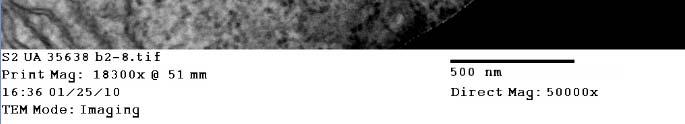

20 TEM Images of Gold Nanoparticles: Incubated with cells Potential uptake of nanoparticles into cells 20
21 Conclusions: Well characterized gold nanoparticles have shown little/ no effect in acute toxicity assays Further testing is scheduled for more cell lines and various types of nanoparticles Data from follow on experiments will be compiled into the database for continued modeling for prediction of relative risk of well characterized materials Disruption of cellular pathways and biomarkers of exposure will be used to develop predictive monitoring tools Acknowledgements: APL: Melissa Betz NIST: Dr. Bryant Nelson, Dr. John Elliot Johns Hopkins University: Microscopy Facility 21
22 References: Atsuya Takagi, Akihiko Hirose, Tetsuji Nishimura, Nobutaka Fukumori, Akio Ogata, Norio Ohashi, Satoshi Kitajima and Jun Kanno: Induction of mesothelioma in p53+/ mouse by intraperitoneal application of multi-wall carbon nanotube : J. Toxicol. Sci., Vol. 33: No. 1, (2008) HEPA Filtration Facts. D. F. Solutions. Minneapolis: 1-2 Maynard, A. and E. Kuempel (2005). "Airborne Nanostructured Particles and Occupational Health." Journal of Nanoparticle Research 7(6): Poland, C. A., R. Duffin, et al. (2008). "Carbon nanotubes introduced into the abdominal cavity of mice show asbestos-like pathogenicity in a pilot study." Nat Nano 3(7): Song, Y., X. Li, et al. (2009). "Exposure to nanoparticles is related to pleural effusion, pulmonary fibrosis and granuloma." European Respiratory Journal 34(3): Wick, P., A. Malek, et al. (2009). "Barrier Capacity of Human Placenta for Nanosized Materials." Environ Health Perspect 118(3) Image Sources