Biomedical Engineering 3D BIOPRINTING OF SILK FOR CELLULAR APPLICATIONS. Martina Ravizza
|
|
|
- Elfreda Nichols
- 5 years ago
- Views:
Transcription

1 Biomedical Engineering 3D BIOPRINTING OF SILK FOR CELLULAR APPLICATIONS Martina Ravizza Supervisor: Prof. Ferdinando Auricchio Correlator: Dott. Michele Conti Sericina Fibroina Academic year 2014/2015 Università degli Studi di Pavia - Structural Mechanics Department


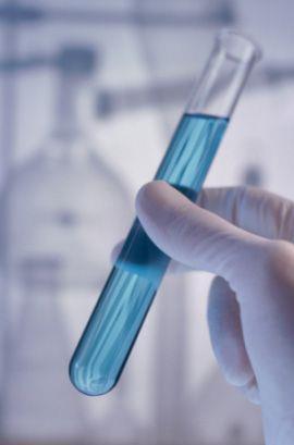
2 The biomaterial of the future: silk Silk origins Natural protein fiber of animal origin (Bombyx mori) Silk structure Two filaments of fibroin (75%) Coating of sericin (25%) which must be removed Sericin Fibroin From cocoons to fibroin solution Cutting of the cocoons Boiling Drying Dissolve Dialysis Fibroin solution


 Secondary")
3 The biomaterial of the future: silk Cocoons Silk fibers Single silk fiber Primary structure of fibroin Fibroin structure Primary structure: hexapeptide Self-assembly of Beta-sheet which gives strength and flexibility Property Biocompatible Biodegradable Sterilizable in autoclave (121 C, 2 atm, 15 min) Secondary structure of fibroin (Beta sheet)
4 Purpose of activity Silk as a biomaterial today Overview of current applications Main technical manufacturing: Electrospinning Example application: 3D silk bone marrow niche for platelets generation ex vivo - Prof. Alessandra Balduini et al (Università degli Studi di Pavia and Tufts University of Boston) Silk as a biomaterial tomorrow 3D Bioprinting of silk-based bioink to produce scaffolds First study in literature: bioink of silk and polyol Our possible implementation

5 Current applications Cardiovascular protheses Films as drug delivery Cartilage tissue Bone tissue Scaffolds


6 Electrospinning Instrumentation Capillary needle or pipette High voltage supplier Grounded collector Method Application of an electric potential Overcome the surface tension and development of the Taylor cone Ejecting of fiber jet Post treatment Crystallization by aqueous methanol at room temperature for min Electrospun Silk Biomaterial Scaffolds for Regenerative Medicine, Xiaohui Zhanga et al Processing of Bombyx mori silk for biomedical applications, B. D. Lawrence

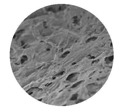
7 Electrospinning to produce 3D scaffolds Method Collector: bath filled with methanol Porogen: NaCl particles with a diameter of μm Salt leaching Results Nano-sized pores at the interstices Pores formed by salt parcles with a diameter of μm Electrospun Silk Biomaterial Scaffolds for Regenerative Medicine, Xiaohui Zhanga et al


8 3D silk bone marrow niche Prof. Balduini Purpose Reproduce the physiology of the bone marrow Produce platelets ex-vivo (hematopoiesis) Hematopoiesis Structure Microtube Sponge Gel spinning Freeze dry Assembly Salt leaching Result Microvasculature Spongy bone Programmable 3D silk bone marrow niche for platelet generation ex vivo and modeling of megakaryopoiesis pathologies, Di Buduo et al

 Programmable 3D silk bone marrow niche for platelet")
9 3D silk bone marrow niche Prof. Balduini Production and collection of platelets Addition of the megakaryocytes and their migration after 16 h Extension of proplatelets after 24 h Blood perfusion for platelets collection into bags containing an anticoagulant (after 6 h) Programmable 3D silk bone marrow niche for platelet generation ex vivo and modeling of megakaryopoiesis pathologies, Di Buduo et al

10 1. Pre - process 2. Process 3. Post - process 3D Bioprinting of silk to produce scaffolds: what we need Process Human resources Cocoons Bioimages Farmes Provide cocoons Fibroin solution Stem cells Elaboration 3D model CAD Medical Research (MD) Provide bioimages, stem cells and ECM components Evaluates the cells function in humans Silk-based Bioink Bioprinter Bioengineers Disegn models Run the printer with appropriate bionks Growth factors Scaffolds Cells Biologist/Biotechnologist Provide bioik of silk Culture of stem cells Evaluates the cell funcion in rats
11 1. Pre - process 2. Process 3. Post - process 3D Bioprinting of silk to produce scaffolds: what we need Process Human resources Cocoons Bioimages Farmes Provide cocoons Fibroin solution Stem cells Elaboration 3D model CAD Medical Research (MD) Provide bioimages, stem cells and ECM components Evaluates the cells function in humans Silk-based Bioink Bioprinter Bioengineers Disegn models Run the printer with appropriate bionks Growth factors Scaffolds Cells Biologist/Biotechnologist Provide bioik of silk Culture of stem cells Evaluates the cell funcion in rats
12 Silk-based bioinks formulation - Tufts University of Boston Pattern Target of measure Method Results Films Solubility PBS at 26 C Phase contrast microscop NS regardless of the printed volume (Glycerol) S for volumes less than 5 μl (Erythritol) Contemporary print for support strucutures Beta-sheet content Spectrometry Beta-sheet 1 solubility Discs Profile Interferometry Linear dependence between disc height and printed layers Low presence of clots Bioinks Viscosity 15% silk, 5% polyol, 26 C Viscometer Low without additives Very high with propanediol Influence of print resolution Drops Buckling height 80% silk, 20% polyol 5 μl, 1 x 3 mm Contact angle Rapid deformation without additives Homogeneous deformation for glycerol and erythritol-based bioinks Contemporary print for support structures Polyol-Silk Bioink Formulations as Two-Part Room-Temperature Curable Materials for 3D Printing, Rod R. et al


13 3D Bioprinter at Tufts University of Boston Positioning of the printhead carriage and estrusion accomplished by 2-phase unipolar 1.8 steppers motor Lead screw: linear movement of 6.53 μm for step Temperature control: catrage heaters and thermistors Layer-by-layer deposition of bioink to produce 3D structures Polyol-Silk Bioink Formulations as Two-Part Room-Temperature Curable Materials for 3D Printing, Rod R. et al


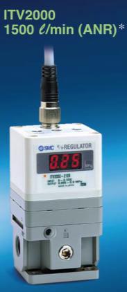

14 Our possible implementation Digital pressure regulator (ITV-2010; SMC, Japan) 2. Microelettrovalve (DFD SMART VALVE) 3. Power supply (0 3,3 V) 4. Pressurized tank 5. 5 or 10 ml disponsable syringes 6. Petri disc
15 Conclusions Silk Excellent biomaterial because of robustness, elasticity, biocompatibility and programmable biodegradability Electrospinning Pros: Nanoscale fibers that mimic the extracellular environment Cons: Inability to control shape of structure and size of pores 3D Bioprinting Novel self-curing silk-polyol blended inks Mechanically robust and insoluble layers for complex 3D geometries Open problem: incorporate stem cells
16 3D Bioprinting of silk for cellular applications Grazie per l attenzione Un ringraziamento particolare per la gentile collaborazione al Dott. Michele Conti, al Prof. Ferdinando Auricchio e alla Prof.ssa Alessandra Balduini
17 Biomedical Engineering 3D BIOPRINTING OF SILK FOR CELLULAR APPLICATIONS Martina Ravizza Supervisor: Prof. Ferdinando Auricchio Correlator: Dott. Michele Conti Sericina Fibroina Academic year 2014/2015 Università degli Studi di Pavia - Structural Mechanics Department