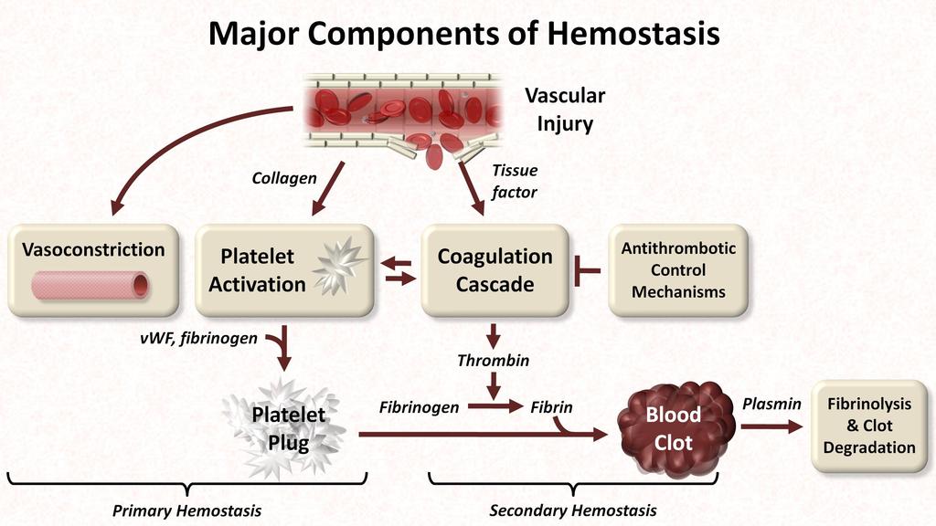
Primary hemostasis. Vascular endothelium Vasoconstriction : local tissue factor, nervous system
|
|
|
- Eric Wilkerson
- 5 years ago
- Views:
Transcription

1

2
3

4 Primary hemostasis Vascular endothelium Vasoconstriction : local tissue factor, nervous system Platelet Plug Platelet Adhesion Platelet Activation Platelet Aggregation Platelet Plug Formation
5 Secondary Hemostasis Cascade / Waterfall Model Activation of coagulation system Extrinsic pathway Intrinsic pathway Common pathway
6 Secondary Hemostasis Cascade / Waterfall Model
7 Secondary Hemostasis

8 Secondary Hemostasis Current concept : Cell-based model of hemostasis Hoffman and Monroe 2001 Hemostatic process 3 phases Initiation Amplification Propagation (thrombin burst)
9 Cell-based model of hemostasis

10 Fibrinolytic Pathway D-dimer
11 PERIOPERATIVE Coagulation monitoring


12 Platelet count Primary hemostasis Reflect quantitative of platelet Normal range : 150, ,000 Turn around time : 1 hour
")
13 Bleeding time Out of Use Assess platelet function Making a puncture and monitoring time for bleeding stop Normal: 2-10 minutes at anterior forearm Delicate, experienced operator Prolongation: thrombocytopenia hypofibrinogenemia severe anemia(hct<30%) vwd


14 Platelet function analyzer PFA-100, PFA-200, Siemens Congenital and Acquired platelet dysfunction

15 Light transmission platelet aggregometry Platelet aggregation assays Congenital and Acquired qualitative platelet disorder
16 Activated Partial Thromboplastin Time (aptt) Intrinsic & Common pathway Activate intrinsic pathway by celite, kaolin, silica Detection at factor concentration below 30%-40% of normal Turn around time : 90 minutes
17 Prothrombin Time (PT) Extrinsic & Common pathway Activate intrinsic pathway by Ca&tissue thromboplastin Turn around time : 90 minutes

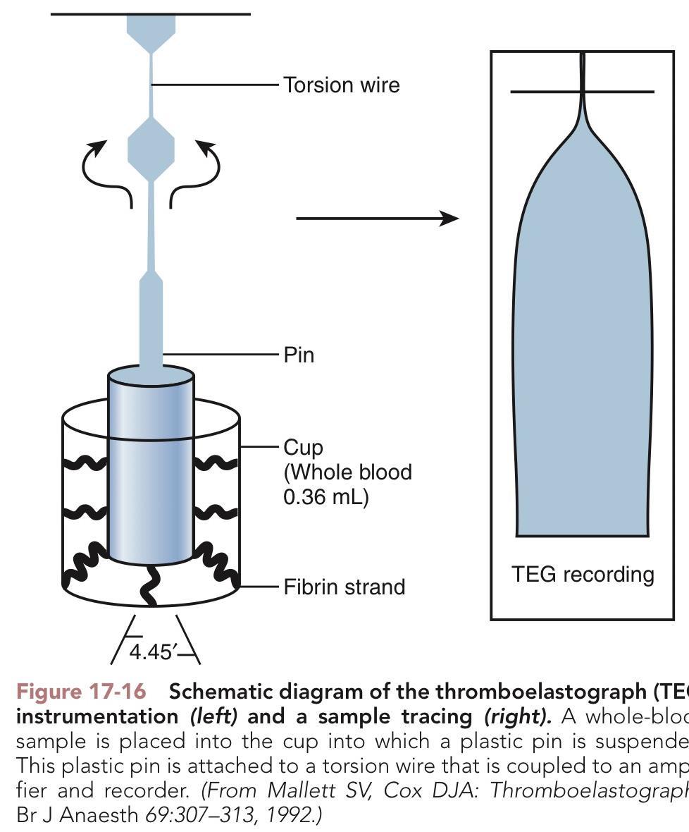
18 Fibrinogen level Fibrinogen is converted into fibrin to stabilize clot Normal value : mg/dl Turn around time : 90 mins

19 POINT-OF-CARE COAGULATION T E S T


20 POINT OF CARE COAGULATION TEST ACT TEG ROTEM
21 Activated Clotting Time Activation of coagulation via the intrinsic [Factor XII] pathway Monitor the anticoagulant effect of heparin Clinical application Cardiopulmonary bypass surgery ECMO support Catheterization laboratory Angiography intervention

22


23

24 Activated Clotting Time Limitation Lack of sensitivity at low heparin concentration Prolong ACT Shorten ACT
25 67-year-old woman was diagnosed severe AR from IE s/p MVR AVR on warfarrin Preoperative lab : INR 1.7 Operation Redo AVR Post CPB: Bleeding from surgical field despite 2 rounds of blood components

26 ThromboElastoGraphy TEG & ROTEM ROtational ThromboElastoMetry
27 Examines entire of hemostasis. Initiation Fibrin formation Platelet - fibrin interaction Clot stability <fibrinolysis> Adventage : Real-time analyse of clot formation and dissolusion Use whole blood & Fast turnaround Guide for specific transfusion Reduce blood product transfusions

28 The amount of transfused RBC FFP and bleeding volume was found to be significant reduce in VHA-guide group


29 Hemostasis Pro-coagulation factor Fibrinolysis Whole blood Fibrinogen level Platelet count Anticoagulant Cellular component Platelet RBC Leucocyte


30


31 Thromboelastography <TEG> CLINICAL USES Cardiac surgery Liver transplantation Major trauma Major obstetric hemorrhage.
32 Thromboelastography <TEG>

33 TEMogram Time Amplitude in mm. The greater amplitude the firmer the clot
34 LY30
35 Angle: Speed of thrombin formation maximum amplitude (MA) : maximums strength of clot LY30 Reaction time : 1st significant clot formation clot kinetics : speed of clot formation Percent lysis in 30 min after MA Point-of-care coagulation testing Continuing Education in Anaesthesia Critical Care & Pain, Volume 13, Issue 1, 1 February 2013, Pages 12 16
36 Thromboelastography <TEG> Initial interpretation R time : Reflects coagulation factor level K & α Angle : Reflects fibrinogen activity MA. : Reflects platelets function and fibrinogen activity LY 30 : Reflects clot stability or fibrinolysis Point-of-care coagulation testing Continuing Education in Anaesthesia Critical Care & Pain, Volume 13, Issue 1, 1 February 2013, Pages 12 16
37 Point-of-care coagulation testing Continuing Education in Anaesthesia Critical Care & Pain, Volume 13, Issue 1, 1 February 2013, Pages 12 16


38 Conventional and near-patient tests of coagulation Continuing Education in Anaesthesia Critical Care & Pain, Volume 13, Issue 1, 1 February 2013, Pages 12 16
39 Thromboelastography <TEG> Limitation : Inability to detect impairment in platelet function induced by anti-platelet agents Poor ability to detect condition affect platelet adhesion e.g.von Willebrand s disease Point-of-care coagulation testing Continuing Education in Anaesthesia Critical Care & Pain, Volume 13, Issue 1, 1 February 2013, Pages 12 16
40 Limitation : TEG Prolong R Narrow amplitude


41 Rotational thromboelastometry<rotem>

42 ROTEM s unique shaft spring and ball bearing technology provides for high level of precision and sensitivity of clot formation
43 ROTEM ASSAY INTEM - HEPTEM EXTEM - FIBTEM - APTEM


44 INTEM EXTEM stable fibrin clot

45 ROTEM assay : HEPTEM Residual heparinization Activation as in INTEM with the addition of heparinase HEPTEM compared to INTEM

46 ROTEM assay : FIBTEM Fibrinogen level & Fibrin net polymerization Activation as in EXTEM with the addition of platelet blocking substance

47 ROTEM assay : APTEM Hyperfibrinolysis Activation as in EXTEM with the addition fibrinolysis inhibitors APTEM compared with EXTEM


48

49 A10 MCF ML CT CFT

50 ROTEM : INTERPRETATION Activation of coagulation Clot firmness Fibrinolysis

51 Activation of coagulation CT prolong DDX -Factor deficiency -Heparin effect Differentiation with HEPTEM

52 HEPTEM compare with INTEM INTEM CT long HEPTEM CT normalization > Heparin effect INTEM CT long HEPTEM CT also long > No Heparin effect > Factor deficiency
53 Clot firmness Narrow A10, MCF DDX -Platelets disorder -Fibrinogen disorder differentiation with FIBTEM


54 FIBTEM compare with EXTEM EXTEM amplitude low FIBTEM amplitude normal > fibrinogen normal > platelets deficiency EXTEM amplitude low FIBTEM amplitude low > fibrinogen deficiency
55 Fibrinolysis ML% >15% :EXTEM&INTEM within 60 mins after MCF indicate premature clot lysis Confirm with APTEM


56 APTEM compare with EXTEM EXTEM: hyperfibrinolysis. ML >100% APTEM: fibrinolysis inhibited. ML <15% > Hyperfibrinolysis
57 ROTEM Limitation : Inability to detect impairment in platelet function induced by anti-platelet agents Poor ability to detect condition affect platelet adhesion e.g.von Willebrand s disease Point-of-care coagulation testing Continuing Education in Anaesthesia Critical Care & Pain, Volume 13, Issue 1, 1 February 2013, Pages 12 16

58
59 67-year-old woman was diagnosed severe AR from IE s/p MVR AVR on warfarrin Preoperative lab : INR 1.7 Operation Redo AVR Post CPB: Bleeding from surgical field despite 2 rounds of blood components


60 Analyze the ROTEM result
61 After Protamine neutralization Clots in surgical field & Operation success After 6 hr Continuous bleeding from the drain!!!!!


62 Analyze the ROTEM result at ICU

63 After Transamine 1 g IV persistent bleeding

64 Set OR Stop bleeddddddddddddddd

65 ไม