BD Accuri C6 Flow Cytometer. Nil Emre, PhD BD Biosciences R&D Manager, Stem Cells and Cell Function
|
|
|
- Thomas Carter
- 5 years ago
- Views:
Transcription
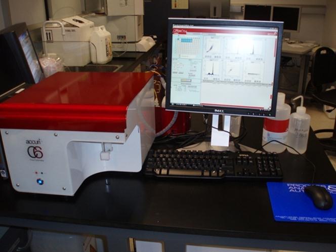
1 BD Accuri C6 Flow Cytometer Nil Emre, PhD BD Biosciences R&D Manager, Stem Cells and Cell Function


2 The BD Accuri C6 Flow Cytometer System An affordable, full-featured, easy-to-use flow cytometer Two lasers and six detectors
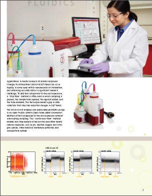
3 The BD Accuri C6 Flow Cytometer Innovations in all the major components of a flow cytometer Fluidics: Peristaltic pumps and pulse dampeners allow miniaturization and direct-volume measurement Optics: Locked-down alignment Signal detection: Broad dynamic range obviates voltage adjustments Software: Intuitive

4 Alignment and Signal Detection are Optimized and Locked Down 488-nm solid state laser SSC 640-nm diode laser PMTs for fluorescence detection FSC Diodes for light scatter detection
5 Advantages of Pre-optimized Detector Settings Greatly reduces the risk of lost data due to improper setup Saves time and sample No specialist training or dedicated operator required Predictable, reproducible analysis relative to the sample type and application Predictable fluorescence spillover
6 Enhanced Sample Handling Direct volume measurement Many types of sample tubes may be used. Flow cytometry tubes Microcentrifuge tubes Ninety-six-well plates with the BD CSampler accessory Open system conducive to kinetic studies BD CSampler accessory for automated sample introduction

7 Intuitive Software Sample Grid Cytometer Status Fluidics Controls Histogram, Dot Plot, and Density Plot Display Area Analysis and Gating Tools Run Criteria Real-Time Updates Plot Statistics
8 Kits and Templates on the BD Accuri C6 As Easy as Cell Analysis Is Going to Get by combining Cell Biology Kits with acquisition and analysis templates. Free downloadable BD Accuri C6 Software Templates are matched to each kit at: Kits and Templates lead to quick and easy set-up and analysis of cell populations.
9 Kits and Templates on the BD Accuri C6 Available Templates:

10 Kits and Templates on the BD Accuri C6 Available Templates: Sample Data:

11 BD Accuri C6 Promotion Note: US Region Only Promotion Period: Oct 1, 2014 Dec 31, 2014
12 For Additional Information If you have further questions: Contact Technical Support (US) at: , Prompt 3, 2 or ResearchApplications@bd.com Please visit our BD Accuri C6 resources site at: Class 1 Laser Product. For Research Use Only. Not for use in diagnostic or therapeutic procedures. BD, BD Logo and all other trademarks are property of Becton, Dickinson and Company BD


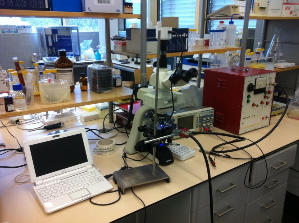
13 Dr. Alfonso Blanco Fernández Scientific Director of Conway Core Facilities Director of Flow Cytometry UCD - Conway Institute University College Dublin Belfield, Dublin 4. IRELAND Real Time alfonso.blanco@ucd.ie Tel: 00353(0) /6947


14 BD FACS440

15 Stop-flow method
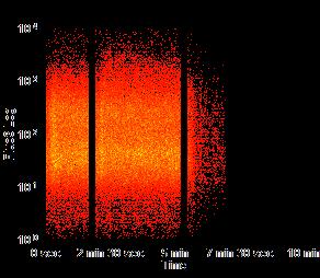
16 Continuous measurement 2009


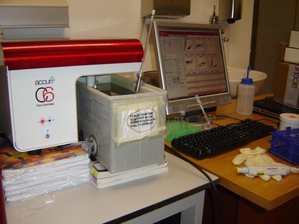
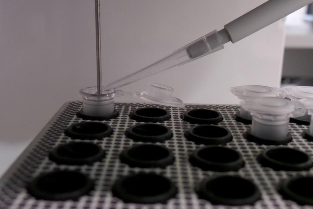
17 BD Accuri C6 Peristaltic Pumps


18 BD Accuri C6 Gel loading tips

19 BD Accuri C6


20 488: 530/30: Fluo-4 488: 530/30: Fluo-4 488: 530/30: Fluo-4 488: 530/40: Fluo-4 488: 530/40: Fluo-4 488: 530/40: Fluo-4 Beckman Coulter Cyan ADP: A23187 EtOH A23187 Thapsigargin A23187 BD Accuri C6:

21 BD Accuri C6. Time limitations? Fluo-3 : 530/30 Fluo-3: 530/30


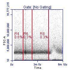
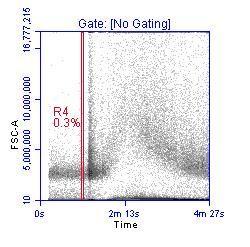
22 Quality Control State of the instrument (QC) Extremely important!! Laser alignment, PMT, laser state Dirty or clean instrument,...


23 SSC-A Alexa647-A SSC-A Alexa647-A SSC-A Alexa647-A Attention!! Scatter Fluorescence 250K K K 100K K Time Ungated Specimen_001_Leish LN.fcs Event Count: Time Ungated Specimen_001_Leish LN.fcs Event Count: K K K 100K K Time Ungated Specimen_001_Leish ears.fcs Event Count: Time Ungated Specimen_001_Leish ears.fcs Event Count: K K K 100K K Time Ungated Specimen_001_Leish ears_001.fcs Event Count: Slide courtesy of ExCyte. Expert Cytometry Time Ungated Specimen_001_Leish ears_001.fcs Event Count:


24 Attention!! Remember! Maximum Number of Events/Well = 10 6 Optimise Number of Events/ sec based on the time of your experiment


25 How much time between addition and analysis? 638 : 675/ : 675/25 Addition Time Analysis Time
26 Do air bubbles affect the flow rate? Fluo-4 : 530/30 Cells Air Ionophore

27 Can you control the addition time?

28 Calcium flux in some mcherry transfected cell lines

29 Real Time Applications


30 Optimization Side Population. Stem Cells Hoechst Blue Murine Bone Marrow - 5µg/ml min 30 min min 90 min Hoechst Red Slide Courtesy of Catherine Simpson

31 Analysis of Algae physiology Kinetics of 4 different algae
32 Analysis of Algae physiology Kinetics of 4 different algae (FCS Express v.4)

33 Platelets
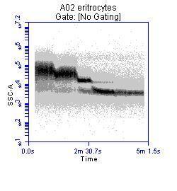
34 Erythrocytes Lysing
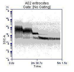


35 Erythrocytes Lysing Beads

36 Nanoparticles uptake 488: 530/30: Yellow - Green NPs NPs

37 Nanoparticles uptake
38

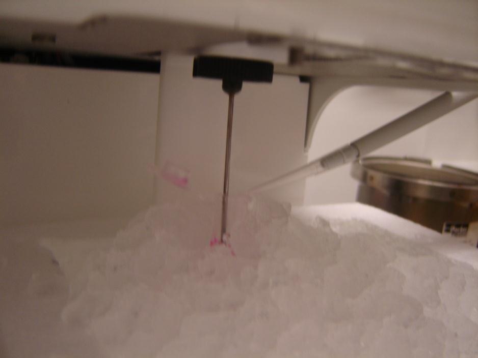
39 What if my sample has to be at 4ºC?
40

41


42 Nanoparticles uptake Fixed cell in suspension covered by nanoparticles, after incubation at 4 C


43 Calcium 488 : 530/30: Fluo-4 Fragmented cells


44 488: 530/30: Fluo-4

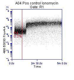

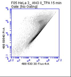
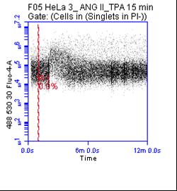
45 Some Calcium profiles


46 WT mcherry Calcium flux in some mcherry transfected cell lines 488: 585/40: Fluo-4 488: 585/40: Fluo-4 488: 675LP mcherry 488: 675LP mcherry 488: 585/40: Fluo-4 488: 585/40: Fluo-4 488: 585/40: Fluo-4 488: 585/40: Fluo-4


47 Calcium flux in some mcherry transfected cell lines


48 Sharing files and Templates

49 Sharing files and Templates


50 Sharing files and Templates Before After


51 488: 585/40: Fluo-4 488: 585/40: Fluo-4 488: 585/40: Fluo-4 488: 585/40: Fluo-4 488: 585/40: Fluo-4 488: 585/40: Fluo-4 Calcium flux Ionophore Thapsigargin Ionophore 2-APB Ionophore Thapsigargin
52 Apoptosis / Viability

53 Apoptosis / Viability


54 Apoptosis / Viability 633: 675/25: DRAQ FSC SSC : 585/40: MitoSox Time


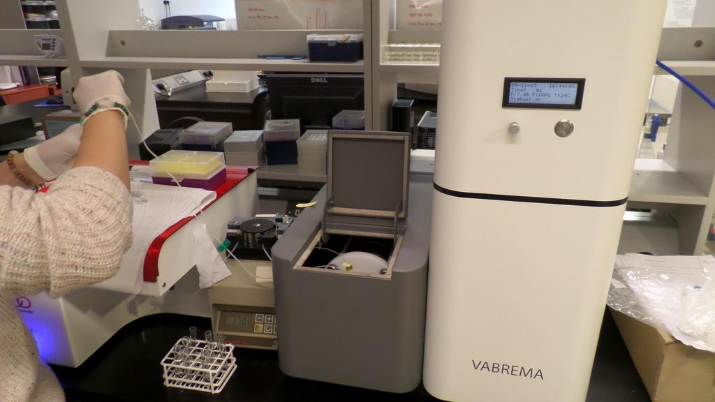
55 Collaborations May 2012 Conference & Tutorials Paper per review White Paper BD Brochure Video in Select Science

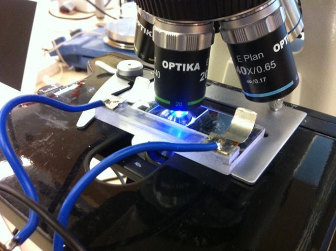
56 Department of Electrical Engineering
 the")
 inducing cell proliferation in a")
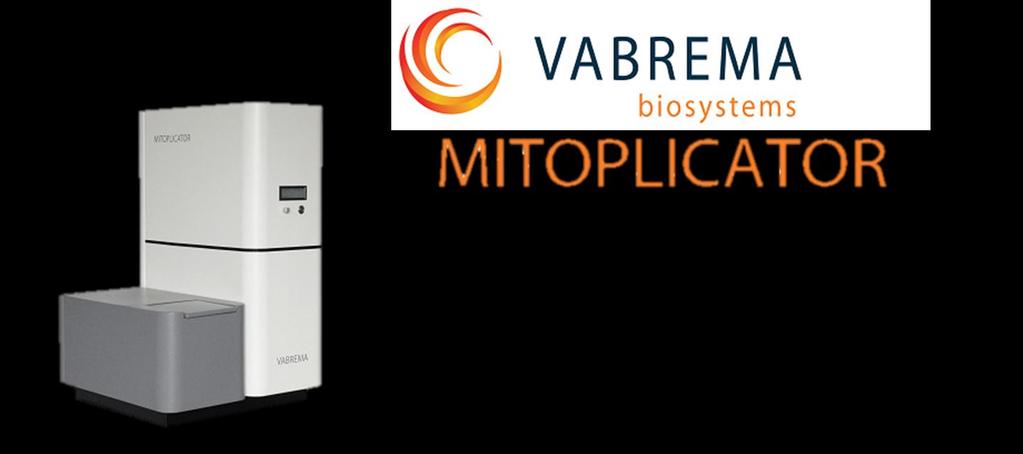
57 Using an electrical antenna (non-contact/inductive system vs electrodes: contact/capacitive) the Mitoplicator generates nanosecond pulsed electrical fields (nspef) inducing cell proliferation in a variety of cells
58


59

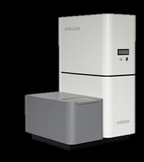


60 Initial Idea BD Accuri C6 Mitoplicator
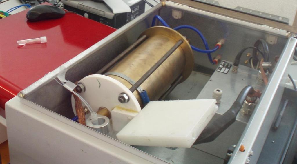
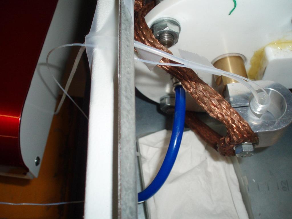


61 Mitoplicator Project Silicone tube (Cyan/HyperCyt) Nov 2012


62 One of the 1 st experiments: Stability 8 Peak beads in tube holder of the Accuri C6 8 Peak beads changing speeds in Mitoplicator
63 Mitoplicator Project


64 Mitoplicator Project CFSE Proliferation Fluo4 Calcium DRAQ7 Viability Mitosox ROS BODIPY Lipids GFP C6 glial HEK CHO Jurkats Platelets
65 Calcium flux in some mcherry transfected cell lines Drug Drug Stimulous


66 Kinetic analysis using FacsKin software
")
 Gate two: Contains 120s baseline and 600s treatment/control (0-720s) (Mitoplicator")
 Analysed parameters: FLUO4,")
67 SSC Kinetic analysis using FacsKin software 488: 530/30: Fluo4 633: 675/25: DRAQ7 Count 488: 530/30: Fluo4 488: 530/30: Fluo4 Gate one: Contains 120s baseline and 600s treatment/control (0-720s) (Mitoplicator on/off) Analysed parameters: DRAQ7 Time (sec) FSC Gate out: DRAQ7+ 633: 675/25: DRAQ7 Time (sec) Gate two: Contains 120s baseline and 600s treatment/control (0-720s) (Mitoplicator on/off) Analysed parameters: FLUO4, FSC SSC Time (sec) Gate three: Contains 100s baseline and 300s quenching time after the addition of 6ul 1mg/ml ionomycin ( s) Analysed parameters: FLUO4, FSC SSC Time (sec)
68 Kinetic analysis using FacsKin software 488: 530/30: Fluo4 633: 675/25: DRAQ7 Mitoplicator OFF Mitoplicator ON Fluo4 DRAQ7
69 Kinetic analysis using FacsKin software Cellular paramete r Mitoplicator treatment Starting value Maximum value Ending value Time to reach maximum Time from maximum to second 50% value Slope at first 50% value AUC I.C. calcium (0-720 s) I.C. calcium ( s) SSC (0-720s) SSC ( s) FSC (0-720s) FSC ( s) Draq 7 (0-720s) ,99 [0,49-1,07] 1,03 [1,02-1,10] 1,42 [1,02-1,81] 1,37 [1,04-1,76] 0,49 [0,00-0,96] 0,78 [0,36-1,00] 138,64 [0, ,00] 110,34 [12,08-255,49] 82,24 [17, ,98] 330,91 [9,53-500,67] 0,11 [0,01-0,69] 0,11 [0,02-0,44] 608,47 [327,72-613,18] 611,61 [601,92-623,05] P= 0,69 0,89 0,49 0,89 0,20 1,00 0,49 1,00 [0,92-1,03] 0,99 [0,97-1,01] 2,35 [1,98-5,02] 1,89 [1,03-3,78] 1,72 [0,58-2,68] 0,94 [0, ,00] 751,69 [748,49-771,29] 752,00 [749,92-754,08] 67,12 [38,06-140,71] 70,97 [29,35-123,40] 0,67 [0,19-7,49] 0,74 [0,52-1,46] 599,74 [549,39-621,62] 591,73 [578,91-607,59] P= 0,89 0,49 0,89 1,00 0,89 1,00 0,89 1,01 [0,94-1,03] 1,02 [0,95-1,04] 1,03 [1,01-1,05] 1,03 [1,02-1,05] 0,96 [0,88-0,98] 0,91 [0,36-0,95] 318,24 [0,00-467,47] 203,21 [15,50-451,66] 166,51 [66,07-265,25] 208,85 [127, ,53] 0,01 [0,00-0,04] 0,15 [0,00-0,26] 597,72 [592,20-603,35] 600,47 [599,39-604,29] P= 0,69 1,00 0,34 0,89 0,34 0,34 0,49 1,00 [0,99-1,01] 0,99 [0,98-1,00] 1,02 [1, ,00] 1,06 [1,04-1,10] 0,99 [0,92-1,06] 1,02 [1,00-1,07] 825,72 [710,93-948,71] 967,99 [841, ,23] 145,97 [1, ,04] 29,84 [1, ,09] 0,09 [0,00-0,21] 0,02 [0,00-0,08] 597,01 [592,13-603,02] 593,56 [588,79-598,02] P= 0,34 0,34 0,20 0,11 0,69 0,34 0,34 1,01 [0,90-1,02] 1,01 [0,93-1,02] 1,04 [1, ,00] 1,02 [1,01-1,04] 0,95 [0, ,00] 0,95 [0,94-0,97] 340,39 [250, ,52] 206,13 [0,00-255,22] 129,13 [0, ,83] 325,43 [0,00-487,41] 0,06 [0,00-0,21] 0,00 [0,00-0,08] 603,04 [583,39-611,28] 599,99 [599,38-600,77] P= 0,69 0,25 1,00 0,06 0,69 0,49 1,00 0,99 [0,99-1,00] 0,99 [0,97-1,00] 1,04 [0, ,00] 1,05 [1, ,00] 0,95 [0,92-1,00] 0,99 [0,98-1,04] 718,60 [0,01-865,07] 842,28 [746,04-955,50] 562,66 [20, ,00] 102,79 [37,45-137,67] 0,17 [0,00-0,52] 0,13 [0,06-,42] 595,18 [592,25-599,96] 595,46 [580,96-01,10] P= 0,89 0,77 0,20 0,34 0,34 0,89 0,89 0,95 [0,89-0,99] 0,95 [0,86-0,99] 1,02 [1,01-1,03] 1,02 [1,00-1,04] 0,98 [0,87-1,02] 0,94 [0,79-0,97] 371,82 [258,03-435,34] 409,59 [332,14-737,63] 167,58 [126, ,25] 533,43 [170, ,91] 0,00 [0,00-0,00] 0,00 [0,00-0,00] 597,86 [584,82-602,63] 595,58 [581,24-601,18] P= 0,88 0,89 0,34 0,49 0,49 0,66 0,69

70 Collaborations C6 glial HEK


71 Collaborations


72 Acknowledge Scholars Program
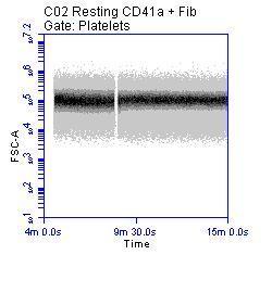
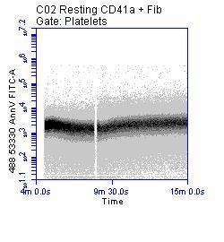
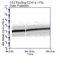
73 Cell Biology Applications of Real- Time Flow Cytometry Platelets Chris Jones ICMR University of Reading

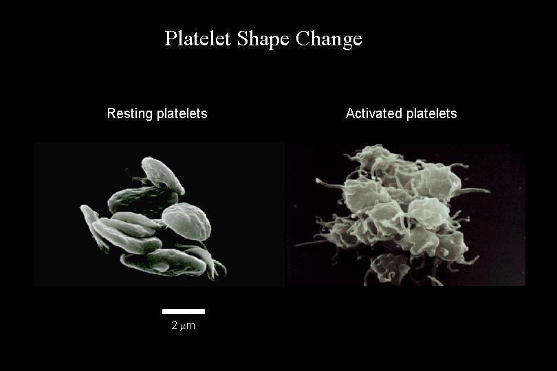
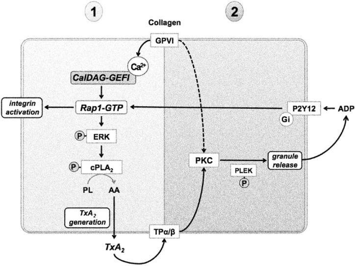
74 Introduction to platelets Platelet function
 -granule Dense granule VWF Aggregation IIb 3 Fibrinogen 2")
75 Introduction to platelets Adhesion (transient tethering) GPIb complex GPVI GPCR Activation (secretion) -granule Dense granule VWF Aggregation IIb 3 Fibrinogen 2 1


76 Introduction to platelets

77 The importance of rate Blood Sep 1;112(5): Blood : Blood Jan 20;117(3):


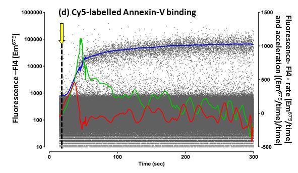
78 The assay 75μl agonist Ca2+ 37 o C 37 o C 225μl Citrated Whole blood (1:100) + Fluo4-NW or FITC-anti-fibrinogen PE-anti-P-selectin Cy5-annexin V no probenicid dialysed
79 The assay FITC-anti-CD42b PE-anti-P-selectin FITC-anti-fibrinogen PE-anti-P-selectin Cy5-annexin V Fluo4-NW

80 The assay FITC-anti-fibrinogen PE-anti-P-selectin Cy5-annexin V
81 Analysis using R FSC-A SSC-A FL1-A FL2-A FL3-A FL4-A FSC-H SSC-H FL1-H FL2-H FL3-H FL4-H Width Time
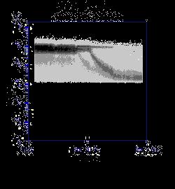
 FL1A (log10 median.ms -1.ms -1 ) Acceleration of LOESS curve Time (100ms)")
82 FL1A (log10 median) Analysis using R Rate of change of LOESS curve Loess curve of median data Raw data Median fluorescence per 100ms FL1A (log10 median.ms -1 ) FL1A (log10 median.ms -1.ms -1 ) Acceleration of LOESS curve Time (100ms)

83 Analysis using R

84 Is the assay robust?
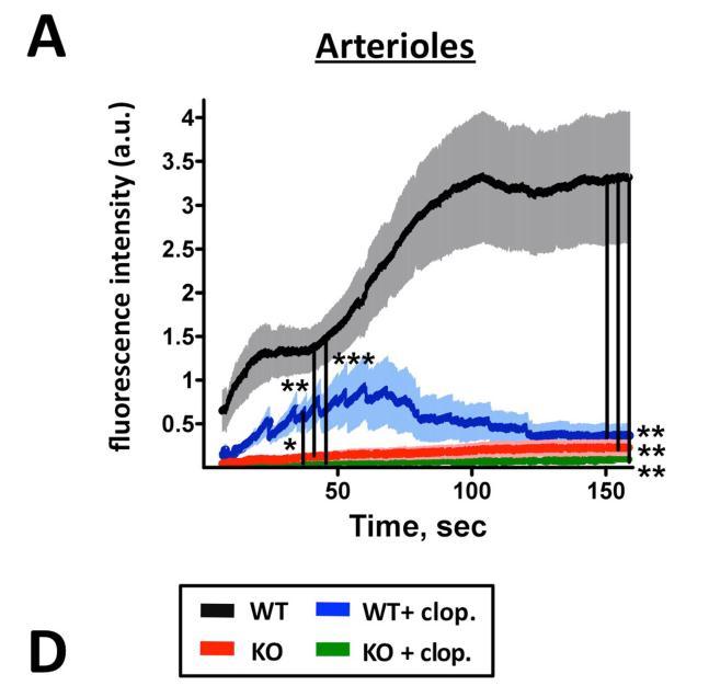
85 Is rate independent of maximum response?
86
87 Is rate independent of maximum response? Integrin-linked kinase regulates the rate of platelet activation and is essential for the formation of stable thrombi. Jones CI, Tucker KL, Sasikumar P, Sage T, Kaiser WJ, Moore C, Emerson M, Gibbins JM. J Thromb Haemost Aug;12(8): doi: /jth Epub 2014 Jul 31.
88 Summary Real time assay for the measurement the calcium flux or fibrinogen binding, P-selectin expression and Annexin V binding of populations of individual platelets Analysis methodology capable of efficiently extracting appropriate data for large studies or identifying interesting subpopulations

89 Acknowledgements Prof. Jon Gibbins Steve Garner This work was funded by the British Heart Foundation