Potential ophthalmological uses of the FEL
|
|
|
- Felix Pitts
- 5 years ago
- Views:
Transcription
1 Potential ophthalmological uses of the FEL Frank Lattanzio Basic Science Director T.R. Lee Center for Ocular Pharmacology Eastern Virginia Medical School Norfolk, VA

2 FEL-multiple wavelengths on demand Laser Focus World. Sept., 2001, pg. 93


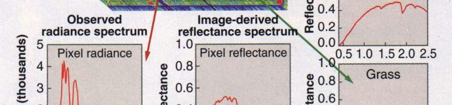
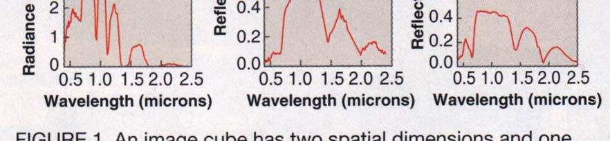
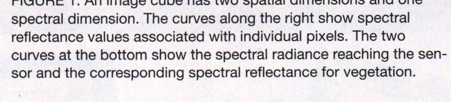
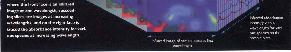
3 Transfer of satellite imaging technology Laser Focus World., May 2004, pg 76
4 Benefits and limitations of the eye as a research model +Made to accommodate entry of light with many of its internal components visible and reasonably accessible +Demonstrates a wide variety of responses to local and systemic pathologies - Vital organ, susceptible to photodamage, ischemia, etc, with limited cellular regeneration - Certain aspects of ocular functionality not easily tested in tissue culture/animal models
5 Please don t look at the laser with your remaining eye
6 Free Electron Laser (FEL) Applications for Ophthalmology The T.R. Lee Center for Ocular Pharmacology is evaluating topical photodynamic (PDT) agents for treatment of pterygium, corneal neovascularization (CNV) and macular degeneration, as well as dermatological cancer models.

, as well as tumor cell disruption CNV")
7 Free Electron Laser (FEL) Applications for Ophthalmology PDT agents permit ablation of blood vessels that compromise corneal clarity (pterygium, CNV) or retinal cell function (macular degeneration), as well as tumor cell disruption CNV Pterygium
8 Benefits of topical preparations of photodynamic agents Reduced systemic toxicity Improved patient compliance/recovery Reduced amounts of drug required
9 Potential uses of FEL Macular degeneration, CNV, Pterygium, Cancer Photodynamic therapy Wide spectrum light source for testing PTD agents and regimens
10 Current photodynamic agents have been designed around existing lasers Development of novel PDT agents would be accelerated by the availability of nonstandard laser light from UV to IR which can be provided by the FEL and its ability to irradiate large numbers of animals for PDT screening
11 Test models Rabbit CNV Rat corneal transplant Primate/human corneal transplant In vitro, in vivo human
12 Potential uses of FEL Uveal cancer Detection TeraHz, N(IR) Corneal transplant Detection of rejection TeraHz, N(IR)

13 Uveal melanoma


14 Hyperspectral scan of chicken tumor Biophotonics Intern. May, 2004


15 TeraHz imaging of basal cell tumor R&D, Sept. 2003, pg. 28

16 Confocal Microscope Uses spinning Nipkow disk to create a 5-7 um optical section Patient s eye is anesthetized and applanated Video data recorded on VCR or digital files (tif, jpeg, etc)
17 TSCM - in vivo Tandem Scanning Confocal Microscopy Furrer et al, J Ocular Pharm & Therapeutics, 1997; 13: 561


18 Cornea Transplant Rejection Normal Rat Corneal Stroma 3 Day Rat Corneal Transplant-Stroma


19 Uveitis Normal Rabbit Endothelium Uveitis-Rabbit Endothelium

20 AC Fibrin & Leukocytes 50 μm

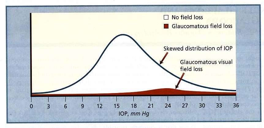
21 ICE Syndrome: Confocal Thickened corneal endothelial cells impair vision in glaucoma patients
22 Free Electron Laser (FEL) Applications for Ophthalmology The T.R. Lee Center has also developed a novel group of free radical scavengers that are known radioprotectants. These agents have a low molecular weight methoxypolyethylene glycol (MPEG) backbone that allows entry of the compounds into cells.
23 Free Electron Laser (FEL) Applications for Ophthalmology These agents can be formulated with different side groups, allowing them to chelate heavy metals responsible for free radical generation, as well as present reducing groups to sustain the cell against ischemic damage. Cellular penetration can be enhanced by the formation of MPEG esters. These agents have therefore potential for treating the free radical component of such ocular conditions as glaucoma, cataracts and macular degeneration, as well as heart attack, stroke and Alzheimer s.
24 MPEG-methoxypolyethylene glycol)
25 Creation of esters to permit ready entry of MPEG derivatives into cells
26 Some MPEG based chelators
27 MPEG attributes Ability to concentrate agent around cellular DNA Selectively protects against radiation damage to normal cells Reduces potential sources of free radicals, as well as detoxifying endogenous free radicals


28 Aqueous humor flow CB cilary body R1 trab/schlemm s R2 Uveoscleral R3 Sclera Pv IOP, episcleral vein
29
30 Glaucoma Test models Rabbit Rat episcleral vein ligation In vitro, in vivo human
31 D ose R esponse of M PD TE 4 2 Change in IOP (mmhg) T 0 T 3 0 T 6 0 T 9 0 T T T T im e (M in.) 3 0 m M M P D T E 8 7 m M M P D T E 1 0 m M M P D T E 3 m M M P D T E 1 m M M P D T E 0.3 m M M P D T E
32 E ffe c t o f d iffe re n t M P E G c o m p o u n d s a t 3 0 m M 4 2 Change in IOP (mmhg) T0 T30 T60 T90 T120 T im e (m in ) MPDTE AVG MPEDE AVG MPIDE AVG MPCDE AVG MPSEDE AVG
33 Am ifostine,a clinical effective free radical scavenger does not low er IOP 6 4 Change in IOP (mmhg) T0 T30 T60 T90 T120 T im e (M in.) 10 m M M PDTE 1 0 m M A m ifo s tin e 1% W IN U n tre a te d e ye 8 7 m M A m ifo s tin e
34 Electroretinogram basics B wave=rods A wave = cones
35 A Wave amplitude Arbitrary units Control Week 0 Weeks after NMDA Week 2 NMDA NMDA +MPDTE NMDA + MPSEDE
36 B Wave amplitude Arbitrary units Control Week 0 Weeks after NMDA NMDA NMDA +MPDTE NMDA + MPSEDE Week 2
37 Potential uses of FEL Glaucoma Shunt and valve function and integration into tissue Structural alterations affecting aqueous humor flow Drug depots and long term drug release Loss of retinal ganglionic cells, measurement of retinal pigments Metabolic changes, production of free radicals, ischemia TeraHz TeraHz TeraHz, N(IR) TeraHz, N(IR) TeraHz, UV-N(IR)


38 TeraHz time domain spectroscopy Industrial Phys., Aug/Sept. 2003, pg 28


39 TeraHz detection of tooth decay Industrial Phys., Aug/Sept. 2003, pg 28

40 Cataract formation UV and cosmic radiation Drugs Trauma Diabetes, Down s, Glaucoma, etc.
41 Current limitations of cataract detection and treatment Slit lamp detects frank cataract formation often years after precipitating events Surgical treatment used to replace lens Necessity to follow early events to monitor efficacy of interventions as well as to track the pathology of disease process
42 Cataract Test models Mouse X-ray, UV, chemical Rat X-ray, UV, chemical In vitro, in vivo human
43 Free Electron Laser (FEL) Applications for Ophthalmology The FEL can be used to generate ultraviolet radiation damage models to test the efficacy of MPEG agents to protect against cataract formation and UV skin damage.
44 Free Electron Laser (FEL) Applications for Ophthalmology The FEL can be used to develop (N)IR and terahertz radiation imaging technology which can be used to detect changes in the hydration of the lens that occur during cataract formation, as well as the presence of tumors in the skin and body, changes in retinal pigments that indicate retinal dysfunction and myloid plaques (Alzheimer s disease).
45 Potential uses of FEL Cataracts Creation of animal models Early detection of capsule and lens changes Detection of IOL rejection UVA, UVB radiation TeraHz, N(IR) TeraHz, N(IR)


46 Ocular Hb oxygenation detection Biophotonics Inter.., May 2004

47 Measurement of Biochemical Parameters Conn, Methods in Enzymology, 1999;307: 540
48 Raman Spectroscopy Pharmacokinetic studies In vivo drug measurements and metabolite levels Localization of drugs diffusion and compartmentalization

49 Biophotonics International, 2001: 48

50 Biophotonics News, 2001: 28


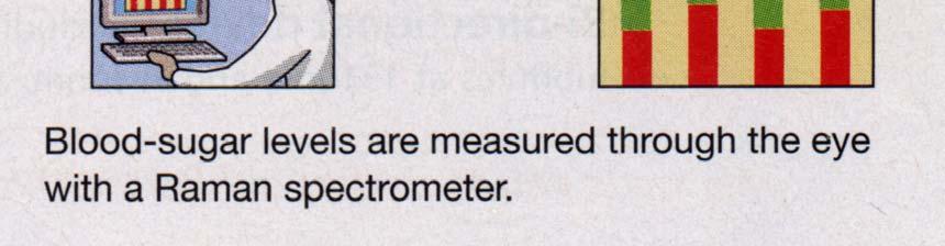
51 Raman Ocular Glucose detector Optoelectronics

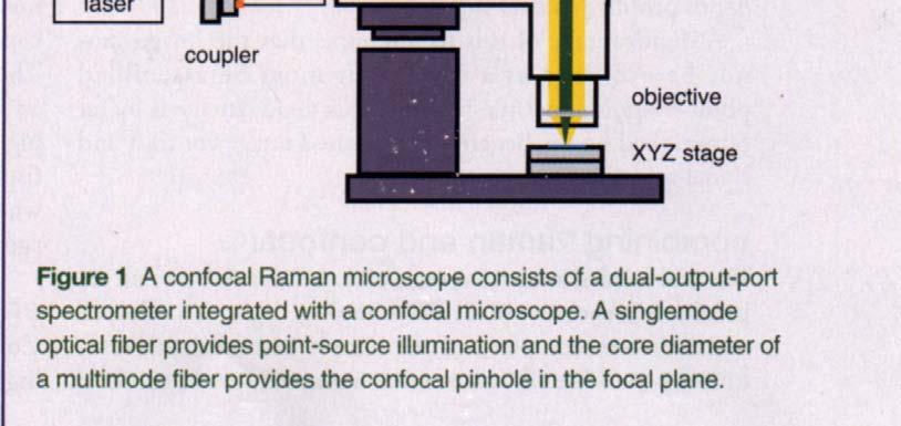
52 Confocal raman microscope OptronicsWorld.


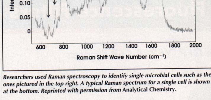
53 Single cell Raman scan Biophotonics Intern., Oct
54 Age related macular degeneration Major cause of vision loss Interventions include PDT, drugs, etc. Interventions have limited efficacy on this progressive disease Future interventions for retinal repair/replacement via stem cells, artificial retina

55 Dresen on retina of AMD patient
56 AMD, Diabetes, Dry eye test models Mouse Rat In vitro, in vivo human
57 Potential uses of FEL Macular degeneration Dry eye Diabetes Detection of dresen, retinal pigments, drug depot function Progressive corneal surface changes Detection of corneal and retinal changes TeraHz, N(IR) TeraHz TeraHz, visible-n(ir)

58 T.R. Lee Center team Patricia Williams, PhD Director Earl Crouch, MD Chairman Ophthalmology John Sheppard, MD, MMSc Clinical Director Frank Lattanzio, PhD Basic Science Director
59 The Beginning
60 Schematic of imaging spectrometer Laser Focus World., May 2004, pg 77

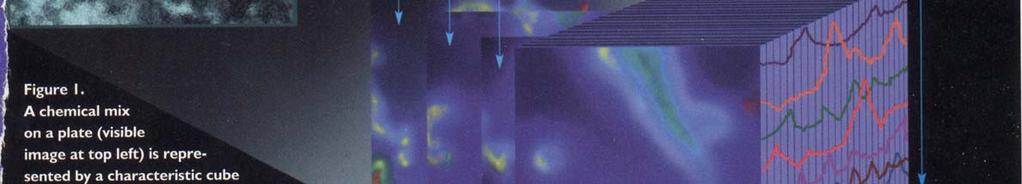
61 IR cell chemical scan Industrial Phys., Oct/Nov. 2003, pg 29

62 Biophotonics International, 2001: 47