ChemiDoc MP Imaging System
|
|
|
- Alannah Horton
- 5 years ago
- Views:
Transcription

1 Imaging ChemiDoc MP Imaging System Confidence in every step of your imaging workflow.

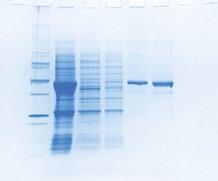
2 ChemiDoc MP Imaging System Because results always matter. Bring a new level of capability and efficiency to your experiments with an imaging system designed for multitasking. The ChemiDoc MP imager is a unique imaging system that enables stain-free operation and lets you visualize proteins at every stage of your blotting experiment. Its flexibility and sensitivity are complemented by simple, intuitive operation that integrates seamlessly into your workflow.

3


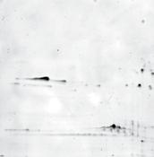
4 Superior Sensitivity Get quantitative, reproducible data without relying on outmoded film processes. The ChemiDoc MP imaging system offers advanced detection technology that creates optimal exposure for even the faintest bands. Rely on it for fast, super-sensitive chemiluminescence and fluorescence detection and for colorimetric gel and blot documentation. Fig. 1. Sensitivity comparison of the ChemiDoc MP system versus X-ray film using blots of serial dilution of transferrin. A, the ChemiDoc imager delivers superior dynamic range and comparable limit of detection to film. B, a 10-second exposure on film reveals a more limited dynamic range than the ChemiDoc MP system. Saturated pixels are highlighted in red. Signal-to-noise ratio Signal-to-noise ratio of transferrin. 10 sec exposure Sample load, ng ChemiDoc MP Imaging System Film Fig. 1A Fig. 1B Film Sample load, ng ChemiDoc MP imaging system Bio-Rad Laboratories, Inc.


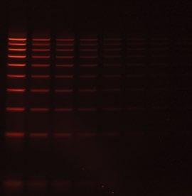
5 superior Sensitivity Signal-to-noise ratio Signal-to-noise ratio of LEDs from a calibrated luminescent test plate. 5 min exposure Fig. 2. A calibrated luminescent target was used to test light collection efficiency and determine overall system sensitivity. A, data from lower limits of detection are graphed to show overall signal-to-noise ratio. The ChemiDoc MP system delivers top-of-the-class chemiluminescence sensitivity against leading multiplexing imagers on the market. B, images of lower limits of detection from a calibrated luminescent target. The ChemiDoc MP system delivers excellent image quality and limit of detection LED well ChemiDoc MP Imaging System Competitor 1 Competitor 2 Competitor 3 Fig. 2A Fig. 2B ChemiDoc MP Competitor Competitor 1 Competitor 3 ChemiDoc MP Imaging System


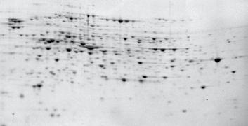
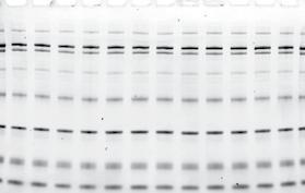
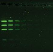
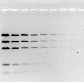
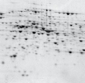
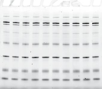
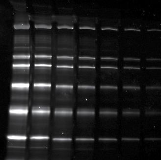
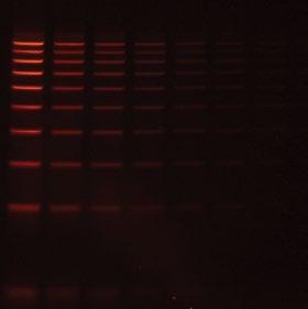
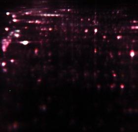
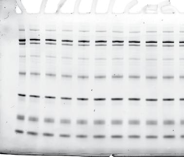
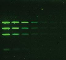
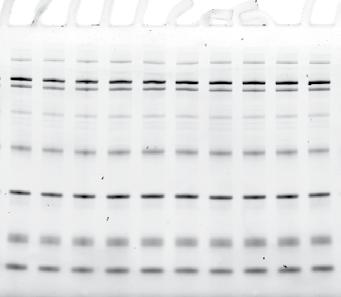
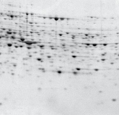
6 Exceptional Image Quality With patented focus calibration technology, images are always in focus at any zoom level. Exceptional dynamic range enables visualization of faint and intense bands on same blot or gel. With Image Lab software you can edit and analyze images on the spot without exporting to other programs. Fig D Coomassie-stained gel. Fig. 4. Chemiluminescent ELISA arrays. Quansys Biosciences Q-Plex array has 16 distinct capture antibodies bound to each well of a 96-well plate. Fig. 5. Fluorescent multiplex blot with DyLight 488, DyLight 549, and DyLight 649 conjugates. Fig. 3 Fig. 4 Fig. 5 Bio-Rad Laboratories, Inc.


7 exceptional Image Quality Minimal cross-talk between blue and green channels. Fluors detected in green channel Fluors detected in blue channel ChemiDoc MP Competitor 1 Cross-talk FAM A % FAM A488 Correct Cy3 A % 100% Cy3 A546 Correct FAM A % 100% FAM A488 Cross-talk Cy3 A % 0.0% Cy3 A546 Fig. 6A. Determination of cross-talk using same multiplex gel. Blue fluors (FAM and Alexa Fluor 488) are detected in the green channel and green fluors (Cy3 and Alexa Fluor 546) in the blue channel using the ChemiDoc MP system and a competitor s system. For competitor 1, the bold percentage values indicate high cross-talk. 12.4% 7.6% 100% 100% 100% 100% 0.2% 0.0% Fig. 6A Competitor 2 Fig. 6B FAM 32.5% A % Cy3 100% A % FAM 100% A % Cy3 9.7% A % Fig. 6B. Visualization of cross-talk signals recorded with a second competitor s instrument. The green and blue channel images show high cross-talk values and are indicated in bold. The blue fluors (FAM and Alexa Fluor 488) are detected in the green channel and the green fluors (Cy3 and Alexa Fluor 546) are detected in the blue channel. Cross-talk Correct Correct Cross-talk FAM A488 Cy3 A546 FAM A488 Cy3 A546 ChemiDoc MP Imaging System


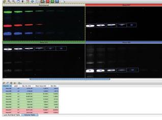
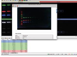
8 Unmatched Application Versatility The ChemiDoc MP imager is the only system you need when your experiments include a variety of sample types or require different detection methods. It is the perfect imager to accompany your protein and DNA electrophoresis runs as well as your western blotting experiments. And it delivers quantitative, reproducible results every time. Fig. 7. Multiplex image file allows multichannel and individual channel view. Image of multiplexed fluorescent blot with Alexa Fluor 488, Alexa Fluor 555, and Alexa Fluor 647. Bio-Rad Laboratories, Inc.

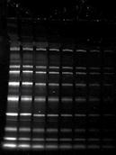


9 unmatched application versatility Fig. 8. Multiple applications of the ChemiDoc MP Imager. DIGE Criterion gel EtBr-stained wide mini ReadyAgarose gel GelRed-stained mini ReadyAgarose gel Silver-stained 2-D Criterion gel Coomassie-stained Mini-PROTEAN TGX gel SYPRO Ruby-stained large-format 2-D gel Flamingo -stained Mini-PROTEAN TGX gel Criterion Stain Free TGX gel SYBR Green-stained wide mini ReadyAgarose gel ChemiDoc MP Imaging System


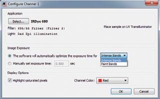
10 Ease of Use The ChemiDoc MP system is designed for productivity. With little or no training, users can acquire publication-quality images in seconds. The system is precalibrated to provide the precise focus for any zoom setting or sample height; automated hands-free operation ensures consistent, reproducible, and high-throughput performance. Fig. 9A. The application selected automatically determines the excitation sources and emission filters. Fig. 9B. The ChemiDoc MP system is always in focus at any zoom level no more out-of-focus gel or blot images. Bio-Rad Laboratories, Inc.


11 Ease of use Autosetup Autofocus Autoexposure Autooverlay Autoanalysis Autoreports Autoprint ChemiDoc MP Imaging System


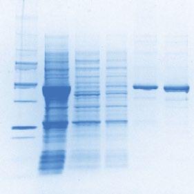
12 Stain-Free Enabled Stain-free technology, offered only by Bio-Rad, eliminates extra steps and unproductive delays in your western blotting experiments. Stain-free technology is the keystone of the Bio-Rad V3 Western Workflow, a portfolio of products that enables researchers to visualize, verify, and validate results at each step of their western blotting experiment. V3 Stain-free gel Stain-free blot Total protein quantification Protein of interest probed with Alexa Fluor 649 Visualize Verify Validate The ChemiDoc MP system will activate and provide immediate visualization of protein separation in all lanes with stain-free gels before blot transfer. The ChemiDoc MP paired with stain-free technology enables instant verification of protein transfer before blot detection. Flexible Image Lab software tools normalize for protein of interest using total protein stain from stain-free blot and provide quantitative blot results. Bio-Rad Laboratories, Inc.
 such as b-tubulin.")

13 Stain-free enabled Stain-Free Normalization: An Alternative to Housekeeping Proteins Traditionally, normalization of western blot signals is performed by probing the membrane with antibodies against a housekeeping protein (HKP) such as b-tubulin. This process usually requires stripping the first antibodies from the membrane prior to reprobing with the HKP antibodies, or performing tedious multiplex immunodetections. Stain-free technology allows you to normalize your western blot data by directly quantifying the total amount of protein bound to the membrane. Benefits of Total Protein Normalization Stain-free normalization is consistently reliable normalization using housekeeping protein (HKP) expression may give different results based on experimental conditions and sample type Stain-free normalization saves time quantitation of total membrane-bound protein using stain-free technology takes only a few minutes Stain-free normalization is a single-step procedure prior probing of the membrane does not affect stain-free total protein detection; there is no need to strip the membrane Tubulin Stain-free total protein 6.0 x 10 7 R 2 = x 10 7 R 2 = Tubulin signal intensity 5.0 x x x x x 10 7 Total protein signal intensity 8 x x x x Protein load, µg Protein load, µg Plot of signal intensity versus protein load. Normalization using total lane signal intensity from stain-free technology exhibits comparable linearity to normalization using the tubulin signal Protein load, µg Protein load, µg A blot of serially diluted untreated HEK293 lysate was probed with an anti-tubulin antibody using Alexa Fluor 649 for the detection of tubulin. The same blot of serially diluted untreated HEK293 lysate was used for total protein detection and quantitation using stain-free technology. ChemiDoc MP Imaging System


14 Instrument Tour Optimized emission and excitation filters for discrete detection of multiplexed visible fluorescence. Red Automatic detection of red fluorescence using dyes such as Cy5, Cy5.5, Alexa Fluor 647, Alexa Fluor 680, DyLight 649, DyLight 680, IRDye 680. Green Automatic detection of green fluorescence using dyes such as Cy3, Flamingo, Krypton, Pro-Q Diamond, Alexa Fluor 546, DyLight 549, Rhodamine. Blue Automatic detection of blue fluorescence using dyes such as Cy2, Coomassie Fluor Orange, Alexa Fluor 488, DyLight 488, Pro-Q Emerald 488, Qdot 523, Qdot 605, Qdot 625, Qdot 705. Bio-Rad Laboratories, Inc.


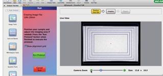
15 Instrument tour Cooled CCD with autofocus for all zoom levels Unmatched sensitivity and image quality Multiplexed fluorescent imaging Multicolor LEDs optimized for quantitative western blot imaging 6-position automated filter wheel Broad application flexibility Sample tray with UV, white, and blue imaging Flexible transillumination source for easy and uniform imaging of gels and blots Touch-pad operation For positioning and band excision Go to to see the complete ChemiDoc MP video. ChemiDoc MP Imaging System

16 Specifications Automation Capabilities Workflow automated selection Workflow automated execution Workflow reproducibility Autofocus (patent pending) Application driven; user selected or recalled by a protocol Controlled by a protocol via application-specific setup for image area, illumination source, filter, analysis, and reporting 100% repeatability via recallable protocols; from image capture to quantitative analysis and reports Precalibrated focus for any zoom setting or sample height Image flat fielding* Dynamic; precalibrated and optimized for every application Autoexposure 2 user-defined modes (intense or faint bands) Hardware specifications Maximum sample size (L x W) 28 x 36 cm Maximum image area (L x W) 26 x 35 cm Maximum image area for 25 x 26 cm standard UV-excited gels (L x W) Excitation source Trans-UV (302 nm included; 254 nm and 365 nm available as options) and epi-white; optional trans-white conversion screen, XcitaBlue conversion screen, and epi-red, -green, and -blue LED modules Illumination control 8 modes available. Trans-UV, epi-white, and no illumination for chemiluminescence are standard; epi-red, epi-green, epi-blue, transwhite, and XcitaBlue conversion screens are optional Detector Supercooled CCD Image resolution 4 megapixels Pixel size (H x V) 6.45 x 6.45 µm Cooling system Peltier Camera cooling temperature 30 C absolute and regulated Filter holder 6 positions (5 for filters, 1 without filter for chemiluminescence) Emission filters 1 included (standard), 4 optional Dynamic range >4.0 orders of magnitude Pixel density (gray levels) 65,535 Instrument size (L x W x H) 36 x 60 x 96 cm Instrument weight 32 kg Operating Ranges Operating voltage 110/115/230 VAC nominal Operating temperature C (21 C recommended) Operating humidity <70% noncondensing * U.S. patent 5,951,838. Ordering Information Catalog # Description ChemiDoc MP Imaging System with Image Lab Software, PC or Mac, includes darkroom, UV transilluminator, epi-white illumination, camera, power supply, cables, Image Lab software Accessories Red LED Molecular Kit, pkg of 2 epi-red LED modules and 1 red emission filter, for use with applications requiring red fluorophore detection Green LED Molecular Kit, pkg of 2 epi-green LED modules and 1 green emission filter, for use with applications requiring green fluorophore detection Blue LED Molecular Kit, pkg of 2 epi-blue LED modules and 1 blue emission filter, for use with applications requiring blue fluorophore detection White Light Conversion Screen, for viewing Coomassie, silver stain, and other colorimetric gel stains XcitaBlue Conversion Screen, includes view goggles; blue conversion screen for viewing SYBR Green, SYBR Safe, GFP, Flamingo, and other fluorescent gel stains XcitaBlue Conversion Screen and Filter, includes view goggles and SYBR Safe filter ( , 560DF50); blue conversion screen for viewing SYBR Green, SYBR Safe, and other fluorescent gel stains Filter 520DF30 62 mm, for SYBR Green/GFP/SYBR Gold/fluorescein nm UV Lamps, pkg of nm UV Lamps, pkg of Standard 302 nm UV Lamps, pkg of Mitsubishi Thermal Printer Mitsubishi Thermal Printer Paper, 4 rolls Gel Alignment Templates, pkg of 3 Software * Image Lab Software, PC or Mac, for automated image capture, optimization, and 1-D data analysis * Included with the imaging system. Scan this QR code to learn more about the ChemiDoc MP imaging system, or visit Alexa Fluor, Coomassie Fluor Orange, Pro-Q, Qdot, SYBR, and SYPRO are trademarks of Life Technologies Corporation. Coomassie is a trademark of BASF Aktiengesellschaft. Cy is a trademark of GE Healthcare Group Companies. DyLight and Krypton are trademarks of Thermo Fisher Scientific. GelRed is a trademark of Biotium, Inc. IRDye is a trademark of LI-COR Biosciences. Mac is a trademark of Apple Inc. Mitsubishi is a trademark of Mitsubishi Companies. Q-Plex is a trademark of Quansys Biosciences. Bio-Rad Laboratories, Inc. is licensed by Life Technologies Corporation to sell SYPRO products for research use only, under U.S. patent 5,616,502. Bio-Rad Laboratories, Inc. Life Science Group Web site USA Australia Austria Belgium Brazil Canada China Czech Republic Denmark Finland France Germany Greece Hong Kong Hungary India Israel Italy Japan Korea Mexico The Netherlands New Zealand Norway Poland Portugal Russia Singapore South Africa Spain Sweden Switzerland Taiwan Thailand United Kingdom Bulletin 6133 Rev C US/EG Sig 1211