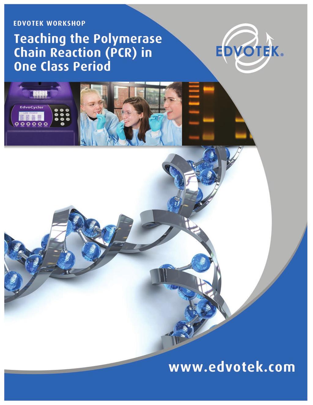
Introduction. Table of Contents Page. Background Information Excerpts from EDVO-Kit 372
|
|
|
- Madison Lawson
- 5 years ago
- Views:
Transcription
1
2 Teaching the Polymerase Chain Reaction (PCR) in One Class Period EDVOTEK WORKSHOP Introduction In this experiment, students will gain an understanding of the traditional three-step Polymerase Chain Reaction (PCR). Using PCR and Agarose Gel Electrophoresis, they will analyze a small section of Lambda DNA in a timesaving two-step process. Table of Contents Page Background Information 2 Experiment Overview 5 Experiment Procedures 6 Experiment Results and Analysis 12 Background Information Excerpts from EDVO-Kit 372 THE POLYMERASE CHAIN REACTION In 1984, Dr. Kary Mullis revolutionized the field of molecular biology when he devised a simple and elegant method to copy specific pieces of DNA. Mullis recognized that he could replicate DNA in vitro using short, synthetic DNA oligonucleotides (known as primers) and DNA polymerase I in a process similar to DNA replication in a cell s nucleus. Because researchers can customize the primers to target a specific gene, this method allows for the rapid amplification of a selected DNA sequence. For the development of this technique, known today as the Polymerase Chain Reaction (or PCR), Mullis was awarded the Nobel Prize in Chemistry in Before performing PCR, template DNA is extracted from a biological sample. Two primers are designed to correspond to the 5 and 3 ends of the target sequence. The template DNA and primers are mixed with buffer, the four free deoxynucleotides (datp, dctp, dgtp, and dttp), and a thermostable DNA polymerase (Taq). Next, the PCR mixture is subjected to sequential heating/cooling cycles at three different temperatures to amplify DNA. In the first step, known as denaturation, the mixture is heated to 94 C to disrupt the hydrogen bonds between the complementarity strands. This causes the target DNA to unzip into single strands (or melt). It is important to use a thermostable DNA polymerase for PCR because this enzyme remains stable at high temperatures. In the second step, known as annealing, the reaction mixture is cooled to 45 C - 65 C. This allows the primers to base pair with the target DNA sequence. In the third step, known as extension, the temperature is raised to 72 C. This temperature is optimal for Taq polymerase to add nucleotides to the 3 end of the primer, synthesizing a new strand of DNA. Together, these three steps - denaturation, annealing, and extension make up one PCR cycle (Figure 1). To simplify this process, a specialized machine, called a thermal cycler or a PCR machine, was created to heat and cool the samples rapidly. Each PCR cycle doubles the amount of the target DNA in less than five minutes. This makes PCR a very sensitive technique, as only a few copies of the template DNA are required to produce a large amount of signal. Mathematically, PCR is described as an exponential relationship if we begin with a starting copy number of m, then after n cycles, we will have m x 2 n copies of our DNA target. For example, if we start with one copy of our target, we will have two copies after the first PCR cycle, four after the second PCR cycle, eight after the third PCR cycle, 2 VISIT for complete experiment details & free student protocols.
3 EDVOTEK WORKSHOP Teaching the Polymerase Chain Reaction (PCR) in One Class Period Target Sequence = Separation of two DNA strands = Primer 1 = Primer 2 Denature 94 C Cycle 1 Anneal 2 primers 40 C - 65 C Extension 72 C Cycle 2 Cycle 3 Figure 1: The Polymerase Chain Reaction (PCR) - Three Cycles CONTACT US? CALL EDVOTEK Fax info@edvotek.com 3
4 Teaching the Polymerase Chain Reaction (PCR) in One Class Period EDVOTEK WORKSHOP and so on. In numbers, cycle 1 equals 1x2 1, cycle 2 equals 1x2 2, cycle 3 equals 1x2 3. After n cycles, we will have 1x2 n copies of our DNA target. In order to produce enough DNA for analysis, twenty to forty cycles may be required. After many cycles (regardless of the quantity of DNA present in the starting material) the amount of DNA produced reaches a maximum amount of product known as the plateau. This is due to depletion of reaction components like primers and nucleotides and the loss of Taq polymerase activity. Because of its ease of use and its ability to amplify DNA rapidly, PCR has become indispensable in medical and life sciences labs, replacing the time-intensive Southern blot as the method of choice. For example, today s research laboratories can quickly create copies of a specific region of DNA for cloning applications. Medical diagnostics use PCR to identify genetic mutations and infectious agents. In addition, because amplification by PCR requires a small amount of starting material, it is ideal for forensic analysis of biological samples or determination of paternity. REINVENTING PCR While PCR is relatively fast and easy compared to techniques like the Southern blot, it still takes several hours to complete the experiment. In response, researchers have devised several strategies to reduce the time necessary to amplify a specific sequence. One timesaving strategy involves designing the primers so that the annealing temperature and the extension temperature are very close. This allows researchers to combine the annealing and extension steps of the traditional PCR cycle. Another timesaving strategy involves reducing the time spent at each temperature. By modifying the PCR program, researchers could reduce the length of each cycle from seconds to 60 seconds or less (Table 1). These changes reduce the time required for this experiment by over 50%. Table 1: Comparison of Traditional and Quick PCR Denaturation (95 C) Annealing (40 C - 60 C) Extension (72 C) TOTAL TIME (30 cycles) Traditional PCR 45s 45s 45s ~70 minutes Quick PCR 15s 0s 15s ~30 minutes In this exploration, we will use quick PCR to analyze genomic DNA isolated from a virus that infects E.coli, known as bacteriophage lambda. Historically, lambda is an important virus for molecular biology. Early studies of the lambda genome contributed to our understanding of DNA replication, transcription, and translation. The 48,500 base pair genome contains information necessary for the virus s entry into the cell, production of new virions, and lysis of the host cell (Figure 2). The primers used in this experiment have been designed to amplify a 500 base pair region of a viral capsid protein. They are engineered to have an annealing temperature of 71 C, which is close to the optimum temperature for Taq s DNA polymerase activity. This allows us to combine the annealing and extension steps of PCR. As a result, the entire amplification can be performed in about thirty minutes, allowing your students to perform PCR in a single lab period. Immunity Head Tail Regulation Regulation Recombination Replication Lysis Kilobases Figure 2: Lambda Phage Map 4 VISIT for complete experiment details & free student protocols.

5 EDVOTEK WORKSHOP Teaching the Polymerase Chain Reaction (PCR) in One Class Period Experiment Overview LABORATORY SAFETY 1. Gloves and goggles should be worn routinely as good laboratory practice. 2. Exercise extreme caution when working with equipment which is used in conjunction with the heating and/or melting of reagents. 3. DO NOT MOUTH PIPET REAGENTS - USE PIPET PUMPS. 4. Exercise caution when using any electrical equipment in the laboratory. Turn off power and then unplug the equipment when not in use. Electrical current from the power source is automatically disrupted when the electrophoresis cover is removed from the apparatus on all EDVOTEK models. However, exercise caution when working with electrophoresis equipment. After electrophoresis is completed, turn off the power, then unplug the power source before disconnecting the leads and removing the cover. 5. EDVOTEK injection-molded electrophoresis units do not have glued junctions that can develop potential leaks. However, in the unlikely event that a leak develops in any electrophoresis apparatus you are using, IMMEDIATELY SHUT OFF POWER. Do not use the apparatus. 6. Always wash hands thoroughly with soap and water after handling reagents or biological materials in the laboratory. Module I - 25 min. Amplification of Lambda DNA Module II min. Separation of PCR Products by Electrophoresis GEL SPECIFICATIONS This experiment requires a gel with the following specifications: Recommended Gel Size: 7 x 7 cm Number of Samples Wells: 6 Placement of the Well-former Template: First set of notches Gel Concentration Required: 1.0% Module III min. Staining with FlashBlue Stain (OPTIONAL) NOTE: Experimental times are approximate. Online Resources Here at EDVOTEK, we ve created Quick Guide manuals, FREE for you to download off our website. We have also filmed several Instructional Videos that show step-by-step procedures. We hope you take advantage of these resources and enjoy teaching and learning with EDVOTEK! Tech Video CONTACT US? CALL EDVOTEK Fax info@edvotek.com 5

 Program Thermal Cycler One day to 30 min. before performing the experiment. One hour before performing the experiment. 30 min. 15 min.")
6 Teaching the Polymerase Chain Reaction (PCR) in One Class Period EDVOTEK WORKSHOP Overview of PreLab Preparation This section outlines the recommended prelab preparations and approximate time requirement to complete each prelab activity. Preparation For: What to do: When: Time Required: Module I: Amplification of Lambda DNA Prepare and aliquot various reagents (Primer, DNA template, ladder, etc.) Program Thermal Cycler One day to 30 min. before performing the experiment. One hour before performing the experiment. 30 min. 15 min. Module II: Separation of PCR Product by Electrophoresis Prepare diluted electrophoresis buffer and SYBR Safe Stain. Prepare molten agarose and pour gel Up to one day before performing the experiment. 45 min. Module III: Staining with FlashBlue (OPTIONAL) Prepare staining components The class period or overnight after the class period. 10 min. 6 VISIT for complete experiment details & free student protocols.
7 EDVOTEK WORKSHOP Teaching the Polymerase Chain Reaction (PCR) in One Class Period Module I: Amplification of Lambda DNA TC SPIN µl Primer Mix 5 µl DNA Template PCR EdvoBead Gently mix REMINDER: At least one negative control should be performed per class. To prepare the control sample, add 20 µl Primer Mix and 5 µl Lambda DNA template to a labeled PCR tube. NO PCR EdvoBead IS ADDED. If your thermal cycler does not have a heated lid, it is necessary to overlay the PCR reaction with wax or mineral oil to prevent evaporation. See for more information. 1. LABEL a PCR tube with the sample and your initials 2. ADD 20 µl Primer mix (orange), 5 µl Lambda DNA Template (blue) and one PCR EdvoBead to a labeled PCR tube. 3. MIX the sample by gently flicking the tube. The solution should be pale green in color, and the PCR EdvoBead should be completely dissolved. NOTE: If the solution is not pale green, the PCR sample has not been correctly assembled. 4. CENTRIFUGE the sample for a few seconds to collect the sample at the bottom of the tube. 5. AMPLIFY DNA using PCR. 6. After PCR, ADD 5 μl of 10x Gel Loading Solution to the sample. PLACE tubes on ice. PROCEED to Module II: Separation of PCR Products by Electrophoresis. OPTIONAL STOPPING POINT: The PCR samples may be stored at -20 C for electrophoresis at a later time. CONTACT US? CALL EDVOTEK Fax info@edvotek.com 7
8 Teaching the Polymerase Chain Reaction (PCR) in One Class Period EDVOTEK WORKSHOP Module II: Separation of PCR Products by Agarose Gel Electrophoresis 1: x Concentrated buffer Flask Distilled water Agarose Caution! Flask will be HOT! 60 C ADD SYBR Safe 60 C 60 C POUR WAIT 20 min. PREPARING THE AGAROSE GEL WITH SYBR SAFE STAIN 1. DILUTE the concentrated (50X) electrophoresis buffer with distilled water to create 1X buffer (see Table A). 2. MIX the agarose powder with 1X buffer in a 250 ml flask (see Table A). 3. DISSOLVE the agarose powder by boiling the solution. MICROWAVE the solution on high for 1 minute. Carefully REMOVE the flask from the microwave and MIX by swirling the flask. Continue to HEAT the solution in 15-second bursts until the agarose is completely dissolved (the solution should be clear like water). 4. COOL the agarose to 60 C by carefully swirling the flask to promote even dissipation of heat. 5. While the agarose is cooling, SEAL the ends of the gel-casting tray with the rubber end caps. PLACE the comb in the appropriate notch. 6. Before casting the gel, ADD the diluted SYBR Safe stain to the cooled molten agarose and swirl to mix (see Table A). 7. POUR the cooled agarose solution into the prepared gel-casting tray. The gel should thoroughly solidify within 20 minutes. The gel will stiffen and become less transparent as it solidifies. 8. REMOVE the end caps and comb. Take particular care when removing the comb to prevent damage to the wells. Wear gloves and safety goggles IMPORTANT: 7 x 7 cm gels are recommended. Place the comb in the first set of notches. If you are unfamiliar with agarose gel prep and electrophoresis, detailed instructions and helpful resources are available at Table A Individual 1.0% UltraSpec-Agarose Gel with SYBR Safe Stain Size of Gel Casting tray Concentrated Buffer (50x) + Distilled Water + Amt of Agarose = TOTAL Volume Add SYBR (Step 6) 7 x 7 cm 0.5 ml 24.5 ml 0.25g 25 ml 25 µl 7 x 14 cm 1.0 ml 49.0 ml 0.50 g 50 ml 50 µl 8 VISIT for complete experiment details & free student protocols.

9 EDVOTEK WORKSHOP Teaching the Polymerase Chain Reaction (PCR) in One Class Period Module II: Separation of Digested Products by Agarose Gel Electrophoresis POUR 1X Diluted Buffer. REMINDER: Before loading the samples, make sure the gel is properly oriented in the apparatus chamber Wear gloves and safety goggles RUNNING THE GEL 9. PLACE the gel (on the tray) into the electrophoresis chamber. COVER the gel with 1X electrophoresis buffer (See Table B for recommended volumes). The gel should be completely submerged. 10. LOAD the entire volume (25 µl) into the well in the order indicated by Table 1, right. 11. CHECK that the gel is properly oriented, then PLACE the safety cover onto the chamber. Remember, the DNA samples will migrate toward the positive (red) electrode. 12. CONNECT the leads to the power source and PERFORM Table 2 electrophoresis (See Table C for time and voltage guidelines). Lane Recommended Sample Name 1 EdvoQuick DNA ladder 13. After electrophoresis is complete, REMOVE the gel and 2 Control DNA* casting tray from the electrophoresis chamber. 3 Student Group #1 OPTIONAL STOPPING POINT: Gels can be stored for several days. Place gel in a watertight plastic bag with 2 ml Student Group #2 Student Group #3 Student Group #4 of electrophoresis buffer and store in the * Optional, or additional student group sample. refrigerator. Table B 1x Electrophoresis Buffer (Chamber Buffer) EDVOTEK Model # M6+ & M12 (new) M12 (classic) M36 Total Volume Required 300 ml 400 ml 1000 ml 50x Conc. Buffer 6 ml 8 ml 20 ml Dilution + Distilled Water 294 ml 392 ml 980 ml Table C Volts Time and Voltage Guidelines (1.0% Agarose Gels) Recommended Times Minimum Maximum 15 min. 20 min. 35 min. 60 min. 20 min. 35 min. 60 min. 90 min. CONTACT US? CALL EDVOTEK Fax info@edvotek.com 9

10 Teaching the Polymerase Chain Reaction (PCR) in One Class Period EDVOTEK WORKSHOP Module II: Separation of Digested Products by Agarose Gel Electrophoresis dh2o VISUALIZING THE SYBR GEL 14. SLIDE the gel off the casting tray onto the viewing surface of the transilluminator and turn the unit on. ADJUST the brightness to the desired level to maximize band visualization. DNA should appear as bright green bands on a dark background. 15. PHOTOGRAPH the results. 16. REMOVE and DISPOSE of the gel and CLEAN the transilluminator surfaces with distilled water. Be sure to wear UV goggles if using a UV transilluminator. Related Products Cat. # 557 TruBlue Blue Light Transilluminator The all-new TruBlu Blue Light Transilluminator is ideal for viewing DNA gels stained with SYBR Safe, thus eliminating the need for UV light or ethidium bromide. It s optimized to fit Edvotek Gels as well as any other agarose gel. The high intensity control and orange lid ensure superior visualization. Developed in concert with the inventor of the technology under license from Clare Chemical Research, Inc. Cat. # 608 SYBR Safe DNA Stain Non-mutagenic and SAFE for the Biotechnology Classroom! More sensitive than ethidium bromide. Excellent gel results! Concentrate-for 750 ml 10 VISIT for complete experiment details & free student protocols.
11 EDVOTEK WORKSHOP Teaching the Polymerase Chain Reaction (PCR) in One Class Period Module III: Staining with FlashBlue Stain (OPTIONAL) FlashBlue Stain is a simple and effective visible DNA stain that can be used as an alternative, or in addition to, UV-reactive DNA stains like SYBR Safe. IF staining with both SYBR Safe and Flash Blue, you must examine and record the SYBR Safe bands before beginning the FlashBlue Staining ml 10X FlashBlue Stain 10x DILUTE 90 ml Distilled water 2. SLIDE gel into small gel-staining tray. 3. 1X FlashBlue Stain Solution 2-3 min. STAIN for 2-3 min. 4. POUR FlashBlue back into flask. RINSE for sec. Pour off water. warm water 5. DESTAIN for 5-15 min. warm water 5-15 min. 6. VISUALIZE with white light system. 1. DILUTE 10 ml of 10X concentrated FlashBlue with 90 ml of distilled water in a flask. MIX well. 2. REMOVE the agarose gel and casting tray from the electrophoresis chamber. SLIDE the gel off the casting tray into a small, clean gel-staining tray. 3. COVER the gel with the 1X FlashBlue stain solution. STAIN the gel for 2-3 minutes. For best results, use an orbital shaker to gently agitate the gel while staining. STAINING THE GEL FOR LONGER THAN 3 MINUTES WILL REQUIRE EXTRA DESTAINING TIME. Wear gloves and safety goggles 4. POUR the 1X FlashBlue back into the flask (the stain can be reused). COVER the gel with warm water (40-45 C). Gently RINSE the gel for seconds. POUR off the water. 5. COVER the gel with clean, warm water (40-45 C). DESTAIN for 5-15 minutes with gentle shaking (longer periods will yield better results). DNA bands will start to appear after 5 minutes of destaining. Changing the water frequently will accelerate destaining. 6. Carefully REMOVE the gel from the destaining liquid. VISUALIZE results using a white light visualization system. DNA will appear as dark blue bands on a light blue background. ALTERNATIVE FLASHBLUE STAINING PROTOCOL: 1. DILUTE 1 ml of 10X FlashBlue stain with 499 ml distilled water. 2. COVER the gel with diluted FlashBlue stain. 3. SOAK the gel in the staining liquid for at least three hours. For best results, stain gels overnight. 4. Carefully REMOVE the gel from the staining liquid. VISUALIZE results using a white light visualization system. DNA will appear as dark blue bands on a light blue background. CONTACT US? CALL EDVOTEK Fax info@edvotek.com 11

 Lane 6 Student Group #4 PCR Reaction (20 cycles) Related Products Cat. #372 Quick PCR For 10 Groups.")
12 Teaching the Polymerase Chain Reaction (PCR) in One Class Period EDVOTEK WORKSHOP Experiment Results and Analysis This PCR experiment will amplify a 500 base pair region of a viral capsid protein coded for by the lambda genome. The control reaction sample will not produce a PCR product because it is missing the PCR EdvoBead. Lane 1 EdvoQuick DNA Ladder Lane 2 Optional control reaction sample (no PCR EdvoBead ) Lane 3 Student Group #1 PCR Reaction (20 cycles) Lane 4 Student Group #2 PCR Reaction (20 cycles) Lane 5 Student Group #3 PCR Reaction (20 cycles) Lane 6 Student Group #4 PCR Reaction (20 cycles) Related Products Cat. #372 Quick PCR For 10 Groups. In this experiment, students will gain an understanding of the traditional three-step Polymerase Chain Reaction (PCR). Using PCR and Agarose Gel Electrophoresis, they will analyze a small section of Lambda DNA in a time-saving two-step process. Cat. # 5067 Classroom PCR LabStation Supports up to 25 Students Includes: 6 Cat. #502/504 M12 Complete Package (7 x 14 cm Tray & 7 x 7 cm Trays (2) 3 Cat. #509 DuoSource 150 (75/150 V, for 1 or 2 units) 6 Cat. #590 Variable MicroPipet (5-50 µl) 2 Cat. #534 Piccolo Microcentrifuge 1 Cat. #541 EdvoCycler (25 x 0.2 ml) 1 Cat. #557 TruBlu Blue Light Transilluminator (14.5 x 18 cm filter) 1 Cat. # L Waterbath 12 VISIT for complete experiment details & free student protocols.