So.. Let us say you have an impure solution containing a protein of interest. Q: How do you (a) analyze what you have and (b) purify what you want?
|
|
|
- Dorothy Byrd
- 5 years ago
- Views:
Transcription
1 So.. Let us say you have an impure solution containing a protein of interest. Q: How do you (a) analyze what you have and (b) purify what you want?
 Note: proteins are usually")
, and a")


2 Polyacrylamide Gel Electrophoresis (PAGE) Note: proteins are usually mixed with a detergent, sodium dodecylsulfate (SDS), and a tracking dye to make the sample. The SDS binds to the protein and gives it size-dependent negative charge and consistent hydrodynamic properties.
, ph at which molecule has net zero charge, determined using computer program for known sequence or empirically (by")
3 Polyampholyte Character of a Tetrapeptide and Isoelectric Points Group pka -NH Glu -COOH 4.2 Lys -NH COOH 2.2 Isoelectric Point (pi), ph at which molecule has net zero charge, determined using computer program for known sequence or empirically (by isoelectric focusing)
4

5 Isoelectric Focusing Electrophoresis through polyacrylamide gel in which there is a ph gradient.
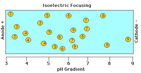
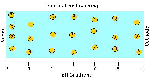
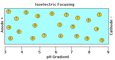
6


7 Two-Dimensional Gel Electrophoresis Separate proteins based on pi in 1st dimension Separate proteins based on molecular weight in 2nd dimension

8 Figure 5.11 The solubility of most globular proteins is markedly influenced by ph and ionic strength. This figure shows the solubility of a typical protein as a function of ph and various salt concentrations.


9 Salting Out : Ammonium Sulfate Precipitation in Protein Fractionation
 Zonal ultracentrifugation (e.g. sucrose-gradient) swinging-bucket rotor Equilibrium-density gradient ultracentrifugation (e.g. CsCl) swinging-bucket or xed-angle rotor")
10 Centrifugation Low-speed, high-speed, or ultracentrifugation: different spin speeds and g forces Centrifugation Methods Differential (Pelleting) simple method for pelleting large particles using xed-angle rotor (pellet at bottom of tube vs. supernatant solution above) Zonal ultracentrifugation (e.g. sucrose-gradient) swinging-bucket rotor Equilibrium-density gradient ultracentrifugation (e.g. CsCl) swinging-bucket or xed-angle rotor
11 Zonal Centrifugation: Sucrose- Gradient Preparative Centrifugation Separates by sedimentation coef cient (determined by size and shape of solutes)

12 Column Chromatography Flow-through Eluate


13 Chromatography Sample containing proteins or peptides Liquid ow Liquid ow Separation according to: -molecular weight/ size -charge -hydrophobicity -af nity Time :
 Negatively-charged acidic residues (E & D) >>> The charged groups, hydrophobic regions, size, and solvation affect the biophysical properties of")
14 Proteins are Amphiphilic Macro-Ions Positively-charged basic residues (K, R, & H) Hydrophobic patch Macromolecular dimensions: ca. 40 Å Ligand binding pocket (active site) Negatively-charged acidic residues (E & D) >>> The charged groups, hydrophobic regions, size, and solvation affect the biophysical properties of the protein and largely determine its puri cation behavior.
15 Different Types of Chromatography Gel ltration/size exclusion - separates by size (molecular weight) of proteins Ion exchange (cation exchange and anion exchange) - separates by surface charge on proteins Cation exchange: separates based on positive charges of solutes/proteins, matrix is negatively charged Anion exchange: separates based on negative charges of solutes/proteins, matrix is positively charged Hydrophobic interaction - separates by hydrophobicity of proteins Af nity - separates by some unique binding characteristic of protein of interest for af nity matrix in column
16 Ion-Exchange Chromatography
17 Ion Exchange (IEX) Chromatography
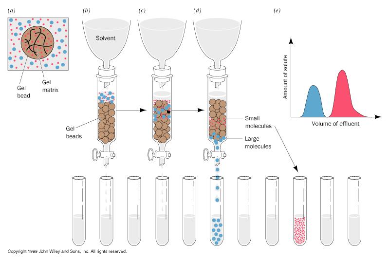

18 Gel Filtration Chromatography
19 Gel Filtration (GF) Chromatography

20 The principle of gel ltration -- excluded volume [Note: gel ltration chromatography is also sometimes called size exclusion chromatography ] V o = void volume V t = bed volume V e = elution volume V i = V t - V o
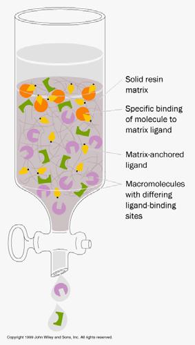

21 Af nity Chromatography
22 Protein Sequencing Frederick Sanger was the first. In 1953, he sequenced the two chains of insulin. Sanger's results established that all of the molecules of a given protein have the same, unique sequence and that the polypeptide chain is unbranched (apart from disul de crosslinks at some cysteines). Proteins can be sequenced in two ways: - real amino acid sequencing (the classic approach) - sequencing the corresponding DNA of the gene (or cdna copy of the mrna), then inferring the protein sequence using the genetic code.

23 Primary Structure of Bovine Insulin First protein to be fully sequenced; Fred Sanger, For this, he won his rst Nobel Prize (his second was for the Sanger dideoxy method of DNA sequencing).
24 Direct Determination of a Protein Sequence An Eight Step Strategy 1. If more than one polypeptide chain, separate. 2. Cleave (reduce) disul de bridges 3. Determine composition of each chain 4. Determine N- and C-terminal residues 5. Cleave each chain into smaller fragments and determine the sequence of each chain 6. Repeat step 5, using a different cleavage procedure to generate a different set of fragments 7. Reconstruct the sequence of the protein from the sequences of overlapping fragments 8. Determine the positions of the disul de crosslinks
25 Comment re previous slide: Obviously, it is easier to just sequence the gene for a protein rather than laboriously sequence the protein itself by direct methods. This is why most (>99%) of protein sequences generated today are from indirect, inferential nucleic acid sequencing data. However, for special cases, e.g., characterizing a new protein isolated from a natural source, direct sequencing is still required. Even in this case, though, often only partial sequencing is required -- enough to generate a unique peptide ngerprint -- then the rest of the sequence can be obtained by looking it up in a database of protein sequences generated from DNA sequencing data from the organism from which the protein was isolated.

26 Example of inferring protein sequence & function from DNA sequence: the human genome data. Figure 5.32 Proteins of the human genome grouped according to their molecular function. The numbers and percentages within each functional category are enclosed in parentheses. Note that the function of more than 40% of the proteins encoded by the human genome remains unknown. Considering those of known function, enzymes (including kinases and nucleic acid enzymes) account for about 20% of the total number of proteins; nucleic acid-binding proteins of various kinds, about 14%, among which almost half are gene-regulatory proteins (transcription factors). Transport proteins collectively constitute about 5% of the total; and structural proteins, another 5%. (Adapted from figure 15 in Venter, J.C., et al., The sequence of the human genome. Science 291: )
27 Cleavage of Polypeptides for Analysis Strong acid (e.g. 6M HCl) - not sequence speci c Sequence-speci c proteolytic enzymes (proteases) Sequence-speci c chemical cleavage (e.g. cyanogen bromide cleavage at methionine residues)
28 Protease Speci cities
29 Cyanogen Bromide Cleavage at Methionine Residues
30
31 Protein Sequencing: Edman Degradation PTC = phenylthiocarbamyl F 3 CCOOH = tri uoroacetic acid PTH = phenylthiohydantion
32
33 Separation of Amino Acids by HPLC
34 Another, more modern way, to sequence proteins is via mass spectrometry. The mass spectrometry method also meshes well with modern proteomics databases where one can have look up tables of peptides based on their mass. Thus mass spectrometry can facilitate peptide ngerprinting and subsequent gene product identi cation. Mass spectrometry can also be done on very small amounts of material (even impure material), which is another advantage.
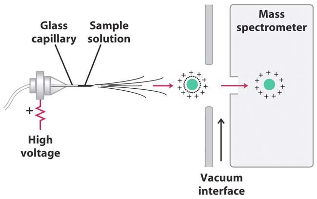

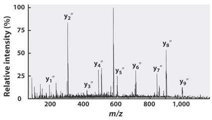
35 Protein/Peptide Identi cation by Mass Spectrometry
36 Locating the Disul de Bonds in Insulin O I O - iodoacetate
37 Determing Primary Structure of an Entire Protein
38 Reactions in Solid-Phase Peptide Synthesis

39 Molecular Biology & Biochemistry 694:407 & 115:511 Methods of Protein Analysis Sept. 16th, 2005, Lecture
40 Special thanks for many slides in this lecture again goes to Dr. Gabriel Fenteany, Dept. of Chemistry, University of Illinois at Chicago (