Characterisation of the osteogenic differentiation of human mesenchymal stem cells using Raman spectroscopy Lindsay L. McManus
|
|
|
- Nathan Shepherd
- 5 years ago
- Views:
Transcription
1 Characterisation of the osteogenic differentiation of human mesenchymal stem cells using Raman spectroscopy Lindsay L. McManus IOM3 Young Persons World Lecture Competition São Paulo, Brazil 29 th September 2011
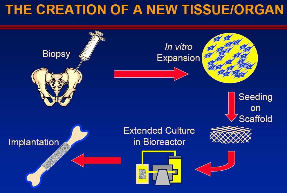
2 Tissue engineering Interested in repairing or replacing human tissue so the body can self-heal 3D scaffold biomaterials Natural polymers (collagen) Synthetic polymers (PLGA, PLA) Ceramics (HA, beta-tcp) Cells Primary cells (osteoblasts) Stem cells (mesenchymal stem cells) Cell line models (U20S) Tissue engineered material Regulators Biochemical factors (growth factors) Mechanical environment Applied strain, fluid sheer
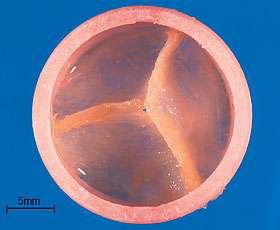

3 Tissue engineering Heart Valve Bone Blood Vessel Cartilage Bladder
4 Bone: composition Organic (30%) Cells (2%) Osteoblasts Osteocytes Osteoclasts + Matrix (98%) Collagen (90-95%) Non-collagenous proteins (5%) Inorganic (70%) Hydroxyapatite (95%) + Magnesium Sodium Potassium Fluoride Chloride


5 Why do we need to replace bone?

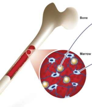
6 Motivation Can be sourced from the bone marrow, blood vessels, adipose tissue, skeletal muscle, synovium, peripheral blood and the prostate glands Can be easily isolated and expanded in culture to generate large numbers of cells Multi-potent Human mesenchymal stem cells
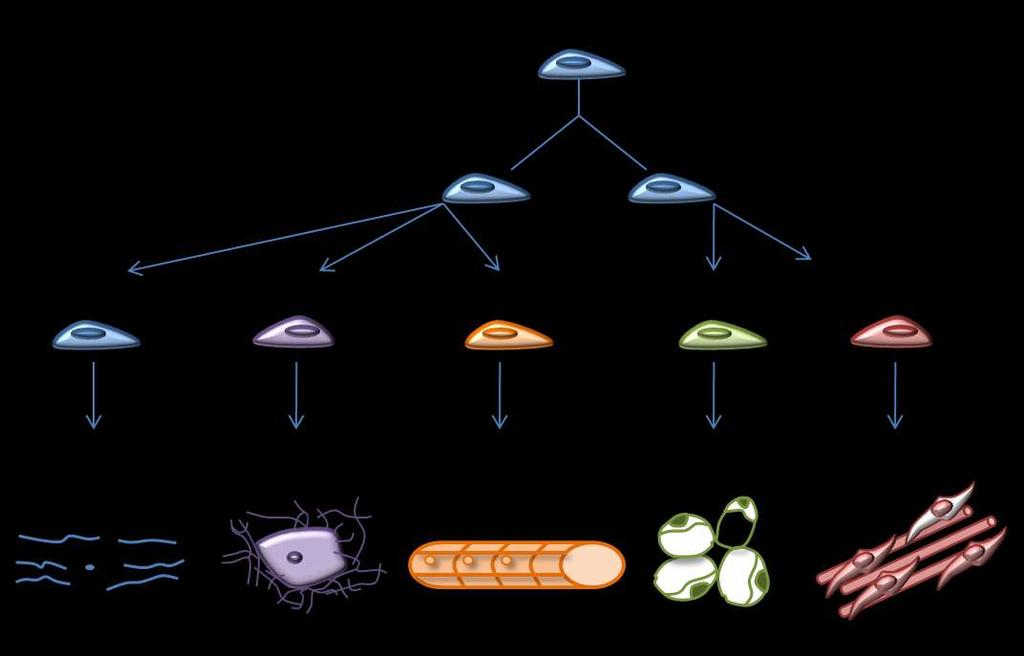
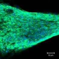
7 Human mesenchymal stem cells


8 hmscs and Raman spectroscopy Expand hmscs in vitro Isolate hmscs from the patient + Dexamethasone + β-glycerophosphate + L-ascorbic acid-2- phosphatase Induce osteogenic differentiation


9 hmscs cultured in osteogenic media Alizarin red staining DAY NON-OSTEOGENIC MEDIA Magnification 100x OSTEOGENIC MEDIA Magnification 100x


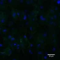
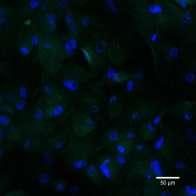
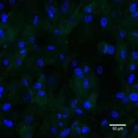
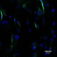
10 Osteogenic differentiation of hmscs A SPARC OPN OCN ALP B C D NON- OSTEOGENIC A B C D E F G H OSTEOGENIC Immunocytochemical localisation of SPARC, osteopontin, osteocalcin and alkaline phosphatase of hmscs cultured in non-osteogenic (A-D) and osteogenic media (E-H) for 28 days
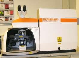
11 Standard methods vs Raman spectroscopy Monitoring of osteogenic differentiation and initial bone formation over various culture times is very time-consuming Destructive techniques are commonly used such as; cell staining for optical microscopy and confocal microscopy; bio-assay based analysis and gene expression analysis There is therefore an existing need to develop an alternative method of monitoring bone formation over various culture times which ideally will be non-destructive to allow real-time monitoring of cell differentiation One such route: RAMAN SPECTROSCOPY
12 How Does it Work? Stokes Ground state molecule Excited state molecule Photon Lower frequency photon Anti-Stokes Excited state molecule Higher frequency photon Photon Ground state molecule

13 Investigation of osteogenic differentiation using Raman spectroscopy The success of tissue engineered material is invariably dependent on the ability of the cells to replicate the functionality of their tissue of origin It has been demonstrated in some studies, that Raman spectroscopy has potential to distinguish between the chemical species that is present in bone - Tarnowski, Gentleman, C, P., E., et et al., al., Mineralisation Comparative of developing materials mouse differences calvaria revealed as revealed in by engineered Raman microspectroscopy. bone as a function J. of Bone cell-specific Min Research, differentiation (6): Nat p Mater, (9): p Huihua K, C., et al., In situ Raman spectroscopic monitoring of hydroxyapatite as human mesenchymal stem cells differentiate into osteoblasts. J. Raman Spec, (5): P MSCs Native bone
14 Peak assignment of mineral and matrix Raman frequency shift (cm -1 ) Peak assignment (intensity) 855 Hydroxyproline ring 876 Hydroxyproline ring PO 4 3- v1 (P-O symmetric stretch) Phenlyalanine 1030 PO 4 3- v3 (P-O asymmetric stretch) CO 3 2- v1 (C-O in-plane stretch) 1245, 1270 Amide III 1445 CH 2 Wag 1665 Amide I Raman probes the molecular and ionic vibrations of the mineralised species such as phosphate and carbonate, as well as many vibrations that arise from the protein matrix
15 hmscs cultured in osteogenic media OSTEOGENIC MEDIA Magnification 100x Raman spectra of hmscs cultured in osteogenic media for 7 days

16 hmscs cultured in osteogenic media OSTEOGENIC MEDIA Magnification 100x Raman spectra of hmscs cultured in osteogenic media for 14 days

17 hmscs cultured in osteogenic media OSTEOGENIC MEDIA Magnification 100x Raman spectra of hmscs cultured in osteogenic media for 21 days

18 hmscs cultured in osteogenic media DAY 28 CONTROL OSTEOGENIC MEDIA Magnification 100x Raman spectra of hmscs cultured in osteogenic media for 28 days
 Mineral/Matrix ratio (PO 4 3-, 960 cm -1 ; Hydroxyproline 850 cm")
19 Band area ratios Carbonate/phosphate ratio (CO 3 2-, 1,070 cm -1 ; PO 4 3-, 960 cm -1 ) Mineral/Matrix ratio (PO 4 3-, 960 cm -1 ; Hydroxyproline 850 cm -1 )
20 Crystallinity Phosphate peak Position (cm -1 ) FWHM (cm -1 ) Day 7 (n=8) - - Day 14 (n=8) ± ± Day 21 (n=8) ± ± 4.86 Day 28 (n=8) ± ± 8.09 Crystallinity of the bone material produced by hmscs after 21 days in osteogenic culture is significantly higher than the crystallinity observed than at day 28.


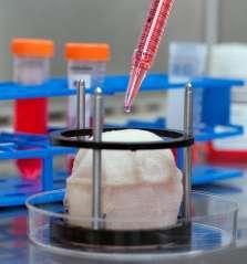
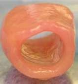
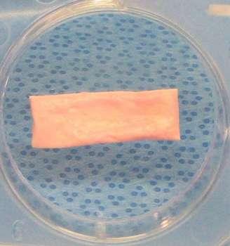
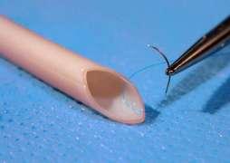
21 Clinical application
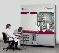
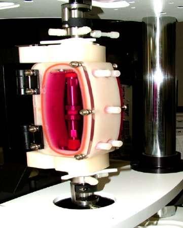
22 Summary Common methods of characterisation are: destructive time consuming provide limited information The results shown here demonstrate potential of Raman spectroscopy for monitoring of key cellular events that can be translated to environments such as bioreactors and large scale automated cell culture system Real-time configuration Non-destructive Rapid diagnostic tool


23 Acknowledgements Professor Brian Meenan Dr George Burke Dr Adrian Boyd Dr Mircea Modreanu Ms Mura McCafferty