Biology 322 Fall 2010 Transfer of genetic information in the bacterium Escherichia coli: Part I
|
|
|
- Erik Heath
- 5 years ago
- Views:
Transcription
1 Biology 322 Fall 2010 Transfer of genetic information in the bacterium Escherichia coli: Part I REQUIRED Reading Assignments: Superbugs on the Hoof Triple threat Microbe Gained Powers from another Bug Review bacterial conjugation in your genetics text and work the dilution problems at the end of this handout. (These problems are for practice not to hand in. Answers are posted on the 322 web site E.coli link) Introduction: E. coli strains can be divided into two groups defined on the basis of conjugational mating properties. F+ or male cells are able to donate chromosomal markers to recipient F - or female cells, if mixed together under the appropriate conditions. F+ cells synthesize long, thin, protein filaments termed F pili, which are required for conjugation. (The F pili also serve as specific adsorption sites for a series of single-stranded DNA and RNA phages. These phages therefore infect only male cells.) All of the properties of the F+ cells are due to the presence of a small circular DNA element (plasmid) termed the F factor (or sex factor). The F factor is member of a class of elements called episomes which can replicate as a free circular genome in the cytoplasm or can be inserted into the main bacterial chromosome and replicate with the chromosome. Strains in which the F factor is stably integrated into the host chromosome are called Hfr strains. Not all Hfr strains are equally stable and many Hfr populations contain revertants in which the F factor is no longer integrated in the chromosome. Rare errors can occur during the excision of the F factor from the chromosome which result in the formation of F' factors carrying bacterial genes, for instance lac or gal. If an F' factor, for instance F'lac+, is transferred to a wildtype strain, then a partial diploid (or merodiploid) for the lac region is created. Transfer of lac + pro + from a F' to an F - strain. Strain Sex Genotype CSH23 F'lac + proa + prob + ara + Δ(lacpro) spc R str S thi - CSH50 F - ara- Δ(lacpro) spc S str R thi - The str R (aka stra) mutation confers resistance to streptomycin; the spc R mutation confers resistance to spectinomycin. A Δ indicates a deletion of the genes in 1
2 parentheses. So Δ(lacpro) means lac - pro -. thi indicates a requirement for thiamine; pro for proline. Strains with a lac mutation cannot utilize lactose as a carbon source; strains with an ara mutation cannot utilize arabinose as a carbon source. In the cross of strain CSH23 with strain CSH50, we wish to transfer an F'lac + pro + from the CSH23 background to the CSH50 strain. This could be done in two ways. In one, a mating mixture could be plated on glucose minimal plates with streptomycin. What would be selected for in this case? Alternatively, we could use lactose minimal streptomycin plates. Again, what would be selected for using these plates? Will either parent survive? PROCEDURE: Day 0 This will be done for you Overnight cultures of the CSH23 and CSH50 will be set up for you in L broth (a rich medium). Day 1 (Thursday 10/28) These cultures will be diluted and grown at 37 o until the donor culture is 2-3 X 10 8 cell/ml. What is the quickest way to quickly determine #cells per ml? (This will be done for you.) Prepare a mating mixture by mixing 1.0 ml of each culture together in a small flask. Rotate at 30 rpms in a 37 o shaking incubator for 60 minutes. At the end of the incubation: Do serial dilutions: Fill 6 tubes with 4.5 ml of sterile saline. Transfer 0.5 ml of the undiluted mating culeture to one of the tubes. This is a 10-1 dilution. Next make serial dilutions of 10-2, 10-3, 10-4, 10-5 & Always change pipets and mix well between dilutions. Plate: 0.1 ml of a 10-3, 10-4 and 10-5 dilution onto minimal + glucose + streptomycin + thiamine. Plate: 0.1 ml of a 10-5 and 10-6 dilution onto a MacConkey + streptomycin plates. [A MacConkey plate is considered a rich media. It has lactose as well as other carbon sources. The phenol red dye is present to differentiate lac + colonies (red) from lac - colonies (white).] 2
3 Controls: Plate: 0.1 ml of a 10-1 dilution of donor (CSH23) cells on minimal + glucose + strep + thiamine plates. Repeat for the recipient (CSH50) cells. Plate: 0.1 ml of a 10-5 dilution of the recipient on a MacConkey + strep plate. Plate: 0.1 ml of a 10-1 dilution of donor on a MacConkey + strep plate. Place all plates at 37 o except MacConkey plates. The former plates will be removed from the incubator the next day and stored at 4 o C. The MacConkey plates will be held at 4 o C until Monday (11/1) afternoon and then placed in at 37 o overnight. NOTE: MacConkey color reactions fade after several days or rapidly in the cold, so plates need to be scored soon after incubation. Day 2 (11/2 Tuesday) 1. Examine the MacConkey plates. What is the genotype of the white colonies? of the red colonies? From the total cell count on the MacConkey plates determine the total number of recipient cells per ml of the mating mixture. 2. Determine the % red colonies on a plate with colonies. Is this a screen or selection for recombinants? 3. Count the number of colonies on the appropriate minimal plate. What is the genotype of the cells on the minimal plates? Do you expect these cells to be lac +? Are lac + cells selected for? Determine the number of recombinants generated per ml of mating mixture. 4. Determine the percentage of cells in the CSH50 population which received the F' factor in the cross. There are two ways in which this % can be determined. Do the results of the two calculations agree? 5. Examine your control plates. What was the purpose of these controls. Be specific. Were there any colonies on your plates? How would you account for colonies on the donor plates? on the recipients plates? Which strain do you think is more likely to give rise to colonies on the control plates? Why? There will be no formal lab report, but all raw data and your complete data analysis should be recorded in your lab notebook. 3
4 STUDY GUIDE FOR THIS LAB EXERCISE 1. Describe and analyze the strategy used for crossing these two strains. Which are the relevant mutations? What genotypes are selected for on each medium? Which markers are unselected? Be sure to indicate whether either parent will survive. 2. What are the results of the mating and the controls? Analyze the data as described above. 3. Be sure to address all of the questions listed in the preceding pages. 4. F' and F factors are lost fairly readily from E. coli cells. How would you maintain the CSH23 strain so that you were certain that only cells with the F' factor grew. 5. F' factors are useful in certain types of genetic analyses. Give an example of one. 6. There is a tremendous amount of horizontal gene transfer between bacterial cells. How is this gene transfer is accomplished? 7. Describe the mode of action of streptomycin. Describe at least three mechanisms by which a cell could acquire resistance to an antibiotic APPENDIX A Guidelines for Handling Microorganisms using Sterile (Aseptic)Technique The aim of sterile technique is to ensure that airborne fungal spores or human-borne bacteria do not contaminate your experimental cultures. 1. Wipe the bench suface with 70% ethanol, which is an effective disinfectant, before starting your experiment. 2. Only open and/or unwrap sterilized equipment (petri dishes, pipets, toothpick packages) the moment you intend to use it. Do not place Petri dish or culture flask lids on the bench surface. Instead, hold the lid in one hand while performing the required manipulation with the other hand and replace the lid immediately. 3. When you remove pipets from the metal container, handle only at the mouth end. If the other end of the pipet touches anything not sterile (other than your E.coli culture), don't use it -- get a new sterile pipet. After using a pipet, place it in the appropriate container. Do NOT place pipets in the sink. The strain of E. coli used in these experiments is not pathogenic. Nevertheless, if you spill any cultures, immediately clean and disinfect the bench surface. Petri dishes containing E. coli cultures should be discarded in the autoclavable bags provided. Liquid cultures should be disposed of in the waste flasks provided. 4
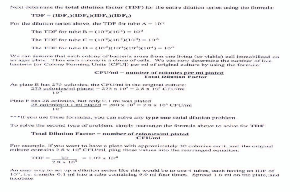
5 Appendix B: Viable Cell Counts The number of microorganisms in an undiluted broth culture after a 24-hour incubation period is usually in the millions. In order to determine the actual number of organisms in the tube or culture flask, it is necessary to dilute the culture to a point where there are a few hundred organisms per milliliter, or to a point where the number or organisms plated onto (or into) an agar medium will be statistically valid. Both spread plates and pour plates are utilized to obtain bacterial counts. A spread plate is made by careful, aseptic pipetting of a known volume of sample (usually 0.1 or 1.0 ml) onto an appropriate culture medium and spreading the liquid with a sterile glass spreader. A pour plate is made by adding a known amount of either the original or diluted sample to a melted (50 C) agar tube called an agar tall, mixing the tube and pouring the mixture into a sterile petri plate. When bacteria are plated on either spread plates or pour plates containing a medium that supports growth, only viable cells will grow, multiply and form colonies. Each of the colonies is presumed to have arisen from only one cell, although this may not be true if pairs, chains or groups of cells are not completely broken apart before plating. For this reason, the results of cell enumeration in the viable plate count are usually given as colony-forming units (CFU)/ml, not as cells/ml. If the number of viable cells on a plate is too great, the colonies will either merge and be impossible to count, or some cells will have insufficient room to form colonies, and the count will be erroneous. At the same time, it is important that the number of cells or colonies not be too few. For statistical validity, it is recommended that only plates with between 30 and 300 colonies be counted. Normally, in order to obtain this number, the original culture must be diluted several fold. Dilutions are usually made ten-fold, hundred-fold or multiples thereof; that is, the most common dilutions are 1/10, 1/100, and 1/1000. As an example, if a ten-fold dilution is to be made, it is feasible to use 0.5 ml of sample in 4.5 ml of diluent or 1.0 ml of sample in 9.0 diluent. The latter gives the fraction: 1 ml of sample 1 ml 1 = = = 1 ml of sample + 9 ml of diluent 1 ml + 9 ml If 100-fold dilutions are to be made, one can use 0.05 ml of a sample in 4.95 ml of diluent, or 0.1 ml in 9.9 ml of diluent, or 1.0 ml in 99 ml of diluent. Generally, a dilution of 1/1,000,000 (10-6 ) is sufficient to decrease the number of viable cells to the point that an appropriate number of colonies will develop on solid medium. A 10-6 dilution can be achieved by making three 1:100 dilutions, or six 1:10 dilutions, or a combination of 100-fold and 10-fold dilutions. 5
6 APPENDIX C: DILUTION PROBLEMS 1. One ml of a sample was mixed with 99 ml of sterile diluent. One ml of this was transferred (using the pour plate method) to nutrient agar. After incubation for 48 h, 241 colonies were present on the plate. How many colony-forming units were present per ml of the original sample? 2. One ml of a sample was mixed with 99 ml of sterile diluent. One-tenth of a ml (0.1 ml) of this was plated (using the spread plate method) on nutrient agar. After incubation for 48 h, 142 colonies were present on the plate. How many colony-forming units were present per ml of the original sample? 3. Given the dilution series outlined below: 0.1 ml 0.1 ml 1.0 ml 0.5 ml 1.0 ml 1.0 ml 1.0 ml 0.1 ml 9.9 ml 9.9 ml 9.0 ml 4.5 ml 1.0 ml A B C D F E G H A. Give the dilution used for each step (A-E). B. What is the total dilution at each step (A-E)? C. If the original broth culture contains 5 X 10 9 cells ml -1, (i) How many bacteria ml -1 in each tube (A-E)? (ii) How many colonies on each plate (F,G, H)? D. If tube C contains 60,000 bacteria ml -1, how many bacteria ml -1 are in the original culture and in the other tubes? How many colonies on each plate? 6
7 7
8 8