Supplementary Materials
|
|
|
- Amberly Strickland
- 5 years ago
- Views:
Transcription

1 Supplementary Materials Supplementary Figure 1. PKM2 interacts with MLC2 in cytokinesis. a, U87, U87/EGFRvIII, and HeLa cells in cytokinesis were immunostained with DAPI and an anti-pkm2 antibody. Thirty cells from each cell line were analyzed. We counted the number of cells with PKM2 localization in the contractile ring. The data represent the mean ± SD of three independent experiments. b, U87/EGFRvIII cells that expressed mcherry-histone H2B (for chromosome staining) were synchronized by thymidine double block (2 mm) and released for 9 h. Doxycycline (500 ng/ml) 1
2 with thymidine was added at the time points indicated in Fig. 1b to induce PKM2 shrna expression. MG132 (25 µm) was added at the time points indicated in Fig. 1b and incubated with the cells for 6 h to sustain them in metaphase. MG132 was then removed, and the cells were imaged using a DeltaVision deconvolution microscope with a 20 lens in a CO 2 environment chamber. Thirty cells from each cell line were analyzed. The data represent the mean ± SD of three independent experiments. The cells that exhibited chromosome segregation defects were quantified separately. c, U87 cells that had been synchronized by double thymidine block (2 mm) for 43 h were unreleased or released for 9 h, followed by MG132 (25 µm) treatment for 1.5 h to arrest cells at metaphase. MG132 was removed for 5 min (upper panel) or 60 min (bottom panel) before immunostaining the cells with the indicated antibodies. Scale bars, 10 µm. d, Quantitative PCR analyses of the indicated GBM cell lines and MEFs with primers against MYL9, MYL12A/B, and MLC2 mrna. The data represent the means ± SD of three independent experiments. e, U87/EGFRvIII cells that had been synchronized by double thymidine block (2 mm) for 43 h were unreleased or were released for 9 h, followed by MG132 (25 µm) treatment for 1.5 h to arrest cells at metaphase. MG132 was removed for 30 min before cell harvesting. Immunoprecipitates with an anti-flag antibody were incubated, with or without calf intestinal phosphatase (CIP) (10 units) and with or without sodium orthovanadate (100 µm), sodium pyrophosphate (100 µm), and sodium fluoride (100 µm), for 30 min at 37 C. The immunoprecipitates were washed with PBS three times. C, cytokinesis; I, interphase (no thymidine release). f, Flag-MLC2-expressing U87 cells, synchronized by double thymidine block (2 mm) for 43 h, 2
3 were released for 10 h (anaphase). These cells were immunostained with an anti-flag and an anti-mlc2 antibodies. Nuclei were stained with DAPI (blue). Thirty cells were analyzed. The data represent the mean ± SD of three independent experiments. g, U87/EGFRvIII cells that expressed MLC2 shrna and mcherry-histone H2B were synchronized by thymidine double block (2 mm) and released for 9 h. MG132 (25 µm) was added at the indicated time points and incubated with the cells for 1 h to sustain the cells in metaphase. MG132 were then removed, followed by imaging analyses using a DeltaVision deconvolution microscope with a 20 lens in a CO 2 environment chamber. Pictures were taken at 4-min intervals. Thirty cells from each cell line were analyzed. The data represent the mean ± SD of three independent experiments. 3
 for 43 h, were released for 10 h (anaphase).")
4 Supplementary Figure 2. Aurora B-phosphorylated PKM2 T45 is required for the interaction between PKM2 and MLC2 in cytokinesis. a, Flag-PKM2-expressing U87 cells, synchronized by double thymidine block (2 mm) for 43 h, were released for 10 h (anaphase). These cells were immunostained with an anti-flag and an 4
5 anti-aurora B antibody. Nuclei were stained with DAPI (blue). Thirty cells were analyzed. The data represent the mean ± SD of three independent experiments. Scale bars, 10 µm. b, U87 and U87/EGFRvIII cells that had been synchronized by double thymidine block (2 mm) for 43 h were unreleased or released for 10 h. Immunoprecipitation analyses with an anti-aurora B antibody were performed. C, cytokinesis; I, interphase (no thymidine release). c, In vitro kinase assays were performed by mixing purified recombinant GST-Aurora B, His- PKM2 proteins, and ATP. Immunoblotting analyses were performed with the indicated antibodies in the presence or absence of a phospho-specific blocking peptide for the PKM2 pt45 antibody. d, U87 cells that had been synchronized by double thymidine block (2 mm) for 43 h were unreleased or were released for 9 h, followed by MG132 (25 µm) treatment for 1.5 h to arrest cells at metaphase. MG132 was removed, and the cells were left untreated or were treated with AZD1152 (100 nm), Y (10 mm), or ML-7 (10 mm) for 30 min before cell harvesting. The kinase activities of immunoprecipitated Aurora B and ROCK2 were measured using HTScan Aurora B and ROCK2 kinase assay kits. The data represent the mean ± SD of three independent experiments. *P<0.01: statistically significant value in relation to untreated U87 cells. MLC kinase activity was determined using immunoblotting analyses with an anti-phospho- S19 of MRLC1 and MYLB12A/B antibody (right panel). e, WT His-PKM2 and His-PKM1 on agarose beads were incubated with purified Aurora B protein for a kinase assay, washed with PBS three times, and incubated with recombinant GST- MLC2. 5
6 f, U87/EGFRvIII cells with expression of Flag-tagged PKM2 proteins were synchronized by double thymidine block (2 mm) for 43 h and released for 10 h (anaphase). Immunoprecipitation analyses with an anti-aurora B antibody were performed. g, U87/EGFRvIII cells with expression of Flag-tagged PKM2 proteins were synchronized by double thymidine block (2 mm) for 43 h and then unreleased or released for 10 h. Immunoprecipitation analyses with an anti-flag antibody were performed. C, cytokinesis; I, interphase (no thymidine release). h, U87/EGFRvIII cells that had been synchronized by double thymidine block (2 mm) for 43 h were released for 10 h (anaphase). Immunoprecipitates with an anti-aurora B antibody were incubated, with or without calf intestinal phosphatase (CIP) (10 units), for 30 min at 37 C and washed with PBS three times. C, cytokinesis; I, interphase (no thymidine release). Immunoblotting analyses were performed with the indicated antibodies. i, U87 cells in cytokinesis were immunostained with an anti-flag or anti-mlc2 antibody. Nuclei were stained with DAPI (blue). Scale bars, 10 µm. 6
7 7

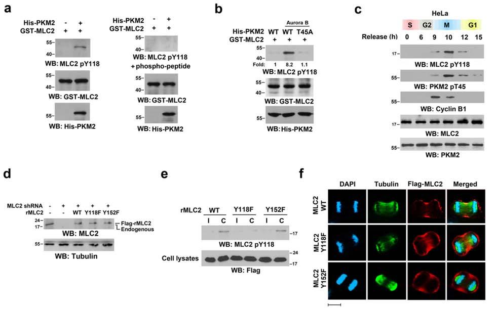
8 Supplementary Figure 3. PKM2 phosphorylates MLC2 at Y118. a, In vitro kinase assays were performed by mixing the indicated GST-MLC2 proteins with Aurora B phosphorylated WT His-PKM2 in the presence of PEP. Immunoblotting analyses were performed with the indicated antibodies in the presence or absence of a phospho-specific 8
9 blocking peptide for the MLC2 py118 antibody. b, In vitro kinase assays were performed by mixing GST-MLC2, PEP, and the indicated PKM2 proteins in the presence or absence of Aurora B. Immunoblotting analyses were performed with the indicated antibodies. c, HeLa cells that had been synchronized by double thymidine block (2 mm) for 43 h were released for the indicated time period. Immunoblotting analyses were performed with the indicated antibodies. I, interphase; C, cytokinesis. d, e, U251 cells, with or without MLC2 shrna expression, were stably transfected with or without a vector that expressed WT rmlc2, rmlc2 Y118F, or rmlc2 Y152F. Immunoblotting analyses were performed with the indicated antibodies. f, U87 cells that expressed rmlc2 WT, rmlc2 Y118F, or rmlc2 Y152F in cytokinesis were immunostained with the indicated antibodies. Scale bars, 10 µm. g, U87 cells that had been synchronized by double thymidine block (2 mm) for 43 h were released for 9 h, followed by MG132 (25 µm) treatment for 1.5 h min to arrest cells at metaphase. MG132 was replaced, with or without AZD1152 (100 nm), for 30 min before cell harvesting. Immunofluorescence analyses were performed with the indicated antibodies. The relative fluorescence intensity of the indicated proteins in equators of 100 cells from each cell line was compared and quantified. Data represent the mean ± SD of three independent experiments. Scale bars, 10 µm. h, U87 (left panel) or U251 (right panel) cells were stably expressed with a vector for control shrna or PKM2 shrna and reconstituted with WT rpkm2 or rpkm2 T45A expression. Immunoblotting analyses were performed with the indicated antibodies. 9
10 i, U251 cells with PKM2 depletion and reconstituted WT rpkm2 or rpkm2 T45A expression were synchronized by double thymidine block (2 mm) for 43 h, followed by no release or release for 10 h. Immunoblotting analyses were performed with the indicated antibodies. j, U87 cells with PKM2 depletion were stably transfected with a vector that expressed WT rpkm2 or rpkm2 T45A. These cells were synchronized by double thymidine block (2 mm) for 43 h and then released for 10 h. Immunofluorescence analyses of cells in cytokinesis were performed using the indicated antibodies. The relative fluorescence intensity of the indicated proteins in equators of 100 cells from each cell line was compared and quantified. Data represent the mean ± SD of three independent experiments. Scale bars, 10 µm. 10
11 11

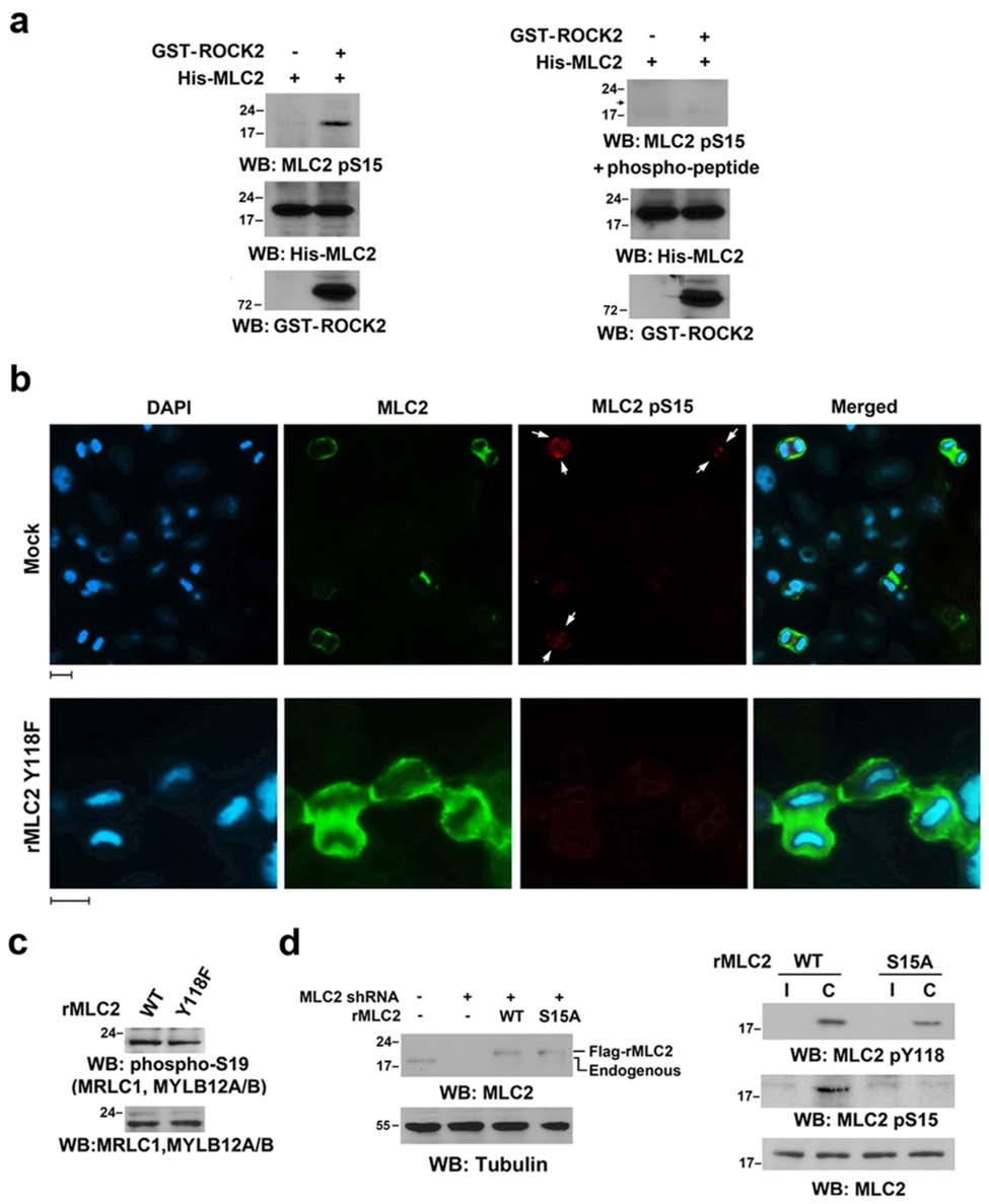
12 Supplementary Figure 4. PKM2-mediated MLC2 Y118 phosphorylation primes ROCK2- phosphorylated MLC2 at S15. a, In vitro kinase assays were performed by mixing purified recombinant active GST-ROCK2, PKM2 phosphorylated His-MLC2, and ATP. Immunoblotting analyses were performed with the indicated antibodies in the presence or absence of a phospho-specific blocking peptide for the MLC2 ps15 antibody. b, U87 cells with MLC2 depletion and reconstituted expression of WT rmlc2 or rmlc2 Y118F were synchronized by double thymidine block (2 mm) for 43 h, with release for 10 h. Immunofluorescence analyses of the cells in cytokinesis were performed using the indicated antibodies. 12
13 c, U87 cells with MLC2 depletion and reconstituted WT rmlc2 or rmlc2 Y118F expression were synchronized by double thymidine block (2 mm) for 43 h, followed by release for 10 h. Immunoblotting analyses were performed with the indicated antibodies. d, U87 cells that expressed MLC2 shrna were reconstituted with WT rmlc2 or rmlc2 S15A expression (left panel). The cells were synchronized by double thymidine block (2 mm) for 43 h, followed by no release or release for 10 h (right panel). Immunoblotting analyses were performed with the indicated antibodies. e, U87 cells with PKM2 depletion and reconstituted expression of WT rpkm2 or rpkm2 T45A were synchronized by double thymidine block (2 mm) for 43 h, with release for 10 h. Immunofluorescence analyses of the cells in cytokinesis were performed using the indicated antibodies. Scale bars, 10 µm. f, U87 cells with MLC2 depletion and reconstituted expression of FLAG-tagged rmlc2 S15A (Supplementary Fig. 4d) were immunostained with the indicated antibodies. Scale bars, 10 µm. g, U87 cells with MLC2 depletion were reconstituted with the expression of FLAG-tagged WT rmlc2, rmlc2 Y118F, or rmlc2 S15A. The cells were synchronized by double thymidine block (2 mm) for 43 h, followed by no release or release for 10 h. Immunoblotting and immunoprecipitation analyses were performed with the indicated antibodies. h, U87 cells that had been synchronized by double thymidine block (2 mm) for 43 h were released for 9 h, followed by MG132 (25 µm) treatment for 1.5 h min to arrest cells at metaphase. MG132 was removed. The cells in the presence or absence of AICAR AMPK activator (10 µm) were treated with or without Compound C AMPK inhibitor (20 µm) for 30 min. The AMPK kinase activity was determined by immunoblotting analyses with an antiphospho-acc S79 antibody. 13
14 14
15 Supplementary Figure 5. PKM2-regulated MLC2 phosphorylation is required for cytokinesis progression. a, U87/EGFRvIII cells were infected with the lentivirus that expressed PKM2 shrna and the lentivirus that expressed WT rpkm2 or rpkm2 T45A (top panel). U87/EGFRvIII cells were infected with the lentivirus that expressed MLC2 shrna and the lentivirus that expressed WT rmlc2, rmlc2 Y118F, or rmlc S15A (bottom panel). Immunofluorescent analyses were performed with the indicated antibodies. Scale bars, 10 µm. b, U87/EGFRvIII cells were infected with the lentivirus that expressed control or PKM2 shrna and the lentivirus that expressed WT rpkm2 or rpkm2 T45A (left panel). U87/EGFRvIII cells were infected with the lentivirus that expressed control or MLC2 shrna and the lentivirus that expressed WT rmlc2, rmlc2 Y118F, or rmlc2 S15A (right panel). Immunoblotting analyses were performed with the indicated antibodies. 15
16 16
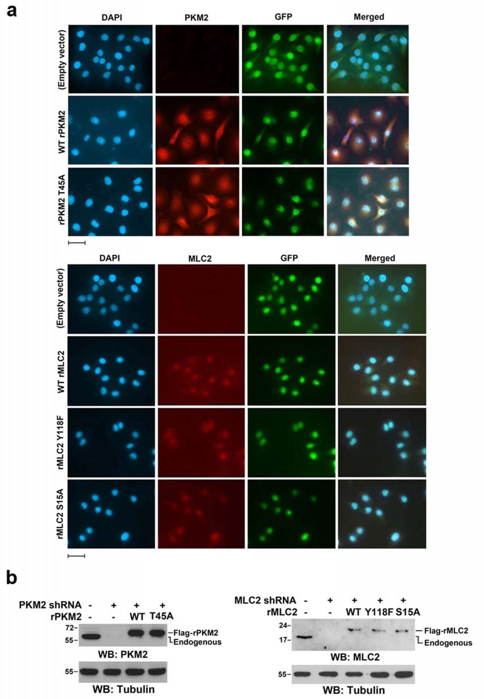
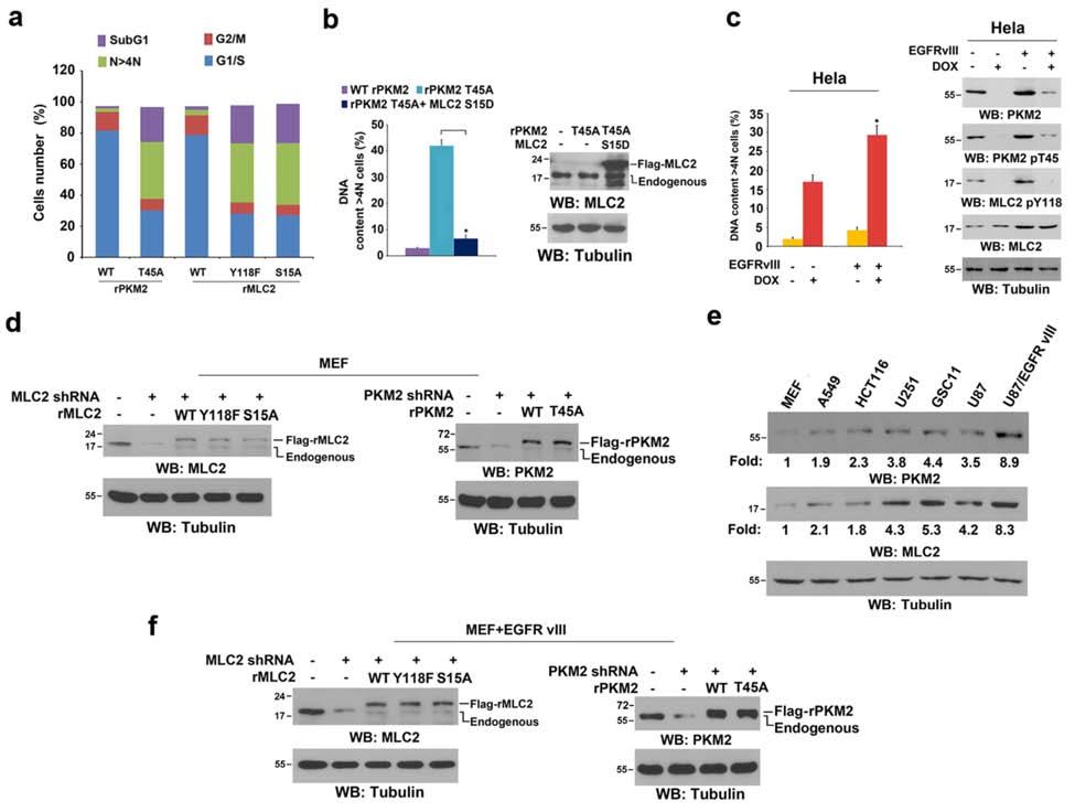
17 Supplementary Figure 6. Expression of EGFRvIII increased cells dependence on PKM2- mediated and MLC2-regulated cytokinesis. a, Asynchronized U87/EGFR cells with reconstituted expression of WT rpkm2, rpkm2 T45A, WT rmlc2, rmlc2 Y118F, or rmlc S15A in DMEM with 10% serum were analyzed by flow cytometry. The data represent the mean ± SD of three independent experiments. b, U87/EGFR cells with reconstituted expression of WT rpkm2 or rpkm2 T45A were overexpressed with or without MLC2 S15D; this was followed by flow cytometric analyses. The data represent the mean ± SD of three independent experiments. Immunoblotting analyses were performed with the indicated antibodies. P<0.01: statistically significant value in relation to 17
18 U87/EGFRvIII cells with PKM2 depletion and reconstituted expression of rpkm2 T45A, Student s t-test. c, HeLa and EGFRvIII-expressing HeLa cells were treated with doxycycline (500 ng/ml) for the indicated time periods, as shown in Fig. 1b, to induce PKM2 shrna. Flow cytometric analyses were performed. The data represent the mean ± SD of three independent experiments. The cell lysates of the indicated cell lines were analyzed by immunoblotting analyses with the indicated antibodies. d, MEFs with depleted PKM2 or MLC2 and reconstituted expression of WT rpkm2, rpkm2 T45A, WT rmlc2, rmlc2 Y118F, or rmlc2 S15A were immunoblotted with the indicated antibodies. e, The cell lysates of the indicated cell lines were studied by immunoblotting analyses of the indicated antibodies. f, EGFRvIII-expressing MEFs with depleted PKM2 or MLC2 and reconstituted expression of WT rpkm2, rpkm2 T45A, WT rmlc2, rmlc2 Y118F, or rmlc2 S15A were immunoblotted with the indicated antibodies. g, A549 and EGFRvIII-expressing A549 cells (left panel) or HCT116 and EGFRvIII-expressing HCT116 cells (right panel) were treated with doxycycline (500 ng/ml) for the indicated time periods, as shown in Fig. 1b, to induce PKM2 shrna. Flow cytometric analyses were performed (top panel). The cells ( ) were plated and counted 7 days after being seeded in DMEM with 2% bovine calf serum (bottom panel). The data represent the mean ± SD of three independent experiments. P<0.01: statistically significant value in relation to the parental cells, Student s t-test. #P<0.05: statistically significant value in relation to the parental cells, Student s t-test. 18
19 h, A549 and EGFRvIII-expressing A549 cells (left panel) or HCT116 and EGFRvIII-expressing HCT116 cells (right panel) were treated with doxycycline (500 ng/ml) for the indicated time periods, as shown in Fig. 1b, to induce PKM2 shrna. The cell lysates of the indicated cell lines were analyzed by immunoblotting analyses with the indicated antibodies. i, HCT116 cells were infected with lentiviruses that expressed MYL9 shrna and then reconstituted with WT rmrlc1 or rmrlc1 T18/S19A expression. Immunoblotting analyses were performed with the indicated antibodies (left panel). These cells were synchronized by double thymidine block (2 mm) for 43 h and released for 36 h. Flow cytometric analyses were performed. The data represent the mean ± SD of three independent experiments (right panel). P<0.01, Student s t-test. j, HCT116 cells, with or without combined or single depletions of MCL2 and MRLC1 and reconstituted expression of the indicated proteins, were synchronized by double thymidine block (2 mm) for 43 h and released for 36 h. Flow cytometric analyses were performed. The data represent the mean ± SD of three independent experiments. P<0.01. Immunoblotting analyses were performed with the indicated antibodies. 19

20 Supplementary Figure 7. PKM2-regulated MLC2 phosphorylation is required for tumor cell proliferation and tumorigenesis. 20
21 a, The relative activity of 0.1 µg of bacterially purified WT PKM2 (set to 1) or PKM2 T45A on PEP was measured using a pyruvate kinase assay. The data represent the means ± SD of three independent experiments. b, Endogenous PKM2-depleted U87/EGFRvIII cells with reconstituted expression of WT rpkm2 or rpkm2 T45A were arrested by double thymidine block (2 mm) for 43 h. The glucose uptake and lactate production were measured. The data represent the means ± SD of three independent experiments. c, U87/EGFRvIII cells with depleted PKM2 and reconstituted expression of WT rpkm2 or rpkm2 T45A were synchronized by thymidine double block (2 mm) with or without release for 9 h. I indicates interphase; M indicates mitosis. Immunoblotting analyses were performed with the indicated antibodies. d, U87/EGFRvIII cells with depleted PKM2 and reconstituted expression of WT rpkm2 or rpkm2 T45A were synchronized by thymidine double block (2 mm) and released for 9 h, followed by MG132 (25 mm) treatment for 1 h (for staining the cells in metaphase). MG132 was then removed for 30 min (for staining the cells in telophase). One hundred cells in mitosis were analyzed for chromosome segregation defects. Data represent the mean ±SD of three independent experiments. e, Serum-starved and endogenous PKM2-depleted U87 cells with reconstituted expression of WT rpkm2 or rpkm2 T45A were treated with EGF (100 ng/ml) for 6 h. Immunoblotting analyses were performed with the indicated antibodies. f, GSC11 cells that expressed control shrna or shrna for PKM2 or MLC2 were reconstituted with WT rpkm2, rpkm2 T45A, WT rmlc2, or rmlc2 Y118F expression. Immunoblotting analyses were performed with the indicated antibodies. 21
22 g, We intracranially injected athymic nude mice with 1) GSC11 cells with PKM2 depletion and reconstituted WT rpkm2 or rpkm2 T45A expression (left panel) or 2) MLC2 depletion and reconstituted WT rmlc2 or rmlc2 Y118F expression (middle panel). The mice were sacrificed and examined for tumor growth. H&E-stained coronal brain sections revealed representative tumor xenografts. Tumor volumes were measured using length (a) and width (b) and calculated using the equation V = ab 2 /2. The data represent the means ± SD of seven mice (right panel). Scale bars, 0.2 cm. 22

23 Supplementary Figure 8. Representative original images of immunoblotting results for Fig

24 Supplementary Figure 9. Representative original images of immunoblotting results for Fig

25 Supplementary Figure 10. Representative original images of immunoblotting results for Fig