Molecules VII. Structures and Functions of Proteins
|
|
|
- Sylvia Hunter
- 5 years ago
- Views:
Transcription
1 Molecules VII Structures and Functions of Proteins
2 The 20 Amino Acids

3 Sequences from Protein Data Bank (PDB) The primary sequence can be indicated very compactly using the one letter abbreviations. This is the first information that one will find in a published sequence or structure, for example when searching sources such as: This is what you might get: (HEWL from 6lyz structure) ORIGIN 1 kvfgrcelaa amkrhgldny rgyslgnwvc aakfesnfnt qatnrntdgs tdygilqins 61 rwwcndgrtp gsrnlcnipc sallssdita svncakkivs dgngmnawva wrnrckgtdv 121 qawirgcrl

4 Secondary Structure in a Folded Protein α-helix β-sheet β-turn Ribbon diagram

5 Class, Architecture and Topology


6 Proteins fold into a compact structure Space filling model Stick diagram
7 Protein Structures who cares? Understanding of catalytic structures Prediction of ligand binding affinity, specificity, maybe even kinetics Comparative genomics, since structures are better conserved than sequences Art gallery fodder? Structure-based drug design!


8 Structure-based Drug Design: HIV-1 Reverse Transcriptase
 gives up to a 8000-fold specificity effect, hence dye-terminators feasible (and uniform).")
9 Basis for dideoxy sequencing A single residue in DNA polymerases of the Escherichia coli DNA polymerase I family is critical for distinguishing between deoxy- and dideoxyribonucleotides. Y to F (one atom) gives up to a 8000-fold specificity effect, hence dye-terminators feasible (and uniform). Tabor & Richardson PNAS :

10 Structure-function: T4 lysozyme

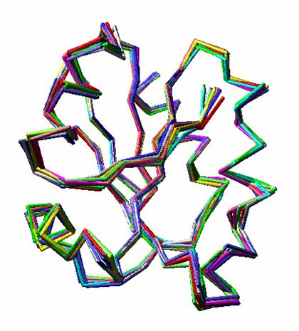
11 Visualizing Function on a Structure Superoxide dismutase Hen egg white lysozyme Nielsen et al., Protein Eng., 12:657 (1999)

12 Cα trace Structure Resolution
13 X-ray Crystallography vs. NMR X-ray crstallography Large structures have been obtained (e.g. ribosome) Crystals are often difficult to obtain Protein structure may be different in crystal vs. solution Refinement parameters are somewhat empirical NMR Can analyze proteins in solution Only smallish structures (< 300 a.a.) can be analyzed
14 Crystallographic refinement Fourier transform relates scattered X-rays, F, to electron density, ρ. Δk is the scattering vector. Minimize Fo-Fc. Linearize with a first order Taylor expansion parameters p (e.g. = x,y,z)
15 Measure Structure Quality R factor = Σ Fo - Fc / Σ Fo < 0.25 good > 0.4 crude Correlation Coefficient > 0.7 RMSD (root mean square deviation) = sqrt[σ (X i1 -X i2 ) 2 ] compare models 1 & 2 i = 1 to n (#atoms) canonical peptide geometry

16 Structural Genomics Initiatives

17 Structure Gallery
 Four")

18 Structure and Function of Hemoglobin Hemoglobin is a tetramer (2 alpha and 2 beta chains) Four sites for binding O 2 employees.csbsju.edu/.../bohreffectcurve.gif

19 Conformational Changes on O 2 Binding Berg et al., Biochemistry, 5 th ed.
20 Bohr Effect Berg et al., Biochemistry, 5 th ed.

21 Ligand-Receptor Binding Non-cooperative L+ R C Cooperative nl + R C
22 Scatchard Plots

23 Hill Plots
24 Artificial Blood RBCs are basically just bags But they are important bags Maintain Hb concentration Maintain circulation time 2,3-DPG affects binding curve (affinity) Updates on clinical trials Hemopure (Biopure) cross-linked bovine hemoglobin Approved in South Africa Phase III results reviewed by FDA; additional information sought Polyheme (Northfield Lab.) polymerized human hemoglobin Pivotal Phase III trial ongoing for trauma centers Hemolink (Hemosol) polymerized human hemoglobin Recently defaulted to creditors
25 Further Reading Tinoco, Jr., K. Sauer, J.C. Wang, and J. Puglisi, Physical Chemistry: Principles and Applications in Biological Systems, 4th ed. Prentice-Hall, Englewood Cliffs, NJ, 2001 Biochemistry text