Infection with Marteilia refringens
|
|
|
- Oliver Gilbert
- 5 years ago
- Views:
Transcription


1 Infection with Marteilia refringens Photo: Ifremer EURL for Mollusc Diseases, Laboratory of Genetic and Pathology of Marine Molluscs, La Tremblade, France (2013)
2 General information Category of the disease notifiable to the OIE and listed in Directive 2006/088/EC Common, generally accepted names of the disease agent Aber disease, Digestive gland disease of the European oyster, Marteiliosis Scientific name or taxonomic affiliation of the causative agent Marteilia refringens, (Grizel 1974) of the phylum Cercozoa and order Paramyxida (Cavalier-Smith & Chao 2003; Feist et al., 2009)
: Genus Hosts Crab Amphipod Oyster Bivalves Polychaeta I ary cell II")
3 Phylum Cercozoa, order Paramyxida Classification (Feist et al. 2009): Genus Hosts Crab Amphipod Oyster Bivalves Polychaeta I ary cell II ary cell Spores
 Possible host species (partly")


4 Wide host range Host species (fully demonstrated) Possible host species (partly demonstrated) Other species Ostrea edulis Mytilus edulis Mytilus galloprovincialis Xenostrobus securis Solen marginatus Chamelea gallina Ostrea angasi, O. Puelchana, O. chilensis, O. denselamellosa Crassostrea virginica Ruditapes decussatus, R. philippinarum Tapes rhomboides, T. pullastra Ensis minor, E. siliqua Argopecten gibbus Saccostrea forskali Tridacna maxima Pinctada margaritifera Crassostrea gigas : mature stages not visible = no release of the parasite? Other Marteilia species? Cardium edule Saccostrea cucullata Scropicularia piperata
5 General information Other Marteilia species : Marteilia sydneyi infects Saccostrea glomerata (= commercialis) and possibly Saccostrea echinata. Marteilia maurini considered as synonymous of M. refringens (Lopez-Florez et al. 2004; Novoa et al. 2005) in Mytilus galloprovincialis and M. edulis in France, Spain and Adriatic sea (Italy and Croatia) Marteilia lengehi in Saccostrea cuccullata reported from Persian Gulf and Western Australia Marteilia christenseni in Scrobicularia plana reported from France


6 Geographical distribution M. refringens IRELAND UK THE NETHERLANDS PCR FRANCE SPAIN ITALY M. sydneyi Marteilia in mussels Marteilia in oysters Marteilia in other species
, discolouration of the digestive gland, cessation of growth, tissue necrosis, and mortalities.")
7 Impact on the host Since 1968, M. refringens has caused serious recurring mortalities with a significant negative impact on the European O. edulis industry. Infection causes a poor condition index with glycogen loss (emaciation), discolouration of the digestive gland, cessation of growth, tissue necrosis, and mortalities. However, Marteilia can occur in some oysters without causing disease.


8 Impact on the host Photo: Ifremer Photo: Ifremer Healthy oyster Diseased oyster


9 Impact on the host
.")

10 Diagnostic techniques Tissue Imprint: Make acetone- (or methanol-) fixed impression smears from digestive gland tissue. Stain with Wright, Wright-Giemsa or equivalent stain (e.g. Hemacolor, Merck; Diff-QuiK, Baxter). The parasite is 5 8 µm in size in the early stages and may reach up to 40 µm during sporulation. The cytoplasm of the cells stains basophilic, the nucleus is eosinophilic. The secondary cells or sporoblasts are surrounded by a bright halo (colour may vary slightly with the stain used) Ifremer Ifremer


11 Digestive gland imprints Ifremer

12 Digestive gland imprints
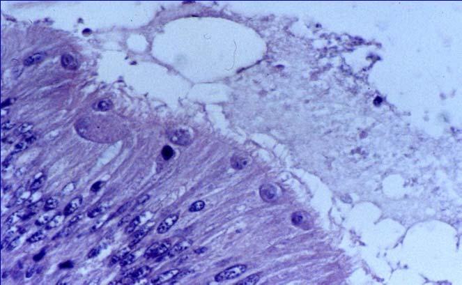
13 Diagnostic techniques Histology: Cross-sections of the digestive gland show the parasite in the epithelial cells of the digestive ducts (basophilic stages) and the epithelial cells of the digestive tubules (acidophilic stages). The unique feature of internal cleavage to produce cells within cells during sporulation differentiates Marteilia spp. from all other protista. A modified staining technique described by Gutiérrez (1977) may enhance the detection of the parasite in paraffin embedded histological sections.


14 Histology Ifremer

15 Histology Ifremer
16 Histology Ifremer


17 Sporulation process


18 Electron Microscopy M. refringens in Ostrea stentina from Tunisia (Elgharsalli et al. 2013) J.-P. Joly J.-P. Joly Sporangiosorus S containing presporongiosora P with immature spores Sp. R: reticulated cytoplasm of sporangium So; Rb: refringent body Almost mature spore with intermediate sporoplasm S2 and innermost sporoplasm S3. S1: outermost sporoplasm containing numerous haplosporosomes H; V: flattened vesicles in the intermediate sporoplasm; W: spore wall
19 Diagnostic techniques Immunological Assay: An immunohistochemistery technique based on monoclonal antibodies was developed by Robledo et al. (1994). However, this technique is very rarely used in diagnostic laboratories. Two clones are of particular interest for their stage specificity: 4/1-1 (sporangia) and 9/1-1 (young plasmodia). No cross reaction with M. sydneyi (Anderson et al., 1994) However, there is a lack of specificity for European isolates (Pernas et al., 2000).

20 Immunological Assay Ifremer Ifremer
21 Diagnostic techniques DNA Probes: Several PCR protocols are available : PCR primers that target the ITS1 (internal transcribed spacer) region (Le Roux et al., 2001) are recommended as they are able to amplify only Marteilia refringens. Some primers targeting the small subunit (SSU) of the rrna gene complex are also available and allow M. refringens and M. sydneyi to be amplified (Le Roux et al., 1999; Berthe et al., 2000) A nested PCR assay targeting the rdna intergene spacer (López-Flores et al., 2004) seems to be more sensitive than other assays
 Conventional PCR (Le Roux et al.")
22 Specificity of PCR assays Marteilia Genus Conventional PCR (Le Roux et al. 1999) IGS ITS1 18S 5.8S 28S Nested PCR (Lopez Flores et al. 2004) Conventional PCR (Le Roux et al. 2001) Marteilia refringens species
.")
23 PCR RFLP Based on a dimorphism in the locus of endonuclease HhaI in the ITS-1 sequence, two types O and M were defined and can be detected by PCR-RFLP (Le Roux et al. 2001). Marteilia refringens type M Marteilia refringens type O Hha I restriction profiles 157 bp bp + 68 bp + 31 bp 226 bp bp + 31 bp Type M Type O
. In addition, it is possible to use primers targeting the ITS-1 to produce a probe able to detect only M.")
24 Diagnostic techniques In situ hybridization: The probe named Smart 2 can detect Marteilia refringens and also M. sydneyi by in situ hybridisation in infected oysters (Le Roux et al. 199; Kleeman et al. 2002). In addition, it is possible to use primers targeting the ITS-1 to produce a probe able to detect only M. refringens by in situ hybridization (SOP available on the EURL website :
25 Methods of control Oysters, mussels, clams from areas known to be infected (currently or historically) should not be transferred to areas with no record of M. refringens. Results of field and experimental studies (Berthe et al. 1998, Audemard et al & 2001, Carrasco et al. 2008, Boyer 2012) provide evidence of an intermediate hosts in the life cycle of M. refringens, the copepod, P. grani. In enzootic areas, control is attempted by curtailing the planting of European oyster seed during the period of transmission (July and August) and by growing European oysters in areas with high salinities (35-37 ppt) to limit the development of M. refringens.
26 The end