Structure. Structural Components of Nucleotides Base. Sugar. Introduction Nucleotide to Cells & Microscopy and Nucleic Acid. Phosphate Glycosidic bond
|
|
|
- Abner Haynes
- 5 years ago
- Views:
Transcription


1 11 Structural Components of Nucleotides Base Sugar Introduction Nucleotide to Cells & Microscopy and Nucleic Acid Structure Phosphate Glycosidic bond H NUCLEOTIDE H Nucleic acid polymer of nucleotides directionality 5 à3 When you write a sequence: ATCG RNA DNA Phosphodiester bonds It is assumed that the 5 -end is on the left and the 3 -end is on the right, unless otherwise labeled. 5 -ATCG-3 3 -GCTA-5 same molecule Composition of DNA? Table 3-1 1


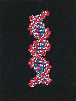
2 Chargaff s Rules DNA is Double Stranded Helix Computer-simulated space-filling model of DNA. Figure 3-8 2

 using Chargaff rules (A=T, G=C).")

3 The crucial piece of evidence for DNA structure came from X-ray crystallography. Wilkins learned how to purify DNA and make regular fiber patterns. Rosalind Franklin performed the X-ray diffraction and deduced there was a helix. Francis Crick saw the data at a seminar Wilkins gave and also deduced there was a helix and the size parameters. James Watson discovered how the bases went together (complementarity) using Chargaff rules (A=T, G=C). Watson & Crick published their structure in Beautiful example of how structure predicted function. Video: Computer-simulated space-filling model of DNA. SUMMARY 12 (34 Å) Introduction Central to Cells Dogma & Microscopyof Molecular Biology sugar phosphate backbone (phosphodiester bonds) Right-handed, antiparallel, doublestranded helix. With the base complementarity, it explains genetic material: Storage of genetic information Information retrival 3
 The Genetic Code Protein Biosynthesis Central Dogma The central dogma of molecular biology Information Flow DNA replication is semiconservative (Meselson-Stahl")
4 From DNA to Protein: Gene Expression Central Dogma: from Genes to Proteins of the genes (DNAàDNA) Transcribing the information (DNAàRNA) Translating the nucleotide sequence into protein sequence (RNAàProtein) The Genetic Code Protein Biosynthesis Central Dogma The central dogma of molecular biology Information Flow DNA replication is semiconservative (Meselson-Stahl Expt) 4
5 DNA Each New DNA Strand Grows from Its 5 End to Its 3 End Arthur Kornberg showed that DNA contains information for its own replication. He combined in a test tube: DNA, the four deoxyribonucleoside triphosphates (dntps monomers), DNA polymerase, salts (Mg +2 ), and buffer. The DNA served as a template for synthesis of new DNA. Each New DNA Strand Grows from Its 5 End to Its 3 End ALL polymerases add nucleotides to the 3 end (Direction is termed 5 g 3 ) Transcription Pyrophosphatase 34 5
6 Central Dogma Central Dogma The central dogma of molecular biology Messenger RNA (mrna) Transfer RNA (trna) Ribosomal RNA (rrna) RNA is key to this process: Messenger RNA (mrna) carries copy of a DNA sequence to site of protein synthesis at the ribosome Transfer RNA (trna) carries amino acids for polypeptide assembly Ribosomal RNA (rrna) catalyzes peptide bond formation and provides structure for the ribosome Central Dogma The central dogma of molecular biology Transcription Transcription Transcription components: A DNA template for base pairings one of the two strands of DNA Nucleoside triphosphates (ATP,GTP,CTP,UTP) as substrates An RNA polymerase enzyme Messenger RNA (mrna) Transfer RNA (trna) Ribosomal RNA (rrna) Transcription process: RNA polymerase unwinds DNA about ten base pairs at a time; reads template in 3 to 5 direction, synthesizes RNA in the 5 to 3 direction. The RNA transcript is antiparallel to the DNA template strand, and adds nucleotides to its 3 end. NTPs incorporate NMP and PP i is a product! 6


7 Transcription Production of mrna transcript by RNA polymerase Transcription: Where to start? 5' Flanking Coding Region 3'-flanking prokaryotes Promoter eukaryotes The consensus sequence for each element in human genes (N is any nucleotide) Transcription Translation 7
 What is the relationship between a DNA sequence and an amino acid sequence?")

8 Central Dogma The central dogma of molecular biology Translation Messenger RNA (mrna) Transfer RNA (trna) Ribosomal RNA (rrna) The Code The Adaptors (trna) The Ribosome (rrna + rproteins) What is the relationship between a DNA sequence and an amino acid sequence? Translation: The Genetic Code Translation: The Genetic Code The genetic code is redundant. The genetic code is universal. The genetic code: Specifies which amino acids will be used to build a protein Codon: A sequence of three bases each codon specifies a particular amino acid. Start codon: AUG initiation signal for translation. Stop codons: UAA, UAG, UGA stop translation and polypeptide is released. 8
 within the molecule. 3 -end is the amino-acid attachment site binds covalently.")

9 Translation: trna Translation: trna trnas must deliver amino acids corresponding to each codon The conformation (three-dimensional shape) of trna results from base pairing (hydrogen bonding) within the molecule. 3 -end is the amino-acid attachment site binds covalently. At the other end (middle of the trna sequence) is the Anticodon site of base pairing with mrna. Unique for each species of trna. Translation: trna Translation: Ribosome Template for mrna read 3 à5 trna anticodon Ribosome: the workbench holds mrna and charged trnas in the correct positions to allow assembly of polypeptide chain. Ribosomes are not specific, they can make any type of protein. N C 9

 has one ribosomal RNA (rrna) (18S) and 33 proteins.")
 and 49 different proteins.")
 (80S).")
10 Translation: Protein Biosynthesis: Ribosome Structure Ribosomes have two subunits, large and small. When not active in translation, the subunits exist separately. The small subunit (40S) has one ribosomal RNA (rrna) (18S) and 33 proteins. The large subunit (60S) has three molecules of rrna (28S, 5.8S, 5S) and 49 different proteins. Ribosomal subunits are held together by ionic and hydrophobic forces (not covalent bonds) (80S). Translation: Ribosome Translation: Protein Biosynthesis; Elongation Translation: Protein Biosynthesis; Elongation ELONGATION Decoding (GTP hydrolysis) EF-Tu GTP Translocation (GTP hydrolysis) Peptidyltransferase 10
11 Central Dogma The central dogma of molecular biology Animated videos of DNA structure and Central Dogma ( Guided_Tour/dnaStructure.html) 11