JB Accepts, published online ahead of print on 16 September 2011 J. Bacteriol. doi: /jb
|
|
|
- Earl Floyd
- 5 years ago
- Views:
Transcription
1 JB Accepts, published online ahead of print on 16 September 2011 J. Bacteriol. doi: /jb Copyright 2011, American Society for Microbiology and/or the Listed Authors/Institutions. All Rights Reserved Revised Version 2 Deciphering morphological determinants of the helix-shaped Leptospira Formatted: Numbering: Continuous Leyla Slamti 1#, Miguel A. de Pedro 2, Emilande Guichet 1 and Mathieu Picardeau 1* 1 Institut Pasteur, Unité Biologie des Spirochètes, 25 rue du Dr Roux, Paris, France 2 Centro de Biología Molecular Severo Ochoa Consejo Superior de Investigaciones Científicas, Universidad Autónoma de Madrid, Facultad de Ciencias, Madrid, Spain # Present address: INRA, Unité MICALIS, UMR-1319, Equipe Génétique Microbienne et Environnement, Domaine de La Minière, Guyancourt, France * To whom correspondence should be addressed: mathieu.picardeau@pasteur.fr Running title: Morphological determinants of Leptospira 1
2 Abstract Leptospira spp. are thin, highly motile, slow-growing spirochetes that can be distinguished from other bacteria on the basis of their unique helical shape. Defining the mechanisms by which these bacteria generate and maintain this atypical morphology should greatly enhance our understanding of the fundamental physiology of these pathogens. In this study, we showed that peptidoglycan sacculi from Leptospira spp. retain the helical shape of intact cells. Interestingly, the distribution of muropeptides was different from the E. coli model, indicating that specific enzymes might be active on the peptidoglycan macromolecule. We could alter the shape of L. biflexa with the broad-spectrum β-lactam antibiotic penicillin G and with amdinocillin and aztreonam that are β-lactams that preferentially target Penicillin-Binding Proteins (PBPs) 2 and 3, respectively, in some species. Although genetic manipulations of Leptospira spp. are scarce, we were able to obtain mutants in genes encoding PBPs, including PBP3. Loss of this protein resulted in cell elongation. We also generated a L. biflexa strain that conditionally expresses MreB. Loss of the MreB function was correlated with morphological abnormalities such as a localized increased diameter and heterogeneous length. A prolonged depletion of MreB resulted in cell lysis, suggesting that this protein is essential. These findings indicate that important aspects of leptospiral cell morphology are determined by the cytoskeleton and the murein layer, thus providing a starting point for a better understanding of the morphogenesis in these atypical bacteria. 2
3 47 Introduction Shape determination and maintenance is a fundamental question in biology (60). This is particularly true for prokaryotic cells that need to maintain their shape to warrant their integrity. Bacteria can take on a variety of shapes but they are, for most of the well-described bacteria, created by manipulating a common essential component: peptidoglycan (PG), a polymer of glycosaminopeptides that forms a rigid exoskeleton (16). Among bacteria with a peculiar morphology are the helix-shaped Leptospira that are 10 to 20μm long with a cell diameter of approximately 0.15μm. These cells belong to the phylum of Spirochetes, an evolutionarily and structurally unique group of bacteria (43). This group also comprises flat wave- or helix-shaped bacteria like Borrelia and Treponema that include the agents of Lyme disease and syphilis, respectively. Pathogenic Leptospira species are responsible for leptospirosis, a zoonotic disease with a worldwide distribution, but particularly prevalent in impoverished regions (28). Leptospirosis is transmitted to humans through contact with water that is contaminated with animal urine (usually rodents). Leptospira then disseminate in the host and cause a systemic infection. It has been shown that Leptospira move faster in viscous environments than other bacteria (12). Although it has never been experimentally proven, this corkscrew motility is undoubtedly an advantage during the infectious process. Leptospira motility depends on the presence of two endoflagella (or periplasmic flagella), each arising at one end of the bacteria. Leptospira biflexa flab mutants cannot form functional endoflagella, but their cell bodies remain intact and helical (45). The endoflagella are therefore not responsible for dictating the helical shape of the cell body in Leptospira spp., as they are in Borrelia burgdorferi (35). It has been shown in other bacterial species, albeit with a different morphology (rodor crescent-shaped) that several factors can be involved in acquiring and maintaining their 3
4 shape (9, 10). Penicillin-binding proteins (PBPs) are involved in PG biosynthesis. PBPs are divided in two categories: high molecular weight (HMW) and low molecular weight (LMW) PBPs. HMW PBPs comprise 2 classes of proteins, class A and B. E. coli cells need two proteins of the latter class, PBP2 and PBP3, to divide and elongate. PBP2 (encoded by pbpa) is involved in lateral PG synthesis and PBP3 (encoded by ftsi) in septal PG synthesis. A pbpa mutant becomes spherical whereas an ftsi mutant produces filaments (51). Antibiotics that affect the function of these PBPs, such as amdinocillin (mecillinam) and aztreonam, induce the same phenotypes (19, 40). LMW PBPs are also involved in shape maintenance by modifying preexisting cell wall (38, 39). The role of PBPs is closely interconnected with proteins of the cytoskeleton, like the tubulin-homolog FtsZ and the actin-homolog MreB, which are also important in bacterial shape determination (11, 42). Other types of cytoskeletal proteins are involved in cell morphology such as crescentin, an intermediate filament responsible for the crescent shape of Caulobacter crescentus (8). Five PBPs have been identified after subcellular fractionation in L. interrogans (22) and the functional homologs of E. coli pona (PBP1a) and ftsi (PBP3) have been isolated in this bacterium (7). Leptospira have a Gram-negative-like cell envelope with their PG layer associated with the cytoplasmic membrane (23). Peptidoglycan from L. biflexa apparently conforms to the A1γ chemotype, with the disaccharide pentapeptide GlucNAc-(β 1 4)- MurNAc-L-Ala-D-Glu-(γ)-mDAP-D-Ala-D-Ala as the basic monomeric subunit (59), but its precise composition is not known. This chemotype is the most common among Gramnegative bacteria, but genera of the Spirochaetacea family are an exception and contain ornithin instead of diaminopimelate (DAP) as the di-amino acid at position 3 of the peptide moiety (59). To explore Leptospira morphology we further investigated PG composition of saprophytic and pathogenic Leptospira strains. We also examined the role of different PBPs 4
5 97 98 that might be involved in maintaining the shape of these bacteria. We then showed that MreB is involved in maintaining certain features of Leptospira morphology Downloaded from on December 3, 2018 by guest 5
6 101 Material and methods Bacterial strains and growth conditions. The sequenced L. biflexa serovar Patoc strain Patoc 1 and L. interrogans serovar Lai strain Lai (National Reference Center for Leptospirosis, Institut Pasteur, Paris, France) as well as L. interrogans serovar Manilae strain L495 (Monash University, Australia) were grown in EMJH liquid medium (17, 26) at 30ºC, under agitation or on plates containing 1% Noble agar. For conjugation experiments, E. coli strain Pi1 was used as host strain for plasmid constructions and E. coli strain ß2163 (15) was used as the donor to mobilize plasmids into L. biflexa. E. coli was grown in Luria-Bertani (LB) medium. Diaminopimelate (DAP) and thymidine (dt) were used at a final concentration of 0.3 mm. IPTG was added to cultures at a final concentration of 1mM. When appropriate, 50 µg/ml spectinomycin, 50 µg/ml kanamycin, or 100 µg/ml ampicillin was added to culture media. Minimal inhibitory concentrations (MICs). MICs were determined by broth microdilution testing as previously described (36). Briefly, after incubation with two-fold serial dilutions of the antibiotics, alamarblue (Trek Diagnostics, Cleveland, OH) which is an oxidation-reduction indicator was added to each well following the manufacturer s instructions. When cells grow, the blue color indicator turns pink. The MIC (aztreonam, 0.5 µg/ml; amdinocillin, 16 µg/ml; penicillin G, 0.12 µg /ml; A22, 4 µg/ml) was defined as the lowest antibiotic concentration that inhibited visible growth (no color change) at the second day of incubation. Plasmid and strain construction. 6
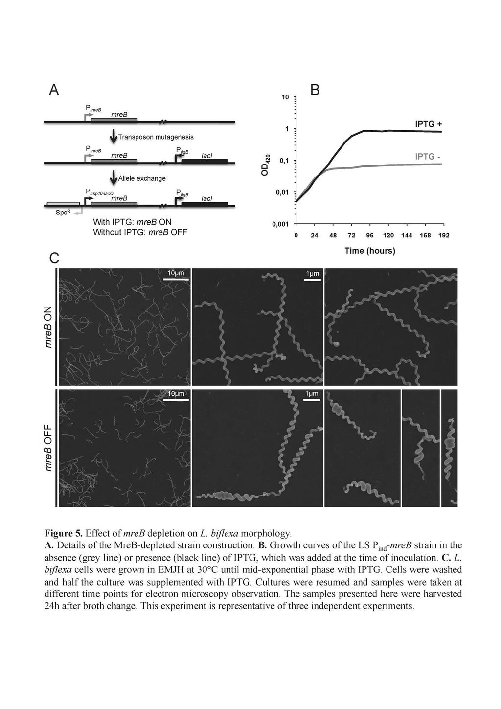
7 The P flgb promoter was amplified from B. burgdorferi chromosomal DNA using primer pair FLG5 and FLG3 (4) and cloned in pcr2.1 (Invitrogen) to create pp flgb. laci was amplified using primers LI1 (5 -CATATGGTGAATGTGAAACCAG-3 ) and LI2 (5 - TCAAACCAGATCAATTCGCG-3 ) and chromosomal DNA from E. coli as template. The amplification product was cloned between the NdeI and NsiI restriction sites of plasmid pp flgb to generate plasmid placi Ec. Transcriptional fusion P flgb -laci Ec, amplified from placi Ec with primer pair Lic1 (5 -TTTGGCGCGCCTAATACCCGAGCTTCAAGGAA-3 ) and Lic2 (5 - TTTGGCGCGCCTCAAACCAGATCAATTCGCG-3 ) was inserted in the AscI site of vector psw29t-tks 2 (44) to create plasmid ptk-laci Ec. The mreb coding sequence was amplified from L. biflexa chromosomal DNA using primers mreb1 (5 -GGAATTCCATATGATATTTGATAACCTTTATG-3 ) and mreb2 (5 - CTAGTCTAGAAAAATGGGAAACCTCGGAG-3 ) and cloned between the NdeI and XbaI sites of plasmid pp hsp10 - LacO (2) to generate plasmid pp hsp10 - LacO -mreb which carries a transcriptional fusion between an IPTG-inducible promoter and the mreb gene. A PCR fragment corresponding to the region upstream from mreb, amplified from L. biflexa chromosomal DNA using primers mreb3 (5 -TCGGGCCCGTTTCGAATGATATAATAAC- 3 ) and mreb4 (5 -CGGAATTCATCAGTTCCTGGTGAAAG-3 ), as well as a spectinomycin resistance cassette, amplified using primers SpcEco3 (5 - ACGGAATTCAACGCGTAAAGTAAG-3 ) and SpcNot5 (5 - ATAAGAATGCGGCCGCAACGCGTCCCGAGC-3 ), were cloned between the NotI and ApaI sites of pp hsp10 - LacO -mreb, creating plasmid pmreb::spc-p hsp10 - LacO. All these fragments were then cloned between the SmaI and XbaI sites of psw29t after digestion of pmreb::spc- P hsp10 - LacO with the same enzymes, thus generating plasmid pp ind -mreb. Strain LS P ind -mreb, that conditionally expresses mreb, was constructed in the following manner. E. coli ß2163 cells harboring ptk-laci Ec were used as donor cells to 7
8 mobilize the vector into L. biflexa as described previously (44). Exconjugants were selected on kanamycin and insertion site of the transposon carrying P hsp10 -laci was verified in a few of these. Strain lic2 with the transposon inserted at position of the large chromosome between LEPBIa3341 and LEPBIa3342 was selected as recipient for conjugation with E. coli ß2163 harboring pp ind -mreb as well as placi Ec to prevent mreb transcription in E. coli. Conjugation was carried out as described except that IPTG was added to the medium. Exconjugants were selected on spectinomycin. Replacement of the native mreb promoter with the spectinomycin resistance cassette and the inducible P hsp10 - LacO promoter by allele exchange was verified by PCR and sequencing. Random insertion mutagenesis was carried out in L. biflexa serovar Patoc strain Patoc 1, L. interrogans serovar Manilae strain L495, and L. interrogans serovar Lai strain Lai with a kanamycin-resistant Himar1 transposon as previously described (37). Among the kanamycin transformants, we identified mutants with an insertion into LA1045, LEPBIa1432, LA3698, and LA3692 (Table 1). Plasmid and chromosomal DNAs were prepared using Qiagen and Promega Maxwell cell purification kits, respectively. Real time quantitative reverse transcriptase PCR (qrt-pcr). These experiments were carried out as previously described (2). The specific primers for L. biflexa mreb (5 -GGTATGGTGATTGCCGAGTC-3 and 5 - TCTGCTGTTCTTTCCCCAAC-3 ) and L. interrogans ftsi (5 - GAACCGAAAGGTGGAACTCA-3 and 5 -AACCGAAATCCCAAGAGGTC-3 ) and LA3697 (5 -TTTAGTTTGTGGGGCTGGTC-3 and 5 -CCTGGGGATCAACTGAGAAA- 3 ), as well as specific rpob primers (5 -GCAAAATGAAAATCGCTGGT-3 and 5 - CAGCATCGAGCATTACCTCA-3 for L. biflexa and 5 -ATGGAGCGGAACGTGTAGTC- 8
9 and 5 -CTTCGTTCGTTCCATGTCCT-3 for L. interrogans) were used for PCR. Relative RNA levels were determined using the comparative C(T) method, also called the 2 (- C(T)) method (31). Peptidoglycan preparation for visualization and analysis. 200mL of L. biflexa or L. interrogans cells were grown until stationary phase and harvested at 5000g for 10min at room temperature. Cells were resuspended in 3mL PBS and dropped in 6mL of a boiling 6% SDS solution while stirring with a magnetic bar. The mixture was stirred for 4h in a boiling water bath, then overnight at room temperature as previously described (14). Sacculi were washed clean of SDS by four successive centrifugations and resuspensions in water using a Beckman TL100 centrifuge equipped with a TLA100.3 rotor at g for 10 min at 18ºC. For electron microscopy, sacculi were observed directly (raw) or after chymotrypsin digestion to remove contaminating protein. In the latter case, once SDS was removed, sacculi were digested overnight at 37ºC with 100 ug/ml chymotrypsin in PBS. After digestion, SDS was added to the suspension to a final concentration of 1% and sacculi were incubated for 2h in a boiling water bath, then cleaned free of SDS as described above. Sacculi were then resuspended into 100µL of water and carbon-formvar coated copper EM grids (200 mesh) were floated onto drops of this suspension. After 10 min, the grids were dried with filter paper and, after a few minutes of air-drying, were floated onto water drops and washed 6 times. Grids were then floated onto 1% uranyl acetate in water for 2min. After removing excess uranyl acetate, grids were washed once again very briefly with water, dried and either observed directly, or subjected to Carbon platinum shadowing at a 10º angle. Observations were made in a JEOL JEM1010 at 60KV, equipped with a TemCam-F416 (TVIPS, Germany) digital camera. 9
10 For composition analysis, PG was digested overnight at 37ºC with 40 μg/ml muramidase (Cellosyl, Hoescht), reduced with NaBH 4 to avoid anomerization of sugars, and subjected to HPLC to separate and purify the muropeptides as previously described (20, 44). Individual components were collected after HPLC separation and subjected to MALDI-TOF mass spectrometry (Autoflex, Bruker Daltonics, USA) to determine the molecular mass of the components. To confirm the proposed structures most muropeptides were further analyzed by electrospray ion-trap mass spectrometry (LCQ Classic, Thermo-Finnigan, USA) to define their amino acid and amino sugar sequences. For the mreb mutant sacculi preparation, LS P ind -mreb cells grown until midexponential phase in the presence of IPTG were harvested, the pellet was washed once in EMJH to remove IPTG and growth was resumed in EMJH for 16 h. Sacculi preparation was then pursued as described above. Scanning electron microscopy. For the analysis of Leptospira morphology, bacteria in exponential growth phase (OD 420 ~ 0.2) were incubated with various compounds (aztreonam, 0.2 µg/ml; amdinocillin, 20 µg/ml; penicillin G, 0.1 µg/ml; A22, 5 µg/ml). Cells were then harvested at 4, 8, 12, 20 or 24h by centrigufation, washed once in PBS, and resuspended in cacodylate buffer (0.1 M; ph7.2) supplemented with 2.5% glutaraldehyde. Samples were then processed for electron microscopy experiments as described previously (49). For cell length measurements, 50 to 100 cells were randomly chosen in the micrographs and distance between two ends was manually calculated using the ImageJ program ( 10
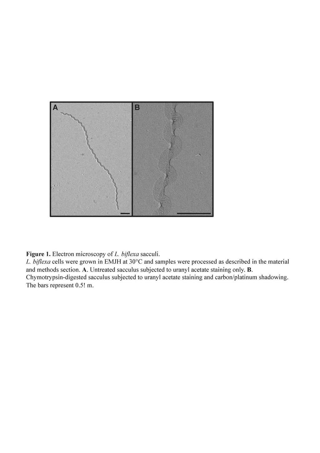
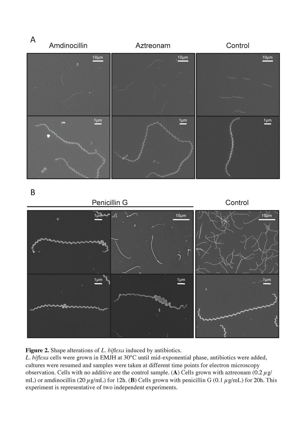
11 224 Results Sacculi from Leptospira retain a helical shape. To study the morphology of Leptospira, we first wanted to examine the shape of their murein sacculi. The PG macromolecules were purified and subjected to electron microscopy as described in the materials and methods section. The micrograph in Fig. 1 illustrates that Leptospira sacculi present a helical shape similar to the shape of the live microorganisms. This shape is intrinsic to the PG macromolecule since they must be devoid of protein after the chymotrypsin treatment to which they were subjected (Fig. 1B). The darker area along the longitudinal axis on Fig. 1B is probably due to the flattening of the sacculi. Purified sacculi of L. biflexa have a helix pitch of 470 ± 40 nm, a helix diameter of 326 ± 12 nm and a sacculus width of 175 ± 10 nm (data from 12 distinct sacculi). Alteration of cell shape by antibiotics or mutagenesis targeting PBPs To understand the role of enzymes involved in peptidoglycan synthesis and modification, we first listed the PBPs found in the genome of all sequenced Leptospira strains. Seven PBPs were identified: 5 class A PBPs, including four PBP1a proteins, and 2 class B PBPs (Table 1). No low molecular weight PBPs were identified. Transposon mutants of genes encoding two PBP1a were subjected to microscopic analysis. No morphological alterations were noted for these mutants compared to the wild-type strain (data not shown). This might be due to a functional redundancy in the Leptospira genomes. We then subjected L. biflexa cells to the action of inhibitors of these PG enzymes. Different antibiotics were chosen according to their spectrum of action in E. coli. Aztreonam preferentially inhibits PBP3, involved in septal peptidoglycan synthesis. Amdinocillin preferentially inhibits PBP2, involved in lateral peptidoglycan synthesis. Penicillin G inhibits all PBPs. In E. coli, 11
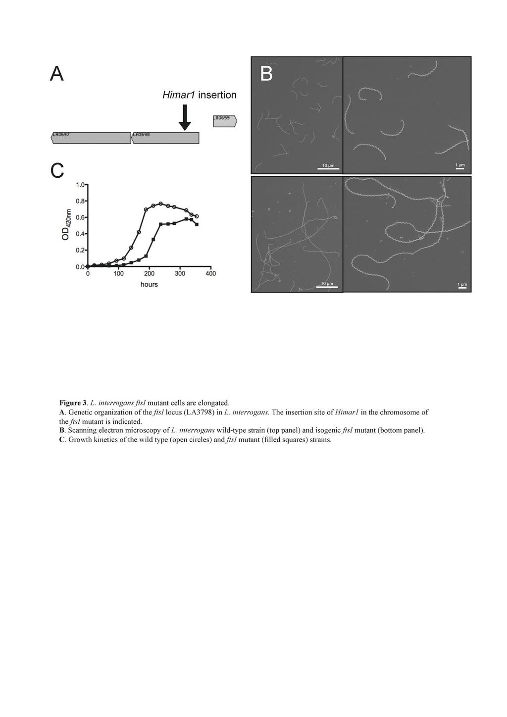
12 aztreonam induces the formation of elongated cells whereas addition of amdinocillin leads to round cells. The micrographs on figure 2A show that the majority (~87%) of L. biflexa cells incubated with 0.2 µg/ml aztreonam, which is a concentration below the MIC (0.5 µg/ml), were longer than the control sample (19.21 ± 5.60 µm vs ± 2.27 µm in the treated and untreated samples, respectively). The growth rate was comparable to that of the control cells (doubling time of 4 hours) until 8 hours after the addition of the antibiotic. Shape changes appeared after 4 hours, and the growth rate decreased to 70% of the control until the end of the experiment. Further, a mutant with a transposon insertion in LA3698 (ftsi) encoding PBP3 in L. interrogans serovar Manilae strain L495 (Fig.3A) presented an elongated-cell phenotype (25.11 ± µm, in comparison to 6.44 ± 0.93 µm for the parental strain) with ~16% of the cells being aberrantly elongated ( 40 µm) (Fig. 3B). The mutant showed poor growth in EMJH, compared to the parental strain (Fig. 3C), but was viable even after prolonged incubation times. In other bacteria, with the exception of cyanobacteria (34), ftsi is included in a gene cluster involved in cell division (3, 56). Similar to the organization in other Leptospira, ftsi in L. interrogans is not included in such a cluster and is adjacent to a gene encoding a protein of unknow function (LA3697) and to a gene encoding an arsenate reductase (LA3699). The mrna level of LA3697 was decreased in the mutant, in comparison to the parental strain (data not shown), suggesting that disruption of ftsi exerted a polar effect on the downstream gene and that we cannot rule out an effect of this gene in the morphological alterations observed. Contrary to E. coli, L. biflexa cells incubated with amdinocillin at concentrations around the MIC level also presented an elongated phenotype (22.38 ± 7.22 µm). Morphological changes appeared after 4 hours. The growth rate was comparable to the control 12
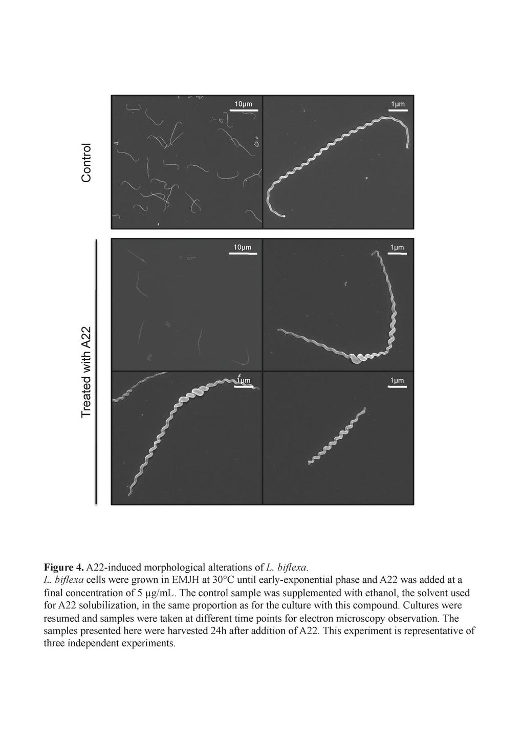
13 cells until 8 hours after addition of the antibiotic; then the cells stopped growing and started lysing 12 hours after addition of amdinocillin. Interestingly, when penicillin G was added to the culture at concentrations around the MIC level, cells presented a different morphology. There was heterogeneity in cell length but also a localized increase of cell diameter at various points along the longitudinal cell axis in ~ 43% of the cells (Figure 2B). Shape changes appeared after 12 hours. The growth rate was comparable to that of the control cells until 8 hours after addition of the antibiotic; it then decreased to 75% of the control and was 50% at the end of the experiment. MreB is involved in maintaining the diameter and length of the cells. We then examined the role of MreB, a protein involved in maintaining the proper morphology of several rod-shaped bacteria. MreB is present in all sequenced Leptospira strains. In the pathogens, but not in the saprophyte L. biflexa, mreb is genetically linked to the mrec and mred genes, as it is also the case in E. coli. L. biflexa MreB shares 50 to 60% identity with MreB from E. coli or C. crescentus. To understand the role of MreB, we first used the A22 compound that has been shown to affect the function of this actin-like homologue and to mimic loss of the protein in several Gram-negative microorganisms (5, 20, 41). Fig. 4 shows that cells treated with A22 at concentrations around the MIC level presented a phenotype similar to what we observed when they were subjected to penicillin G. Morphological changes appeared after 8 hours. The growth rate was comparable to the control cells until 8 hours after addition of the compound; then the cells stopped growing and started lysing 12 hours after addition of A22. Cells were of different lengths and there was a localized increase in cell diameter in various points along the longitudinal cell axis in ~ 95 % of the cells. A22-treated cells seem to present a less homogeneous helix pitch than the control cells. However the cells remained helical. 13
14 Since we did not succeed in disrupting the mreb gene by allelic exchange (our unpublished data), we constructed strain LS P ind -mreb carrying laci from E. coli and mreb under the control of a promoter containing LacO operator sequences (Fig. 5A). In the presence of IPTG, the mreb mrna level of LS P ind -mreb is comparable to the wild-type strain. In the absence of IPTG the LacI regulator binds to LacO and represses transcription of mreb. Transcript level of mreb decreased approximately 14 fold 1h after IPTG has been removed from the culture compared to the mreb mrna level in cells grown with IPTG (data not shown). The growth curve presented in figure 5B shows that in the absence of IPTG the culture reached a plateau at an OD 420 0,05 in 36 hours whereas the cells grew until they reached stationary phase at an OD after 90 hours in the presence of IPTG. Length of most of the cells grown without IPTG was shorter (5.92 ± 1.84 µm vs 8.58 ± 1.1 µm in absence of IPTG) and 16 % of the cells were smaller than 4 µm. 38% of the cells of the mutant also exhibited an increase diameter in localized regions along the longitudinal axis (Fig. 5C), as was observed with A22-treated cells. Further, after a longer incubation period cells started lysing. This observation and the fact that we could not disrupt the mreb gene suggest that MreB is essential for the growth of L. biflexa in standard laboratory conditions. Composition of L. biflexa peptidoglycan The morphological peculiarities of L. biflexa, in particular its extremely small diameter and helical geometry, might well impose constraints in the organization of the sacculus. Therefore, the muropeptide composition of its PG was determined as previously done in other microorganisms (21, 47). The HPLC elution pattern obtained is shown in Fig. 6A. Each peak should correspond to a specific PG subunit. The results presented in Table S1, allowed for the unambiguous identification of the major components resolved by HPLC and whose structures are displayed in Fig 6B. Identified peaks accounted for more than 80% of 14
15 the total peak area as calculated from the integration of the HPLC results shown in Fig 6A. The major components, peaks 1 and 10, were identified as the disaccharide tripeptide GlucNAc-MurNAc-L-Ala-D-Glu-(meso)-DAP (M3) and the cross-linked dimer GlucNAc- MurNAc-L-Ala-D-Glu-(meso)-DAP-D-ala (meso)-dap-d-glu-l-ala-murnac-glucnac (D4-3), respectively. An unusual feature of the HPLC elution pattern was the identification of two well-separated peaks (9 and 10) as the same component D4-3. The components in both peaks had virtually identical masses (Table S1). Furthermore, LC-MS/MS analysis supported identical sequences for both (data not shown). Peak 5 corresponded to the disaccharide pentapeptide GlucNAc-MurNAc-L-Ala-D-Glu-(meso)-DAP-D-Ala-D-Ala (M5) the canonical basic subunit of A1γpeptidoglycan (50), and peak 4 is the same component without the terminal D-Ala, which is usually the major muropeptide in Gram-negative bacteria. We identified 1 6 anhydromuramic acid residues in peak 15. These residues are important because they occupy the C-1 terminal position on the glycan chain and their relative abundance is an indication of the average length of PG strands (57). Most remarkable was the presence of a series of muropeptides (Fig 6, peaks 4, 6, 7, 8, 11, and 13) derived from D4-3, which might represent intermediate steps of a specific PG remodeling process. M4-3, M4-3L, D4-3(-GNA) and D4-3(-GNA)(-Ac) (Fig 6, peaks 4, 6, 7 and 8, respectively) are seldom detected in macromolecular PG in other species. Muropeptides in peaks 2, 8, 11 and 13 were also quite interesting because of specific chemical modifications; de-acetylation of the GlucNAc residue in M3(-Ac), D4-3(-Ac) and D4-3(-GNA)(-Ac); and amidation of the DAP residues in muropeptide D4-3(NH 2 ) 2. Neither modification is particularly rare, but both require specific enzymatic activities. Once the nature of the major muropeptides was defined, it was possible to quantify the relative abundance of each in molar terms (Table 2). Taking the well-known PG of E. coli as a comparative standard, L. biflexa PG is as cross-linked (31.6 %) as that of E. coli (30-35 %) 15
16 and other Gram-negative bacteria. Based on anhydro muropeptide proportion, L. biflexa PG seems made of longer glycan strands, averaging 125 disaccharides/strand, than E. coli PG which is made of disaccharides/strand (21). About 29% of the total muropeptides exhibited some alteration over the basic structure of M3 and D4-3. Most abundant were de-glucosaminidated and de-acetylated muropeptides which accounted for about 10 and 13 % of total muropeptides, respectively. The presumptive products of MurNAc-L-Ala amidases and etherases, M4-3 and M4-3L, accounted for about 2% of the total each, a rather significant fraction in structural terms. Such a large-scale postinsertional modification should certainly have consequences on PG properties, and suggests an elaborate processing of macromolecular PG in L. biflexa. The proposed structures can be explained as naturally derived from the basic disaccharide pentapeptide subunit as the consequence of known biosynthetic and degradative enzyme activities. However, we could not suggest a sound structure even after MS/MS for X1 and X2 that compose peaks 12 and 14 which seem to be modified muropeptides as both contain residues of GlucNAc and MurNAc. Treatment of L. biflexa PG with pronase-e had no detectable effect on the HPLC elution pattern (data not shown), suggesting absence of covalently PG- bound proteins equivalent to Braun s lipoprotein of E. coli (21). We also investigated PG composition of a representative pathogenic strain, L. interrogans serovar Manilae strain L495. The results show that overall PG composition of L. interrogans was similar to that of L. biflexa. However some differences were noted. L. interrogans showed a low proportion of cross-linked muropeptides (D4-3) and consequently of the muropeptides that derive from D4-3. There is also a lower proportion of 1 6 anhydromuramic acid residues, which indicates that the glycan chains might be longer in L. interrogans than in L. biflexa. 16
17 Effect of impaired MreB functionality on L. biflexa PG composition. To study whether the morphological modifications observed on cells subjected to alterations of the function or level of MreB could be associated to specific changes on PG composition, sacculi isolated from A22-treated cells, or from a strain depleted of MreB were subjected to HPLC analysis. The results presented in Table 2 show that the presence of A22 caused a dramatic rise in the proportion of M4 and a concomitant drop in M3. In addition, A22 treatment caused a strong reduction in the proportion of de-acetylated muropeptides, which dropped to 60% of the control, and a moderate decrease in the proportion of crosslinked muropeptides. Decrease of MreB level showed both similarities and important differences on the PG composition of these cells compared to cells treated with A22. The effect on cross-linking was more pronounced than that of A22, and was accompanied by a marked reduction in D4-3(-GLA) muropeptides. De-acetylated muropeptides were reduced to a similar proportion as in the drug treated cells. The more intriguing observation was the effect on the relative levels of M4 and M3, which was the opposite to the effect of A22 causing a further reduction of M4 instead of an accumulation (Table 2). It has to be noted that while A22 acts fast and inhibit all MreB molecules simultaneously, MreB depletion is probably a progressive and slow process in particular in an organism with such a long generation time as Leptospira. This difference could explain why the effects on PG composition were not identical although the trends were similar. 17
18 Discussion Cell shape determination and maintenance have been studied until now mainly in rodshaped organisms such as E. coli and B. subtilis, or in the crescent-shaped C. crescentus. Spirochetes have a unique morphology, ranging from helix- to flat wave-shaped cells (12). It is not clear why some spirochetes form helices and others flat waves. It has been shown that endoflagella is sufficient to determine the flat-wave morphology of B. burgdorferi. In contrast to B. burgdorferi, the endoflagella in Leptospira do not cover the entire cell length and mutants deficient in endoflagella retain their helical morphology (45). The saprophytic and pathogenic Leptospira strains share the same morphological features except that pathogenic strains are usually shorter in length than saprophytic strains (18). Our results show that the helical morphology of Leptospira is intrinsic to the PG macromolecule, unlike in B. burgdorferi (35). Similar to Leptospira spp., purified sacculi from Treponema pallidum examined by electron microscopy indicated that the PG contributes to the spiral cell shape (48, 55). Our work shows that although its PG conforms to a common Gram-negative chemotype, the constituents of this macromolecule are peculiar. For example, the fact that muropeptides with terminal D-Ala residues were so scarce in L. biflexa and L. interrogans PG is quite unusual and suggests the presence of specific peptidases such as the tandem action of DD- and LD-carboxypeptidases, or an LD-endopeptidase able to accept M5 as a substrate. Interestingly, a similar occurrence has been documented in the Gram-positive bacterium B. subtilis (1). Detection of M4-3, M4-3L, D4-3(-GNA) and D4-3(-GNA)(-Ac) is rare in the PG of other species. Enzymes potentially able to produce each of them from D4-3 subunits in the sacculus have been described in other systems and are indicated in Fig 7. Muropeptide M4-3 is normally the product of a MurNac-L-alanyl-amidase. These enzymes are often found in Gram-negative bacteria where they play an important role in division, but their products rarely accumulate in macromolecular PG (58). Interestingly amidase activity seems to be up- 18
19 regulated in Salmonella typhimurium following colonization of non-phagocytic cells (46). Generation of M4-3L from D4-3 would require the activity of an etherase. Such an enzyme designated as MurQ has been described in E. coli. However this enzyme is cytoplasmic, and is only active on previously phosphorylated muropeptides in the course of PG recycling (54). Therefore, the putative enzyme responsible for generation of M4-3L might represent a new kind of PG hydrolase. Cleavage of the GlucNAc-MurNAC glycosydic bond by an N- Acetylglucosaminidase is the likely origin of muropeptides lacking a GlucNAc residue (peaks 7 and 8). Such enzymes are widespread among bacteria. However their role in Gram-negative bacteria is often restricted to PG recycling and requires soluble muropeptides as substrates (58). Interestingly, all the unusual muropeptides discussed above require cleavage of preexisting glycan strands or peptide bridges in macromolecular PG and therefore could cause local variations on the physical properties of PG. Peptidoglycan relaxation by hydrolytic enzymes has been found to promote helical shape in Helicobacter pylori (53). A similar process of modification of the organization of the network of peptidoglycan subunits may play a predominant role in the generation of the helical cell shape of Leptospira spp. (24). Subtle and notable differences were found in the muropeptide composition of the PG in the environmental species L. biflexa and the pathogen L. interrogans. The very low cross-linking in L. interrogans may reflect an adaptation to isoosmotic conditions. Genome-wide analysis revealed that several shape-related proteins such as FtsZ, MreBCD and RodA are present in Leptospira spp. We showed that MreB, one of the most abundant cytoplasmic protein of L. interrogans with 2500 copies per cell (32), is involved in maintaining specific features of Leptospira morphology, such as its length, diameter in localized areas and homogeneity of the pitch of the helix. This phenotype was similar to what we observed with penicillin G treated cells. These observations suggest that the role of MreB in Leptospira is linked to at least one PBP. The localized increase in diameter could suggest 19
20 that there is accumulation of PG synthesis in that area. B. subtilis mreb mutants form elongated and wider cells with bulging poles. It has been proposed that this phenotype was due to mislocalization of PBP1 in these cells and to its accumulation at the poles (27). It would be interesting to examine the localization of MreB and PBPs in healthy cells and in cells treated with various shape-altering compounds. Among a library of random mutants (37), we identified transposon mutants exhibiting an insertion in genes encoding PBP1a and PBP3 (Table 1). Morphological alteration was only observed in the PBP3 mutant. The numerous HMW PBPs belonging to the class A that are present in leptospires may compensate for the loss of PBP1a production. Like in E. coli (13), the L. interrogans PBP3 mutant forms filamentous cells, suggesting its involvement in cell division. However, unlike E. coli, FtsI does not appear to be essential in Leptospira (52). Surprisingly, LMW PBPs that generally act as carboxypeptidases or endopeptidases were not found in the Leptospira genomes which suggests that other enzymes should undertake these functions as it has recently been shown in H. pylori (6). Among the antibiotics used to assess the role of various PBPs in the morphology of Leptospira, amdinocillin did not induce the expected phenotype and cells were not affected in lateral peptidoglycan synthesis. It is possible that amdinocillin does not target the same PBP in Leptospira as in E. coli, or that this PBP does not have the same role in this species. The antibiotics that we tested did not really affect the overall helical shape of the bacterium. Other proteins might be involved in the determination of this feature. Cytoplasmic filaments have been visualized using tomography in several spirochetes, including Leptospira spp. (29, 30, 32). However, little is known about the function of these elements. Previous findings in Treponema denticola suggest that they might be involved in cell division, structural integrity, motility and/or chromosome structure and segregation (25). Genome-wide analysis of Leptospira spp. allowed for the identification 20
21 of several putative coiled-coil proteins (33) that might be involved in shape determination similarly to CreS in C. crescentus. In conclusion, our data provide the first description of the muropeptide content of Leptospira strains. We also showed that inactivation of mreb or ftsi resulted in mutants with morphological abnormalities such as aberrant diameter or length of the cells, therefore demonstrating that these proteins are important components of the morphology of Leptospira cells. Our results provide a starting point for a better understanding of the involvement of PBPs and cytoskeletal proteins in the morphogenesis of these atypical bacteria. Acknowledgments We are grateful to Stéphanie Guadignini and Marie-Christine Prevost at the Plateforme de Microscopie Ultrastructurale for their help with the electron microscopy. We would also like to thank Gustavo Cerqueira for preliminary attempts to inactivate L. biflexa mreb. This work was supported by the Institut Pasteur, Paris, France and the French Ministry of Research ANR-08-MIE-018. M.A.P. was supported by Ministry of Education and Science, Spain (MEC, BFU ) and Fundación Ramón Areces. 21
22 References 1. Atrih, A., G. Bacher, G. Allmaier, M. P. Williamson, and S. J. Foster Analysis of peptidoglycan structure from vegetative cells of Bacillus subtilis 168 and role of PBP 5 in peptidoglycan maturation. J. Bacteriol. 181: Aviat, F., L. Slamti, G. M. Cerqueira, K. Lourdault, and P. M Expanding the genetic toolbox for Leptospira species by generation of fluorescent bacteria. Appl. Environ. Microbiol. 76: Ayala, J., C. Goffin, M. Nguyen-Distèche, and J. M. Ghuysen Site-directed mutagenesis of penicillin-binding protein 3 of Escherichia coli: role of Val-545. FEMS Microbiol. Lett. 121: Bauby, H., I. Saint Girons, and M. Picardeau Construction and complementation of the first auxotrophic mutant in the spirochaete Leptospira meyeri. Microbiology 149: Bean, G. J., S. T. Flickinger, W. M. Westler, M. E. McCully, D. Sept, D. B. Weibel, and K. J. Amann A22 disrupts the bacterial actin cytoskeleton by directly binding and inducing a low-affinity state in MreB. Biochemistry 48: Bonis, M., C. Ecobichon, S. Guadagnini, M. C. Prevost, and I. G. Boneca A M23B family metallopeptidase of Helicobacter pylori required for cell shape, pole formation and virulence. Mol. Microbiol. 78: Brenot, A., D. Trott, I. Saint Girons, and R. Zuerner Penicillin-binding proteins in Leptospira interrogans. Antimicrob. Agents Chemother. 45: Cabeen, M. T., G. Charbon, W. Vollmer, P. Born, N. Ausmees, D. B. Weibel, and C. Jacobs-Wagner Bacterial cell curvature through mechanical control of cell growth. EMBO J. 28: Cabeen, M. T., and C. Jacobs-Wagner Bacterial cell shape. Nat. Rev. Microbiol. 3: Cabeen, M. T., and C. Jacobs-Wagner Skin and bones: the bacterial cytoskeleton, cell wall, and cell morphogenesis. J. Cell Biol. 179: Carballido-Lopez, R., and A. Formstone Shape determination in Bacillus subtilis. Curr. Opin. Microbiol. 10: Charon, N. W., and S. F. Goldstein Genetics of motility and chemotaxis of a fascinating group of bacteria: the spirochetes. Annu. Rev. Genet. 36:
23 Curtis, N. A., R. L. Eisenstadt, K. A. Turner, and A. J. White Inhibition of penicillin-binding protein 3 of Escherichia coli K-12. Effects upon growth, viability and outer membrane barrier function. J. Antimicrob. Chemother. 16: de Pedro, M. A., J. C. Quintela, J. V. Höltje, and H. Schwarz Murein segregation in Escherichia coli. J. Bacteriol. 179: Demarre, G., A. M. Guerout, C. Matsumoto-Mashimo, D. A. Rowe-Magnus, P. Marliere, and D. Mazel A new family of mobilizable suicide plasmids based on broad host range R388 plasmid (IncW) and RP4 plasmid (IncPalpha) conjugative machineries and their cognate Escherichia coli host strains. Res. Microbiol. 156: den Blaauwen, T., M. A. de Pedro, M. Nguyen-Disteche, and J. A. Ayala Morphogenesis of rod-shaped sacculi. FEMS Microbiol. Rev. 32: Ellinghausen, H. C., and W. G. McCullough Nutrition of Leptospira pomona and growth of 13 other serotypes: fractionation of oleic albumin complex and a medium of bovine albumin and polysorbate 80. Am. J. Vet. Res. 26: Ellis, W. A., K. Hovind-Hougen, S. Möller, and A. Birch-Andresen Morphological changes upon subculturing of freshly isolated strains of Leptospira interrogans serovar hardjo. Zentralbl. Bakteriol. Mikrobiol. Hyg. 255: Georgopapadakou, N. H., S. A. Smith, and R. B. Sykes Mode of action of azthreonam. Antimicrob. Agents Chemother. 21: Gitai, Z., N. A. Dye, A. Reisenauer, M. Wachi, and L. Shapiro MreB actinmediated segregation of a specific region of a bacterial chromosome. Cell 120: Glauner, B., J. V. Höltje, and U. Schwarz The composition of the murein of Escherichia coli. J. Biol. Chem. 263: Haake, D. A., E. M. Walker, D. R. Blanco, C. A. Bolin, M. N. Miller, and M. A. Lovett Changes in the surface of Leptospira interrogans serovar grippotyphosa during in vitro cultivation. Infect. Immun. 59: Holt, S. C Anatomy and chemistry of spirochetes. Microbiol. Rev. 42: Huang, K. C., R. Mukhopadhyay, B. Wen, Z. Gitai, and N. S. Wingreen Cell shape and cell-wall organization in Gram-negative bacteria. Proc. Natl. Acad. Sci. U.S.A. 105:
24 Izard, J., W. A. Samsonoff, and R. J. Limberger Cytoplasmic filamentdeficient mutant of Treponema denticola has pleiotropic defects. J. Bacteriol. 183: Johnson, R. C., and V. G. Harris Differentiation of pathogenic and saprophytic leptospires. J. Bacteriol. 94: Kawai, Y., R. A. Daniel, and J. Errington Regulation of cell wall morphogenesis in Bacillus subtilis by recruitment of PBP1 to the MreB helix. Mol. Microbiol. 71: Ko, A. I., C. Goarant, and M. Picardeau Leptospira: the dawn of the molecular genetics era for an emerging zoonotic pathogen. Nat. Rev. Microbiol. 7: Kurner, J., A. S. Frangakis, and W. Baumeister Cryo-electron tomography reveals the cytoskeleton structure of Spiroplasma melliferum. Science 307: Liu, J., J. K. Howell, S. D. Bradley, Y. Zheng, Z. H. Zhou, and S. J. Norris Cellular architecture of Treponema pallidum: novel flagellum, periplasmic cone, and cell envelope as revealed by cryo electron tomography. J. Mol. Biol. 403: Livak, K. J., and T. D. Schmittgen Analysis of relative gene expression data using real-time quantitative PCR and the 2(-Delta Delta C(T)) methods. Methods 25: Malmström, J., M. Beck, A. Schmidt, V. Lange, E. W. Deutsch, and R. Aebersold Proteome-wide cellular protein concentrations of the human pathogen Leptospira interrogans. Nature 460: Mazouni, K., G. Pehau-Arnaudet, P. England, P. Bourhy, I. Saint Girons, and M. Picardeau The scc spirochetal coiled-coil protein forms helix-like filaments and binds to nucleic acids generating nucleoprotein structures. J. Bacteriol. 188: Miyagishima, S. Y., C. P. Wolk, and K. W. Osteryoung Identification of cyanobacterial cell division genes by comparative and mutational analyses. Mol. Microbiol. 56: Motaleb, M. A., L. Corum, J. L. Bono, A. F. Elias, P. Rosa, D. S. Samuels, and N. Charon Borrelia burgdorferi periplasmic flagella have both skeletal and motility functions. Proc. Natl. Acad. Sci. U.S.A. 97: Murray, C. K., and D. R. Hospenthal Broth microdilution susceptibility testing for Leptospira spp. Antimicrob. Agents Chemother. 48:
25 Murray, G. L., V. Morel, G. M. Cerqueira, J. Croda, A. Srikram, R. Henry, A. I. Ko, O. A. Dellagostin, D. M. Bulach, R. Sermswan, B. Adler, and M. Picardeau Genome-wide transposon mutagenesis in pathogenic Leptospira spp. Infect. Immun. 77: Nelson, D. E., A. S. Ghosh, A. L. Paulson, and K. D. Young Contribution of membrane-binding and enzymatic domains of penicillin binding protein 5 to maintenance of uniform cellular morphology of Escherichia coli. J. Bacteriol. 184: Nelson, D. E., and K. D. Young Penicillin binding protein 5 affects cell diameter, contour, and morphology of Escherichia coli. J. Bacteriol. 182: Neu, H. C Penicillin-binding proteins and role of amdinocillin in causing bacterial cell death. Am. J. Med. 75: Noguchi, N., K. Yanagimoto, H. Nakaminami, M. Wakabayashi, N. Iwai, M. Wachi, and M. Sasatsu Anti-infectious effect of S-benzylisothiourea compound A22, which inhibits the actin-like protein, MreB, in Shigella flexneri. Biol. Pharm. Bull 31: Osborn, M. J., and L. Rothfield Cell shape determination in Escherichia coli. Curr. Opin. Microbiol. 10: Paster, B. J., F. E. Dewhirst, W. G. Weisburg, L. A. Tordoff, G. J. Fraser, R. B. Hespell, T. B. Stanton, L. Zablen, L. Mandelco, and C. R. Woese Phylogenetic analysis of the spirochetes. J. Bacteriol. 173: Picardeau, M Conjugative transfer between Escherichia coli and Leptospira spp. as a new genetic tool. Appl. Environ. Microbiol. 74: Picardeau, M., A. Brenot, and I. Saint Girons First evidence for gene replacement in Leptospira spp. Inactivation of L. biflexa flab results in non-motile mutants deficient in endoflagella. Mol. Microbiol. 40: Quintela, J. C., M. A. de Pedro, P. Zollner, G. Allmaier, and F. Garcia-del Portillo Peptidoglycan structure of Salmonella typhimurium growing within cultured mammalian cells. Mol. Microbiol. 23: Quintela, J. C., E. Pittenauer, G. Allmaier, V. Aran, and M. A. de Pedro Structure of peptidoglycan from Thermus thermophilus HB8. J. Bacteriol. 177:
26 Radolf, J. D., C. Moomaw, C. A. Slaughter, and M. V. Norgard Penicillinbinding proteins and peptidoglycan of Treponema pallidum subsp. pallidum. Infect. Immun. 57: Ristow, P., P. Bourhy, S. Kerneis, C. Schmitt, M. C. Prevost, W. Lilenbaum, and M. Picardeau Biofilm formation by saprophytic and pathogenic leptospires. Microbiology 154: Schleifer, K. H., and O. Kandler Peptidoglycan types of bacterial cell walls and their taxonomic implications. Bacteriol. Rev. 36: Spratt, B. G Distinct penicillin binding proteins involved in the division, elongation, and shape of Escherichia coli K12. Proc. Natl. Acad. Sci. U.S.A. 72: Spratt, B. G Temperature-sensitive cell division mutants of Escherichia coli with thermolabile penicillin-binding proteins. J. Bacteriol. 131: Sycuro, L. K., Z. Pincus, K. D. Gutierrez, J. Biboy, C. A. Stern, W. Vollmer, and N. R. Salama Peptidoglycan crosslinking relaxation promotes Helicobacter pylori's helical shape and stomach colonization. Cell 141: Uehara, T., K. Suefuji, T. Jaeger, C. Mayer, and J. T. Park MurQ Etherase is required by Escherichia coli in order to metabolize anhydro-n-acetylmuramic acid obtained either from the environment or from its own cell wall. J. Bacteriol. 188: Umemoto, T., T. Ota, H. Sagawa, K. Kato, H. Takada, M. Tsujimoto, A. Kawasaki, T. Ogawa, K. Harada, and S. Kotani Chemical and biological properties of a peptidoglycan isolated from Treponema pallidum kazan. Infect. Immun. 31: Vicente, M., M. J. Gomez, and J. A. Ayala Regulation of transcription of cell division genes in the Escherichia coli dcw cluster. Cell Mol. Life Sci. 54: Vollmer, W., D. Blanot, and M. A. de Pedro Peptidoglycan structure and architecture. FEMS Microbiol. Rev. 32: Vollmer, W., B. Joris, P. Charlier, and S. Foster Bacterial peptidoglycan (murein) hydrolases. FEMS Microbiol. Rev. 32: Yanagihara, Y., K. Kamisango, S. Yasuda, S. Kobayashi, I. Mifuchi, I. Azuma, Y. Yamamura, and R. C. Johnson Chemical compositions of cell walls and polysaccharide fractions of spirochetes. Microbiol. Immunol. 28:
27 Young, K. D The selective value of bacterial shape. Microbiol. Mol. Biol. Rev. 70: Downloaded from on December 3, 2018 by guest 27
28 Table 1. Penicillin-binding-proteins present in all Leptospira sequenced strains. Gene number Name Class L. interrogans L. borgpetersenii L. biflexa PBP1a A LA3692 a LBL_0402 LEPBIa0847 PBP1a A LA1221 LBL_2123 LEPBIa1010 PBP1a A LA1009 LBL_0498 LEPBIa0637 PBP1a A LA1045 a LBL_2793 LEPBIa1432 a PBP1c A LA2187 LBL_1576 LEPBIa0534 PBP2 B LA2755 LBL_1957 LEPBIa3075 PBP3 B LA3698 a LBL_2647 LEPBIa0257 a mutants harboring a transposon in these genes are available. Localization of Himar1 insertion: LA3692 (808 amino acids), insertion after aa 66 ; LA1045 (827 aa), insertion after aa 122 ; LA3698 (602 aa), insertion after aa 96 ; LEPBIa1432 (850aa), insertion after aa 437. Downloaded from on December 3, 2018 by guest 28
29 Table 2. Muropeptide composition of peptidoglycan purified from Leptospira strains Molar fraction x L. biflexa L. interrogans Muropeptide 2 Control A22 MreB wild-type M ,2 M3(-Ac) ,6 M M ,3 M ,6 M4-3L ,5 D4-3(-GNA) ,5 D4-3(-GNA)(-Ac) D D ,9 D4-3(-Ac) X ,9 D4-3(NH 2 ) ,7 X ,4 D4-3-Anh ,5 Groups 3 Monomers ,1 Dimers ,6 Anhydro ,5 Amidase ,3 Etherase ,5 Glucosaminidase ,5 Amidation ,7 Deacetylase ,6 1 Calculated as defined in (21). 2 The detailed structure of each muropeptide is shown in Fig 5B. 3 Muropeptides grouped by common characteristics. Italics indicate that the muropeptides are potential products of the indicated enzymes or reactions. 29
30 673 Figure Legends Figure 1. Electron microscopy of L. biflexa sacculi. L. biflexa cells were grown in EMJH at 30 C and samples were processed as described in the material and methods section. A. Untreated sacculus subjected to uranyl acetate staining only. B. Chymotrypsin-digested sacculus subjected to uranyl acetate staining and carbon/platinum shadowing. The bars represent 0.5µm. Figure 2. Shape alterations of L. biflexa induced by antibiotics. L. biflexa cells were grown in EMJH at 30 C until mid-exponential phase, antibiotics were added, cultures were resumed and samples were taken at different time points for electron microscopy observation. Cells with no additive are the control sample. (A) Cells grown with aztreonam (0.2 µg/ml) or amdinocillin (20 µg/ml) for 12h. (B) Cells grown with penicillin G (0.1 µg/ml) for 20h. This experiment is representative of two independent experiments. Figure 3. L. interrogans ftsi mutant cells are elongated. A. Genetic organization of the ftsi locus (LA3798) in L. interrogans. The insertion site of Himar1 in the chromosome of the ftsi mutant is indicated. B. Scanning electron microscopy of L. interrogans wild-type strain (top panel) and isogenic ftsi mutant (bottom panel). C. Growth kinetics of the wild type (open circles) and ftsi mutant (filled squares) strains. Figure 4. A22-induced morphological alterations of L. biflexa. L. biflexa cells were grown in EMJH at 30 C until early-exponential phase and A22 was added at a final concentration of 5 µg/ml. The control sample was supplemented with 30
31 ethanol, the solvent used for A22 solubilization, in the same proportion as for the culture with this compound. Cultures were resumed and samples were taken at different time points for electron microscopy observation. The samples presented here were harvested 24h after addition of A22. This experiment is representative of three independent experiments. Figure 5. Effect of mreb depletion on L. biflexa morphology. A. Details of the MreB-depleted strain construction. B. Growth curves of the LS P ind -mreb strain in the absence (grey line) or presence (black line) of IPTG, which was added at the time of inoculation. C. L. biflexa cells were grown in EMJH at 30 C until mid-exponential phase with IPTG. Cells were washed and half the culture was supplemented with IPTG. Cultures were resumed and samples were taken at different time points for electron microscopy observation. The samples presented here were harvested 24h after broth change. This experiment is representative of three independent experiments. Figure 6. L. biflexa muropeptides. A. A sample of purified and muramidase-digested L. biflexa PG was subjected to HPLC. The eluate was monitored measuring A 204. Peaks were collected and subjected to MS for further analyses. Peak numbers correlate with Fig. 6B and Table S1. B. Composition and structure of L. biflexa muropeptides as deduced from HPLC and MS data presented in A and table 1. The proposed abbreviations stand for: First letter M monomer, D dimer; first number indicates number of amino acids in the donor stem peptide, second number indicates number of amino acids in the acceptor stem peptide; (-GNA) lacking a residue of N-acetylglucosamine; (-Ac) lacking an acetyl group at position 2 of N-acetylglucosamine; (NH 2 ) amidated at the D- carboxyl of DAP; -Anh with a residue of (1 6) anhydro muramic acid. 31
32 Figure 7. Representation of target sites in macromolecular PG for the enzymes proposed as origin of L. biflexa modified muropeptides. x and y indicate two glycan chains of undefined length Downloaded from on December 3, 2018 by guest 32
33
34
35
36
37