Comprehensive Single-Shot Proteomics with FAIMS on a Hybrid Orbitrap Mass Spectrometer
|
|
|
- Julius Flowers
- 5 years ago
- Views:
Transcription
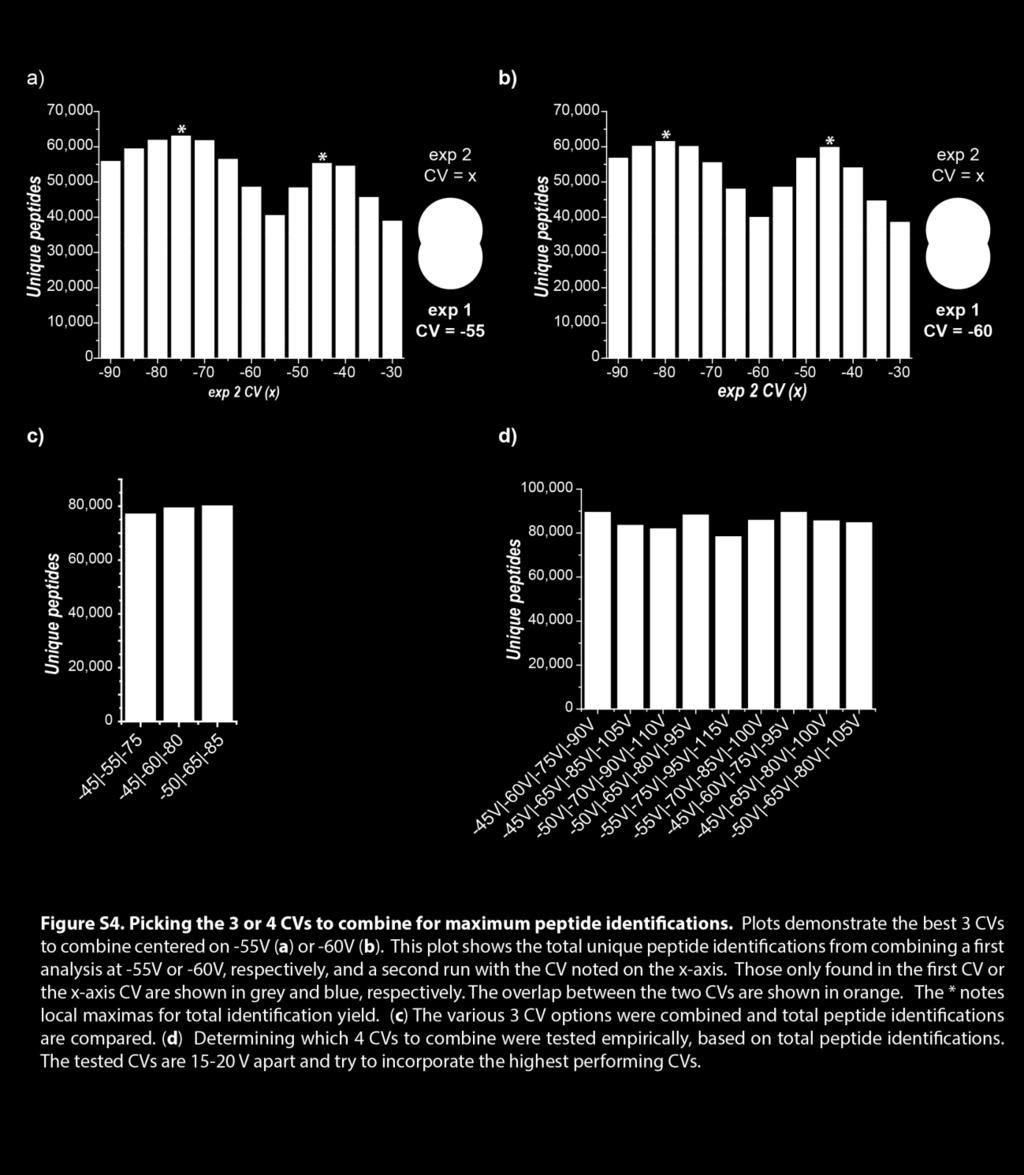
1 Supporting Information Comprehensive Single-Shot Proteomics with FAIMS on a Hybrid Orbitrap Mass Spectrometer Alexander S Hebert 1,2, Satendra Prasad 3, Michael W Belford 3, Derek J Bailey 3, Graeme C McAlister 3, Susan E Abbatiello 4, Romain Huguet 3, Eloy R. Wouters 3, Jean-Jacques Dunyach 3, Dain R. Brademan 5, Michael S. Westphall 1, and Joshua J. Coon 1,2,5,6,7* 1. Genome Center of Wisconsin, University of Wisconsin-Madison, Madison, WI DOE Great Lakes Bioenergy Research Center, University of Wisconsin-Madison, Madison, WI Thermo Fisher Scientific, San Jose, CA Thermo Fisher Scientific, Cambridge, MA Department of Chemistry, University of Wisconsin-Madison, Madison, WI Department of Biomolecular Chemistry, University of Wisconsin-Madison, Madison, WI Morgridge Institute for Research, Madison, WI *Corresponding author: jcoon@chem.wisc.edu Figure S1. Characterization of peptides over range of tested CVs. Figure S2. Unique peptides per protein comparison with and without FAIMS, and LC fractions. Figure S3. FAIMS inner electrode temperature effect on peak capacity. Figure S4. Selecting the 3 or 4 CVs to combine for maximum peptide identifications. Figure S5. Instrument method diagrams and External vs internal CV stepping. Figure S6. Peptide intensity reproducibility and cycle time. Figure S7. Beneficial characteristics of FAIMS analyses. Supplemental Materials and Methods


2


3


4

5


6

7


8
9 Supplemental Materials and Methods Tryptic digestions. Cells (E. coli, yeast, or K562) and tissue (whole rat brain) were suspended or homogenized (probe sonication) in 6 M guanidine, 50 mm tris ph 8, respectively. Cells were lysed and proteins were precipitated by addition of methanol to 90% final concentration. Samples were centrifuged at 10,000xg for 5 min. Supernatants were discarded and protein pellets were dissolved in 8 M urea, 100 mm Tris ph 8.0, 40 mm chloroacetamide, and 10 mm TCEP. LysC (Wako, Japan) was added to each sample at an estimated 100:1, protein:enzyme, ratio and incubated at room temperature for 4 hours. Each sample was diluted with 50 mm tris ph 8.0 to a final urea concentration of 2 M. Trypsin (Promega, Madison, WI) was added at an estimated 50:1, protein:enzyme, ratio and incubated overnight (~16 hours) at room temperature. Resulting peptides were desalted over a Strata-x PSDVB cartridge (Phenomenex, Torrance, CA), dried, and re-suspended in 0.2% formic acid. Peptide concentrations were determined by nanodrop absorbance at 205 nm (Thermo Fisher Scientific, Madison, WI). GluC, Chymotrypsin, and LysC digestions. K562 cells were lysed and the protein pellet was re-suspended as described above. Each sample was diluted with 50 mm tris ph 8.0 to a final urea concentration of 2 M. Either LysC, GluC (Promega, Madison, WI), or chymotrypsin (Promega, Madison, WI) was added at an estimated 50:1, protein:enzyme, ratio and the samples were incubated overnight (~16 hours) at room temperature. Resulting peptides were desalted over a Strata-x PSDVB cartridge, dried, and re-suspended in 0.2% formic acid. Peptide concentrations were determined by nanodrop absorbance at 205 nm. High ph reversed phase fractionation. For fractionation experiments 0.5 mg of tryptic K562 peptides were gradient separated over a 4.6 mm x 150 mm 3.5 µm bridged ethylene hybrid particles C18 column (Waters, Milford, MA) using an Ultimate 3000 (Thermo Fisher Scientific) with absorbance at 210 nm. Mobile phase A was 20 mm ammonium bicarbonate and mobile phase B was 20 mm ammonium bicarbonate in 80% methanol. For each experiment twice the number of desired fractions were collected and concatenated down by half. Fractions were further pooled to the final desired number by combining equivalent numbers of adjacent fractions. Fractions were dried and then peptides were dissolved in 0.2 % formic acid. NanoLC conditions - All analyses, except where noted, were performed with 2 µg of peptides injected on column. For fractionation experiments this was estimated based on the amount fractionated and the number of fractions the peptides were divided into. Separations were performed over 75 µm inner diameter x 360 µm outer diameter columns with an integrated electrospray emitter (New Objective, Woburn, MA) packed 30 cm long with 1.7 µm C18 bridged ethylene hybrid particles (Waters, Milford, MA) 8. Separation times noted in the text include loading time in 100% A (0.2% formic acid), gradient elution in increasing % B (0.2% formic acid/70% acetonitrile), and re-equilibration times in 100% A. An additional overhead of ~3 minutes for loading the sample into the sample loop is not included. All separations were performed with a Thermo Dionex Ultimate 3000 RSLC-nano liquid chromatography instrument and an in house fabricated column heater to conduct separations at 50 C.