Workshop in Diagnostic Immunohistochemistry Aalborg University Hospital, September 2018
|
|
|
- Lisa Logan
- 5 years ago
- Views:
Transcription
1 Workshop in Diagnostic Immunohistochemistry Aalborg University Hospital, September 2018 The technical test approach Pre-Analytical - Analytical (I & II) - Post Analytical phase Michael Bzorek Histotechnologist Department of Surgical Pathology University Hospital, Region Zealand, Denmark

2 The total test paradigm: Key elements in the IHC procedure The Analytic phase : Begins with dewax of the cut slides and is completed with the coverslipping of the stained slides. Unlike the pre-analytic factors, analytic factors (excentric to the tissue block) can be modified and controlled within the immunohistology laboratory.

3 Immunohistochemistry A simple technique? M External Quality Assurance programs Staining quality varies greatly between different laboratories depending on the individual selection of methods and the technical expertise


 Can we use the")
4 Calcitonin optimization (data sheets?) Ventana/ Cell Marque Optimizing an assay can be confusing (Vendor recommendations) Can we use the recommendations provided by the manufactures spec sheets?


5 Omnis, Flex+ Calcitonin optimization (data sheets?) Spring B recommendation : No pretreatment Thyroid medullary carcinoma Ventana recommendation: HIER in alkaline buffer
6 Optimization of the IHC assay issues to be addressed Purpose and/or fit-for-purpose of the IHC test How to establish best practice protocol of the IHC test (Calibration of the IHC assay with focus clone, antigen retrieval, titer & detection system) How to validate (technical) the IHC-test - Is the IHC test reproducible/robust (preanalytic conditions) - Evaluation of the analytical sensitivity and specificity Identification of most robust controls providing information that the established level of detection is obtained in each test performed in daily practice. Tissue materials are essential for these processes (calibration, validation and controls)
7 Purpose What do we want to detect and what is the intended use of the assay? Fit-for-purpose Describes an assay that has been successfully validated for the intended use at the time the assay was developed combining both laboratory and clinical definitions. In other words: An assay that is fit-for-purpose is good enough to do the job it was designed to do Expectations of the biomarkers/assays: It may or will improve diagnosis It may or will define disease subsets that may differ in response to therapy. It may or will provide early clues regarding response to therapy. It may or will define individual variability in the drug s molecular target
8 Immunohistochemistry: Calibration of a biomarker/antibody may vary depending on IHC-type (1&2) IHC-type 1 markers (Diagnostic) Often calibrated to produced the highest level of sensitivity and specificity (positive versus negative) IHC-type 2 markers (Disease screening, predictive treatment & prognosis) Predictive markers: The assays are calibrated to provide information of which patients will or will not benefit from a specific treatment (HER-2, PD-L1.)
9 IHC: Technical considerations to intended use and fit-for-purpose approach Do we have the right antibody (IHC type markers 1 & 2) can it provide appropriate sensitivity and specificity Does the antibody work on the chosen automatic platform(s) Does the automatic platform come with appropriate reagents fulfilling purpose and intended use of the IHC assay - Appropriate Antigen Retrieval solutions (enzymes and HIER buffers) - Appropriate antibody diluents and wash buffers - Appropriate detection and visualization products - Appropriate protocol library Do we have access to appropriate tissue, reflecting the range of different antigen expression levels, both for the optimization process but also for the laboratory`s internal quality assurance program (control tissue) monitoring specificity and sensitivity of the assays
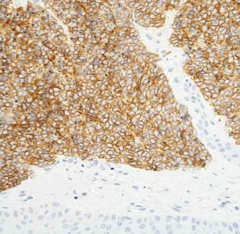
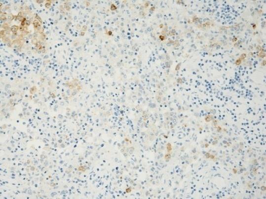
10 An assay should be calibrated so it fit-for-purpose Malignant melanoma Optimal results (NQC run 49) MART1/Melan A (MLA) IHC-Type 1 marker mab A103 mab BS52 mab M2-7C10 rmab EP43 Steroid producing tumors (e.g. Granulosa cell tumor of the ovary) Optimal results (NQC run 49) mab A103
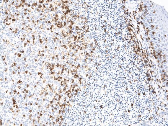
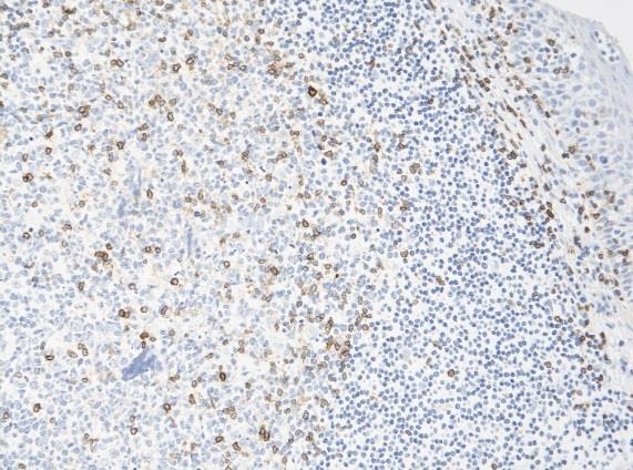
11 MART1/Melan A NQC results (Run 49) conclusions and challenges Melan A clone A103: Optimal result is difficult to obtain on the platforms Dako Omnis or Ventana Benchmark (HRP conjugated detection systems)? RTU product mab A103 (IS/IR633,Dako) developed for the Autostainer was used on the Omnis - 13 % suff. (2 of 15) mab A103 MLA RTU system ( Ventana): UltraView-AP as detection system = pass rate of 7% (recommended protocol settings by the vendor) UltraView-AP with amplification = pass rate of 100%. The recently introduced rmab clone EP43 showed promising performance as optimal results were seen on both the Ventana Benchmark and Dako Omnis platforms steroid producing tumors? Control material mab A103 versus rmab EP43, mab BS52 & M2-7C10? Other melanosome producing tumours (melanotic neural crest derived tumours e.g. melanotic neurofibroma)?
")


12 RTU IR/IS633 (Autostainer) RTU IR/IS633 (Omnis) Vendor recommend protocol settings
 (ALK-NPM")
, HIER high ph 20`, Flex+")
13 Anaplastic lymphoma kinase (ALK) Anaplastic large cell lymphoma (ALCL) (ALK-NPM rearrangement) D5F3 (1:200), HIER high ph 20`, Flex+ ALK1 (1:10), HIER high ph 20`, Flex+ Anything wrong?

14 IHC-Type 2 marker Lung tumors Low concentration of fused protein = require a high sensitive antibody for detection Intended use & fit-for-purpose MCC ALK,D5F3 = 94% pos ALK,5A4 = 88% pos ALK, ALK1 = 13% pos
 were assessed as sufficent, none were")
15 Don`t use clone ALK1 to detect ALK rearranged lung adenocarcinomas It does not fit-for-purpose D5F3, 5A4, OTI1A4 35 protocols were based on ALK1: Only one protocol (3%) were assessed as sufficent, none were optimal
 ALK,")


16 ALK, D5F3 (1:200) ALK, 5A4 (1:50) ALK, ALK1 (1:10) Adenocarcinoma Lung ALK-EML4 Merkel cell carcinoma Skin HIER in high ph buffer 20`, Flex+ Clone ALK1 provides low sensitivity

 ALCL")


17 HIER in high ph buffer, Flex+ ALK, D5F3 (1:200), ALK, ALK1 (1:10) ALCL Clone ALK1 provides low sensitivity icaps : Ganglion and peripheral nerve cells? Appendix
18 Optimization of the IHC assay issues to be addressed Purpose and/or fit-for-purpose of the IHC test How to establish best practice protocol of the IHC test (Calibration of the IHC assay with focus clone, antigen retrieval, titer & detection system) How to validate (technical) the IHC-test - Is the IHC test reproducible/robust (preanalytic conditions) - Evaluation of the analytical sensitivity and specificity Identification of most robust controls providing information that the established level of detection is obtained in each test performed in daily practice. Tissue materials are essential for these processes (calibration, validation and controls)
19 How to establish best practice protocol of the IHC test - parameters to consider Use a Test battery approach (pre-treatment and dilution range) Test more than one antibody clone against an antigen of interest before implementation in the routine Test with robust, specific & sensitive detection system Test/validate on normal and tumor tissue material with broad spectrum of antigen densities (specificity/sensitivity) Compare results with external quality assurance programs, literature or colleagues No antibody should be acquired without the basic knowledge of its performance characteristics and expected expression pattern Hadi Yaziji and Todd Barry Adv Anat Pathol Vol13, Number 5, September 2006
20 Technical aspects of IHC and pitfalls Analytical phase Concentrated antibodies - Dept. of Surgical Pathology, Region Zealand, Denmark Omnis (app. 240 Abs) Antibody Performance Testing ( Test Battery approach ) Dil. 1 Dil.2 Dil.3 A None None None B Enzyme (1) 5 min. Enzyme (1) 5 min. Enzyme (1) 5 min. C HIER TRS Low ph 6.1 (30`) HIER TRS Low ph 6.1 (30`) HIER TRS Low ph 6.1 (30`) D HIER TRS High ph 9.0 (24`) HIER TRS High ph 9.0 (24`) HIER TRS High ph 9.0 (24`) E TRS Low (20`) + Pep (12`) TRS Low (20`) + Pep (12`) TRS Low (20`) + Pep (12`) F HIER TRS High ph 9.0 (48`) HIER TRS High ph 9.0 (48`) HIER TRS High ph 9.0 (48`) G Pep 6 & 10 min + TRS High * Pep 6 & 10 min + TRS High Pep 6 & 10 min + TRS High H Pepsin 20 min. Pepsin 20 min Pepsin 20 min Protocol A: 0.5 % Protocol B: 2.0 % Protocol C: 10.0 % Protocol D: 83.5 % Protocol E: 1.0 % Protocol F: 3.0 % Protocol G: 0 % Protocol H: 0 % Off board enzymatic pre-treatment Identify the protocol that discriminate between the desired (specific) positive staining and any unwanted (non-specific) background staining
: 10 high, 10 low and 5 non-expressors) How many")
: 10 positive and 10 negative cases including high")
21 Technical aspects of IHC and pitfalls Analytical phase Analytical Validation - Evaluation of sensitivity and specificity - Tissue is the key element Goldstein NS et al : Appl Immunohistochem Mol Morphol 2007 Mar; 15 : tissue samples (Non-predictive markers/ IHC-type I): 10 high, 10 low and 5 non-expressors) How many tissue samples are needed for the analytical validation process? Fitzgibbons PL et al : Arch Pathol Lab Med 2014;138: tissue samples (Non-predictive markers/ihc-type I): 10 positive and 10 negative cases including high & low expressors 40 tissue samples (predictive markers/ihc-type 2): 20 positive and 20 negative cases
 systematically")
 of a test, through test")
.")

22 Article sequence (part 1-4) published in Appl Immunohistochem Mod Morphol (2017) systematically describing/defining all aspects of the IHC test from purpose (fit-for-purpose) of a test, through test performance characteristics (analytical sensitivity, analytical specificity, preanalytical reproducibility ). Importance of validation with focus on the technical part and the use of tissue tools for Quality assurance in immunohistochemistry. Full technical validation
23 TMA Normal Tissue Protocol set-up: Evaluate analytic sensitivity and specificity Normal tissue including fixation and decalcification controls Tonsil 6h Tonsil 24-72h Tonsil h Liver Identification of the best practice protocol (clone, titer, retrieval etc.) SOX10, BS7; HIER High ph 24`; 1:350 RR; Flex+Mouse linker Colon 6h Colon 24-72h Colon Placenta Establishing robustness of the IHC assay / pre-analytic parameter`s? SOX10, BS7; Robust to both fixation time in NBF and decalcification Breast Pancreas Salivary Gl. Skin Identification of robust controls SOX10, BS7; High, Low & Non-expressors? Kidney Adrenal Gl. Prostate Brain Cervix Testis Skeletal Colon/Tonsil Muscle 24h decalc. EDTA Decalc Fixation LE HE HE

24 TMA Mixed tumors TMA Malignant Melanomas Liver Mamma Ductal carcinoma Mamma Ductal carcinoma Mamma Lobular carcinoma Lung Adeno carcinoma SOX10 Lung Adeno carcinoma Lung Squam. carcinoma Diagnostic potential Analytical validation Colon Adeno carcinoma Colon Adeno carcinoma Kidney Clear cell carcinoma Kidney Clear cell carcinoma Thyroid Follicular carcinoma Thyroid Medullar carcinoma Melanoma 1 Kidney Ovary Serous carcinoma Ovary Serous carcinoma Ovary Clear cell carcinoma Ovary Endom. carcinoma Ovary Endom. carcinoma Uterus Carc. Sarcoma Melanoma 2 Melanoma 3 Melanoma 4 Tonsil Testis Seminoma Testis Seminoma Prostate Adeno carcinoma Prostate Adeno carcinoma Colon Carcinoid Melanoma 5 Melanoma 6 Melanoma 7 Lymph n. Melanoma Skin Melanoma Lymph n. Melanoma Pancreas Adeno carcinoma Bladder Uroth. cell carcinoma Bladder Uroth. cell carcinoma Melanoma 8 Melanoma 9 Colon GIST Uterus Leiomyo sacrcoma Testis Lipo Sarcoma Hodgkin L. Classic Hodgkin L. Mixed Diffuse L. B Cell lymphoma Melanoma 10 Melanoma 11 Melanoma 12 Diffuse L. B Cell lymphoma CLL Follicular B Cell lymph. Mantle Cell Lymph. Pheriph. T Cell lymph. Large C. Anaplastic T Cell lymph.


25 Slide (modified) and information kindly provided by Søren Nielsen, Aalborg, DK
26 Main causes of insufficient staining reactions are related to: The choice of antigen retrieval method The choice of primary antibody (Concentrate or RTU) a) Calibration of the antibody dilutions b) Stainer platform dependent antibodies The choice of detection system 83 % of insufficient results 89 markers assessed during the period and several markers have been assessed several times. Seven runs for HER2 ISH (more than slides assessed)
27 Problems related to the choice of antigen retrieval method : Using non-alkaline HIER buffer (low ph buffer) Using inefficient / too short HIER period 27% Using no or enzymatic pre-treatment instead of HIER Using excessive retrieval procedure impaired morphology False positive or false negative results The purpose of antigen retrieval is to unmask antigen epitopes /restore antigenic determinants and recover immuno-reactivity
28 The purpose of antigen retrieval is to unmask antigenic determinants (epitopes) and recover immuno-reactivity Antigen retrieval procedures for formalin fixed tissue: Heat Induced Epitope Retrieval (HIER) Tissue digestion using proteolytic enzymes
29 Shi et al. demonstrated that : Enzyme pre-digestion of tissue could be omitted. Incubation time with primary antibodies could be reduced, or dilutions of primary antibodies could be increased. Staining could be achieved on long-term formalin fixed that failed to stain with conventional methods. Certain antibodies which where typically unreactive with formalin-fixed tissue gave excellent staining.
30 Several hypothesis in regard of the mechanism of HIER has been proposed, but the mechanism of action of HIER is not completely understood. Heating tissue sections in an appropriate buffer may unmask epitopes by : Hydrolysis of methylene cross-links formed by formalin fixation Extraction of diffusible blocking proteins Precipitation of proteins Rehydration of the tissue section allowing better penetration of the antibody Removal of tissue-bound calcium ions by chelating substances Other mechanism s? Reviewed by Ramos-Vara JA: Vet Pathol 42: , 2005
31 Efficient HIER depends on: ph of the HIER buffer Temperature Time Elementary nature of the HIER buffer (e.g. Citrate; TRIS; EDTA; TE) Fixation time in formalin Less sensitive to routinely fixed tissue (formalin) compared to enzymatic pre-treatment > 95% of all commonly used antibodies require HIER
32 Shi SR et al. J Histochem Cytochem : Efficient HIER - Influence of ph A: CD20 (clone L26) B: Ki-67 (clone MIB1) C: MSA (clone HMB45) Demonstrated that the performance of monoclonal antibodies were highly influenced by ph of the Antigen Retrieval buffer (AR). Also, the results indicate the advantage of using an AR solution of higher ph value (8-9).
 MUM-1, MUM1p")
 Autostainer: Flex+")
 Tonsillar")
33 Efficient HIER - Influence of ph CD79, JCB117 (1:300) MUM-1, MUM1p (1:400) HIER in TRS ph 6.1 (20 min at 97 C) Autostainer: Flex+ as the detection system HIER in TRS ph 9 (20 min at 97 C) Tonsillar tissue fixed in 10% NBF (48h).

 1:300")
.")
34 Efficient HIER - Influence of ph HIER in TRS ph 9 CD79, JCB117 (1:300) HIER in TRS ph 6.1 CD79, JCB117 (1:50) 1:300 Tonsillar tissue fixed in 10% NBF (48h). Flex+ as detection system
")


")
35 Efficient HIER - Influence of ph BCL-6, LN22 (1:100) CD163, MRQ-26 (1:200) HIER in TRS ph 6.1 (20 min at 97 C) FN Autostainer: Flex+ as the detection system HIER in TRS ph 9 (20 min at 97 C) Tonsillar tissue fixed in 10% NBF (48h).
/Flex+ HIER in TRS ph 6.")

36 Efficient HIER - Influence of ph HIER in TRS ph 9 CD163, MRQ-26 (1:200)/Flex+ HIER in TRS ph 6.1 CD163, MRQ-26 (1:25)/Flex+ 1:200 For app % of the epitopes, HIER in buffers at ph 8-9 is preferable to ph6
37 Efficient HIER - Influence of time and temperature Taylor CR et al : Applied Immunohistochemistry 1996; 4(3) : Temperature and time are inversely related : Similar strong intensity of staining could be generated by the following heating conditions: 100 C for 20 min = 90 C for 30 min = 80 C for 50 min = 70 C for 10 h Balaton AJ et al : Applied Immunohistochemistry 1996; 4(4) : Optimal staining intensity could be generated by the following heating conditions: MWO at 100 C for 20 min = Pressure cooker at 120 C for 3 min


 10 min")


38 Efficient HIER - Influence of time and temperature CD79, JCB117 (1:300) 10 min 20 min 80 min HIER at 80 C TRS ph 9, Flex+ HIER at 97 C TRS ph 9, Flex+ Tonsillar tissue fixed in 10% formalin (48h).


")


39 HIER buffer - Influence of time and temperature 10 min CD163, MRQ-26 (1:200) 20 min 80 min HIER at 80 C TRS ph 9, Flex+ HIER at 97 C TRS ph 9, Flex+ Tonsillar tissue fixed in 10% formalin (48h).


40 Length of formalin fixation and HIER time HIER / TRS ph 9/ 10 min HIER / TRS ph 9/ 20 min HIER/ TRS ph 9/ 40 min Tonsil / NBF 6h MUM-1, MUM1p HIER in TRS High ph 9 at 97 C Tonsil / NBF 24h Tonsil / NBF 168h Best performance: Efficient HIER time ~ min at C
 or Diva decloaker (Biocare) Overall best performance: HIER in EDTA ph 8.0 (compare with Tris-HCL ph 8.0)")
41 ph8 ph8 Chemical composition of the HIER buffer`s Standard low ph buffer`s (e.g. citrate based ph 6.0) Standard high ph buffer`s (e.g. TE based ph 8-10) Modified low ph buffers ph : S1699/S1700 (Dako) or Diva decloaker (Biocare) Overall best performance: HIER in EDTA ph 8.0 (compare with Tris-HCL ph 8.0)

 or Diva Decloaker ph 6.")

42 Modified low ph buffers CD21, clone 1F8 CD21, clone 1F8 CD21, clone 2G9 TRS ph 9 TRS ph 6.1 (S1699/S1700) TRS ph 9 HIER time 20`/ Flex+ Markers requiring the TRS Low ph 6.1 (Dako, S1699/S1700) or Diva Decloaker ph 6.2 (Biocare, DV2004) : EP-CAM (clone EP-4 or MOC-31 or VU-1D9 ); GP200 (clone SPM 314 or 66.4.C2); CD21 clone 1F8; CD61 clone Y2/51; NGFR clone MRQ-21; Desmoglein-3 clone BC11 and. Mandatory for : CD7 clone CBC 3.7; CD30 clone ConD6/B5; CD5 clone Leu1


")
")
")
")
43 Modified low ph buffers TRS ph 9 (Dako) TRS ph 6.1 (Dako S1700) Diva Decloaker (Biocare) PT / 99 / 20 min PT / 99 / 20 min PT / 99 / 20 min CD30, ConD6/D5 (1:50) (Hodgkin Lymphoma) Desmoglein-3, BC11 (1:25) (Skin) EP-CAM, MOC-31 (1:20) (Small cell carcinoma)


44 Modified low ph buffers CD30 clone ConD6/B5 Tonsil Hodgkin lymphoma Embryonal carcinoma HIER buffer, TRS ph 6.1 (Dako S 1700) HIER buffer, Low ph (LabVision TA-999-DHBL)
45 The purpose of antigen retrieval is to unmask antigen epitopes /restore antigenic determinants and recover immuno-reactivity Antigen retrieval procedures for formalin fixed tissue: Heat Induced Epitope Retrieval (HIER) Proteolytic enzymes cleave more or less specific amino acid sequences within peptide chains and not covalent cross-links formed in tissues during formalin fixation. Tissue digestion using proteolytic enzymes Improves penetration of reagents into the tissue structures and restore the immunodominant conformation of epitopes of interest.
 47% Enzymatic")
 were based on enzymatic pre-treatment AE1/AE3: App.")

46 Enzymatic digestion? mab clone Ks20.8 Sufficient result Optimal result HIER in Alkaline buffer 92% (91 of 99) 47% Enzymatic pre-treatment 75% (9 of 12) 25% As concentrate: App. 10 % of the protocols (12 of 126) were based on enzymatic pre-treatment AE1/AE3: App. 6 % of all protocols (44 of 742) were based on enzymatic pre-treatment (seven NQC Runs). Problem A significant proportion of Labs still uses enzymatic digestion for a wide range of markers requiring HIER for optimal performance Only few markers require enzymatic digestion as the solitary pre-treatment procedure for routine purpose
47 Optimal enzymatic digestion depends on: Enzyme type Concentration Time Temperature Fixation type & time Tissue type Most common Enzymes Proteinase K Pronase XIV Pronase XXIV Pepsin Trypsin Difficult to control and to standardizes within routine LAB Short time formalin fixation = gentle proteolysis Long time formalin fixation = prolonged proteolysis 2% of all commonly used antibodies require enzymatic (or no) pre-treatment
48 Enzyme Typical working conc. Activation Temperature Typical Incubation time Cleavage nature Proteinase K 0.1%, ph C 5-10 min. Broad - all amino acids Trypsin %, ph C 10 min. Arginin / Lysin Pepsin %, ph C 5-20min. Broad,favor peptides with aromatic aminogroups Protease XXIV %, ph C 5-10 min. Broad - all amino acids Protease XIV %,pH C 10-30min. Broad, favor peptides with aromatic residues Markers requiring enzymatic pretreatment : FVIII (poly), LMV CK (CAM 5.2), PAN CK (MNF116), EGFR (various), TCR-β (8A3).. Extracellulare matrix proteins (COLL-III (poly), Laminin (poly) and COLL-IV (CIV-22)...


 40`")


49 Choice of proteolytic enzyme Trypsin (Biocare, RTU) 40` Pepsin (ZytoVision, RTU-H) 15` Proteinase K (Dako, RTU) dil. 1:4 / 5` TCR-β, 8A3, 1: 200 RR Tonsil Ubiquitin, Ubi-1 1: 750 Liver/ Mallory bodies Neutrophil Elastase, NP57 1: 1000 Tonsil


50 Proteolytic enzyme & digestion time? Tonsil NBF 48h Digestion temp. 32 C 5 min 10 min 20 min 40 min Proteinase K (RTU S3020, Dako) Proteinase K dil. 1:4 (RTU S3020, Dako) Trypsin (RTU, Biocare) TCR β clone 8A3 (1:200 RR) / Flex+ (Omnis)

 / Flex+ Trypsin Digestion temp.")
 Trypsin Digestion temp.")
51 Proteolytic enzyme & digestion temperature? Tonsil NBF 48h TCR β clone 8A3 (1:200 RR) / Flex+ Trypsin Digestion temp. 4 C (10 `) Trypsin Digestion temp. 24 C (10`) Trypsin Digestion temp. 37 C (10`) Increased intensity of TCR β positive T-cells

")

 10")
 20 min")

52 Enzymatic digestion (Influence of fixation time) EP-CAM, clone MOC-31, dilution 1:20 Pepsin / (Dako, S3002) 10 min/37 C HIER, Low ph (S1700) 20 min / 97 C NBF 24 h NBF 48 h NBF 120h Adenocarcinoma (Breast) fixed in 10% Formalin
53 Problems related to the choice of antigen retrieval method : Using excessive retrieval procedure impaired morphology False positive or false negative results
54 Excessive retrieval: Proteolytic pretreatment - over digestion (not calibrated to the fixation time in NBF) HIER - using too high temperature for too long time especially in alkaline retrieval buffers Antigen Retrieval using standard HIER procedures - on fragile tissue/cell material (cell clot s )
 Omnis, CD20 RTU TRS (3-1) / 30 min at 97")

55 CD20 clone L26 Bone Marrow Coagulum/Clot (fixed for 24h in NBF) Omnis, CD20 RTU TRS (3-1) / 30 min at 97 C BOND, CD20 BERS2 / 20 min at 100 C x200 x200 HIER settings: Recommendations given by the manufacturer`s
")
")

56 Excessive antigen retrieval related to the PT-module (Dako) Influence of pre-heat temperature (65 C versus 85 C) CD138, B-A38 P/E 65 P/E 85 Bone marrow clot, NBF 96h PT, High ph (3-1) 95ºC, / 20 min


 Recommended")
")
57 Bone marrow clot AS: PT-Link, High ph buffer s at 97 / 20` CD5 clone SP19 CD34 clone QBEND-10 High ph (3-1) (Dako) Recommended settings: 65 HIER buffer H (LabVision) Recommended settings: 85
 Glycophorin A clone")


58 Bone marrow clot (NBF 24h) Glycophorin A clone JC159 (1:500) Flex+ BOND, BERS-2 / 20 min at 100 C OMNIS TRS (3-1) ph 9/ 30 min at 97 C OMNIS TRS ph 9/ 10 min at 97 C OMNIS TRS ph 9/ 20 min at 97 C

 CD138,")


59 Chemical composition of the HIER buffer morphology? Bone Marrow cloth (NBF 24 h) CD117, EP10 (1:25 RR) CD138, B-A38 (1:1000) TRS (3-1) High ph 9, 24` at 97C, Agilent/Dako Omnis: Flex+ HIER buffer H, 24` at 97C Thermo S./ LabVision
60 Pause

61 Parameters related to the primary Ab affecting antibody-antigen reactions in tissue Antibody choice Sensitivity/Specificity Antibody Titer Antibody performance related to the chosen automated platform Antibody diluents Incubation time Incubation temperature Sensitive to endogenous peroxidase blocking Storage of concentrated primary antibodies Storage of diluted primary antibodies Provided that efficient antigen retrieval has been performed and a sensitive detection system has been used
62 37% Problems related to: The choice and use of the primary antibody (Concentrate or RTU) Inappropriate primary antibody - Provide low sensitivity/specificity Appropriate primary antibody - Inapp. titre (too low or too high concentration) Stainer platform dependent antibodies - Provide low sensitivity / specificity False positive or false negative results

 clone 9FY ALK clone ALK1")


63 Problem: Primary antibody provides low sensitivity Primary antibodies providing low sensitivity (NordiQC results/latest run) ERG (Ets-Related-Gene) clone 9FY ALK clone ALK1 GATA3 clone HG3-31 CEA clone II-7 CGA clone DAK-A3 P63 clone 7JUL ERG, 9FY prostate adenocarcinoma TMPRSS2-ERG gene fusion? Focus on clones giving optimal results and use app. tissue control material
 protein?")
64 Detection of ERG using clone 9FY in prostate adenocarcinomas - antibody raised against the N-terminal part of the ERG (wt) protein? TMPRSS2-ERG rearrangements often encodes N-terminal truncated ERG proteins
 Is MLA, A103 the best primary Ab for detection of melanomas and does it")
65 Melan A /MART1 Melan A (MLA) / MART-1: 238 participants ~ 93% used clone A103 (single or in cocktail antibody solutions) Is MLA, A103 the best primary Ab for detection of melanomas and does it fit-for-purpose?


66 Melan A /MART1 MLA, A103 1:25 AutoStainer MLA, A103 1:25 Omnis MART1, EP43 1:30 Omnis Melanoma (Sentinel node) Melanoma (Lymph node) Adrenal gland
 Focus on clones giving optimal results and use app.")
67 MUM1 Primary antibodies providing low specificity and/or poor signal-to-noise ration (NordiQC results/latest run) MUM1 clone MRQ-43 & BC5 CK-HMW clone 34βE12 PR clone 1E12 ECAD clone EP700Y PAX5 clone SP34 Many pabs (e.g. P40 and SOX10) Focus on clones giving optimal results and use app. tissue control material (colon and tonsil) MUMp1, EAU32 & EP190


68 Problem: Primary antibody provides low specificity and/or poor signal-to-noise ration Tonsil MUMp1, optimal BC5, aberrant cytoplasmic staining result Clones providing optimal results: MUMp1, EAU32 & EP190 MRQ-43, aberrant cytoplasmic staining result MRQ-43, false positive


")
 Ovary Carcinoma")


69 Problem: Primary antibody provides low specificity and/or poor signal-to-noise ration Renal Cell Carcinoma (CC) Thyroid Carcinoma (Pa) Ovary Carcinoma (Se) Which antibody? Pax-8 / CM / Dil 1:2000 / Clone MRQ-50 - Mab Pax-8 / BC / Dil 1:150/ Clone BC12 - Mab
70 Problem: Primary antibody provides low specificity and/or poor signal-to-noise ration Liau J-Y et al.: Appl Immunohistochem Mol Morphol Jan;24(1):57-63 Demonstrated that neuroendocrine tumors (NET s) from a large variety of organs were immuno-reactive with the two less specific antibodies (pab Proteintech & mab MRQ-50) - cross-reacting with other PAX proteins Also, all NET`s were immuno-negative with the two monoclonal antibodies raised against the C-terminal part of PAX8 protein (PAX8R1 & BC12) Moretti L et al. : Mod Pathol. 2012; 25 : Demonstrated that an N-terminal PAX-8 polyclonal antibody cross-react with N-terminal region of PAX-5 and is responsible for reports of PAX-8 positivity in malignant lymphomas. Also, PAX8 mrna levels were not detected in any of the B-cell lymphoma cell lines studied. These results indicate that benign and malignant B-cells do not express PAX8.


 SCLC (Lung) Pax8,")


 Pax8, ZR1 raised")

71 Problem: Primary antibody provides low specificity and/or poor signal-to-noise ration Papillary carcinoma (Thyroid) Pancreas Carcinoid (Appendix) SCLC (Lung) Pax8, MRQ-50 Pax-8, MRQ-50 most likely cross-reacts with Pax-6 in NET`s Pax8, ZR1 Pax8, MRQ-50 most likely raised against the N-terminal part of the PAX8 protein (cross-reacts with other PAX proteins) Pax8, ZR1 raised against the C-terminal part of the PAX8 protein (no cross-reacting with other PAX proteins)
 ZR1 (lot variations/antibody diluent dependent) EP298 SP348 Cross react with other Pax proteins in the family (e.g.")
72 Problem: Primary antibody provides low specificity and/or poor signal-to-noise ration? BC12 (platform dependent) ZR1 (lot variations/antibody diluent dependent) EP298 SP348 Cross react with other Pax proteins in the family (e.g. PAX5) Question`s: Should we use primary antibodies that cross react with other proteins in the same family? Would we accept cross-reactivity in the family of CD`s and CK`s - e.g. CD20 to CD3 or CK5 to CK8?
 Poorly calibrated primary Ab? Tissue controls are the key element Normal skin is the preferred positive control for GCDFP-15.")

73 Skin Problem: Primary antibody poorly calibrated providing low sensitivity The right primary antibody Gross cystic disease fluid protein-15 (GCDFP-15) The right protocol (AR procedure and detection system) Poorly calibrated primary Ab? Tissue controls are the key element Normal skin is the preferred positive control for GCDFP-15. The epithelial cells of the eccrine sweat glands must show an as strong as possible positive cytoplasmic staining reaction, while all other cells should be negative. Normal breast tissue can also be used as control in which epithelial cells of the ductal glands must show an as strong as possible staining reaction. Background staining may be seen in vicinity of highly positive tissue structures (e.g. eccrine sweat glands)

74 Problem: Primary antibody poorly calibrated providing low sensitivity Estrogen Receptor (ER), NQC Run B24 Optimal Good Borderl. Poor Suff * All Ab clones and protocol settings Total protocols assessed Proportion 71% 36% 6% 2% 92% The most frequent causes of insufficient staining reactions were: - Less successful primary Ab. - Insufficient HIER - too short efficient HIER time and/or use of a non-alkaline buffer - Too low concentration of the primary Ab. Estrogen receptor - Control tissue Normal cervix (high and non-expressors) Breast tumor s x 3 (non, low and high-expressors) Tonsil (Normal tissue low and non-expressors)


75 Problem: Primary antibody poorly calibrated providing low sensitivity ER clone 6F11 / 1:100 ER clone 6F11 / 1:200 ER clone 6F11 / 1:400 Cervix Tonsil Breast tumor High ph 20`, Flex+Mouse Staining indicators are extremely important - helping us to calibrate the IHC assay correctly


76 Problem: Primary antibody poorly calibrated providing low sensitivity ER, SP1/ 1:200 ER, SP1/ 1:400 ER, SP1/ 1:800 ER, SP1/ 1:1600 Cervix Tonsil Breast Ca High ph 24`, Flex+Rabbit Reduced intensity and proportion of cells expected to be stained


77 Problem: Primary antibody poorly calibrated providing low sensitivity Liver Hepatocellular carcinoma Appendix Too diluted ARG1, EP261 1:800 Optimal ARG1, EP261 1:50 HIER High ph 24`; Flex+ Rabbit linker In collaboration with Ole Nielsen, Department of Pathology, Odense
78 IHC: Technical considerations to intended use and fit-for-purpose approach Do we have the right antibody (IHC type markers 1 & 2) can it provide appropriate sensitivity and specificity Does the antibody work on the chosen automatic platform(s) Does the automatic platform come with appropriate reagents fulfilling purpose and intended use of the IHC assay - Appropriate Antigen Retrieval solutions (enzymes and HIER buffers) - Appropriate antibody diluents and wash buffers - Appropriate detection and visualization products - Appropriate protocol library Do we have access to appropriate tissue, reflecting the range of different antigen expression levels, both for the optimization process but also for the laboratory`s internal quality assurance program (control tissue) monitoring specificity and sensitivity of the assays
79 Technical aspects of IHC and pitfalls Analytical phase HIER buffers used by NordiQC laboratories In house Dako Roche Ventana Low ph buffers Leica Microsystems Biocare Thermo S LAB Vision Citrate buffer ph 6 / ph6.7 TRS Low ph 6.1 CC2 ph 6 BERS-1 ph 6 Diva Decloaker ph 6.2 High ph buffer EDTA/EGTA ph 8 TRS High ph 9 CC1 ph 8.5 BERS-2 ph 9 Borg Decloaker ph 9.5 HIER buffer H ph 9 Tris-EDTA/EGTA ph 9 TRS High (3-in-1) ph 9 Tris-HCL ph 9 App % of all pretreatment protocols Challenges: The instrumentation / platforms dictates the choice of HIER buffers For some antigens, the HIER buffers dictate`s the choice of primary Ab

80 Optimal results with HIER in High ph buffers e.g. CC1 (Ventana) (with or without gentle enzymatic digestion performed after HIER) No optimal results with HIER in High ph buffer CC1 (Ventana) or proteolytic pretreatment Optimal results with HIER in mod. Low ph buffers (Dako) BS14 could be an alternative to Ber-EP4 on platforms excluded from the use of modified low ph buffers e.g. Diva ph 6.2 (Biocare) or TRS ph6.1 (Dako)


 /")


81 EPCAM clone EP4 or BS14 Proteinase K 1:4 (Dako) / 5` Citrate buffer ph 6 (Dako) / 20` High ph buffer (Dako) / 20` TRS Low ph (Dako, S1699/S1700) /20` EP4 BS 14 Kidney

 / TRS ph 9.")
 is a")


82 Omnis Kidney Hepar Breast tumor EPCAM, BS14 (1:500) / TRS ph 9.0 EPCAM, BS14 (Nordic Biosite) is a better alternative than EPCAM MOC31 or Ber-EP4 for automated platforms not offering the possibility to use mod. low ph buffers. EPCAM, MOC31 (1:25) / TRS ph 6.1


 HIER High ph 48`,")
83 Primary antibodies sensitive to the chosen platform Autostainer PAX8, BC12 1:50 Omnis PAX8, BC12 1:50 Omnis PAX8, ZR1 1:50 RR Kidney: Clear cell carcinoma HIER High ph 20`, Flex+ (10+20) HIER High ph 48`, Flex+ (10+20)


 Pancreatic")
84 Primary antibodies sensitive to the chosen platform Autostainer Omnis Omnis SMAD4, B8 1:400 SMAD4, B8 1:400 SMAD4, EP168Y 1:2000 RR HIER High ph 20`, Flex+ (10+20) HIER High ph 24`, Flex+ (10+20) Pancreatic Adenocarcinoma
85 Platform dependent antibodies (NordiQC results/latest runs): Marker Clone ASMA 1A4/BS66 BCL2 124 /E17 CD3 F7.2.38/LN10 CD4 4B12/EP204 CD23 1B12/DAK-CD23 CD56 123C3 & 123C3.D5/MRQ-42 CDX2 DAK-CDX2/EPR2764Y or EP25 CEA II-7/CEA31 CK (LMW) 5D3/EP17/EP30 Marker Clone CR DAK-Calret1/CAL6 Desmin D33/BS21 EPCAM EP4/BS14 Melan A A103/EP43 OCT 3/4 C-10/MRQ-10 or N1NK PAX8 MRQ-50/SP348 or EP298 Podop D2-40 WT1 6F-H2/D817F or EP122 Alternative antibodies: Antibody clones applied on the Omnis (Dept. of surgical Pathology, Region Zealand, Denmark) Go to the NordiQC website for information of the individual markers in relation to the chosen platform


86 Primary antibodies sensitive to the chosen platform Implementing a new platform has been a challenge ALK clone D5F3 or 5A4 HCL, clone DBA44 GATA3, clone L MART-1/Melan A, clone 103 PAX 8, clone BC12 SMAD4, clone B8 WT1, clone WT49 MMR ASMA, 1A4. Changing the primary Ab Changing Ab-Ag reaction microenvironment (Diluent) Low affinity primary antibodies
87 Antibody diluents Demonstrated that: ph of the Ab-diluent had a high impact on the IHC result Addition of NaCL (increasing the ionic strength) to the diluent negates most of the sensitivity gained through Antigen Retrieval (Table 3). Antibody diluent formulations can significantly alter stability and binding properties of antibodies affecting both epitope specificity and non-specific interactions


 / PK ~")

 diluted")
88 Antibody diluents PAX8, ZR1 1:50 Dako Dil. ph7.3 PAX8, ZR1 1:50 Renoir R ph 6.2 4x Clear Cell Carcinomas (Kidney ) PK (2` at RT/ off-board) + HIER (Dako, S2367 ph9) (30` at 97 C) / PK ~ Proteinase K Solution RTU (Dako cat.no.s3020) diluted 1:10 in TBS ph7.6 /Flex+ Rabbit (10+20`)


 Renoir")
89 Antibody diluents Thymus Tonsil TdT, SEN28 1:50 Dako dil. ph 7.3 Omnis: HIER/HIGH ph 24`, Flex+ Mouse (10+20`) TdT, SEN28 1:50 Renoir Red ph 6.2


90 Antibody diluents ALK, 1A4 1:300 Dako Dil. ph 7.3 ALK, 1A4 1:1200 Renoir R ph 6.2 ALK, 1A4 1:1200 Dako Dil. ph7.3 MCC Omnis: HIER/HIGH ph 24` at 97 C, Flex+ Mouse (10+20`) App.


91 Omnis: HIER/HIGH ph 24`, Flex+ Rabbit (10+20`) The technical test approach Analytical phase Antibody diluents HHV8, 13B10 1:100 Renoir Red ph 6.2 HHV8, 13B10 1:100 Dako dil ph 7.3 Case 1 Renoir Red is not always the best antibody diluent Remember to use a antibody diluent test battery Case 2 Immunodeficient patients Kaposi's sarcoma, Castleman's disease, Primary effusion lymphoma...
 Kidney HIER TRS")

92 Antibody diluents WT1,EP122 1:25 Renoir Red (Biocare) WT1,EP122 1:25 Background Sniper (Biocare) Kidney HIER TRS ph9 (24` /97 C) + Pep (3`) The choice of antibody diluent has a high impact on unwanted / unspecific background staining
93 Antibody diluents Omnis (Department of Surgical Pathology, Region Zealand, Denmark) Markers benefitting from dilution in Renoir Red ph 6.2 (improving signal): ALK (1A4), CR (CAL6), CD4 (EP204), CD5 (SP19), CMYC (EP121), GATA3 (L20-823), GPC3 (1G12), IMP3 (69.1), MLH1 (ES05 & GM011), MSH2 (G ), MSH6 (EP49), NKX 3.1 (poly), SALL4 (6E3), PAX8 (ZR1), PMS2 (EP51), SOX10 (EP268), SOX11 (C1 & MRQ58), TdT (SEN28 & EP266), UP-II (BC21), WT1 (WT49) and. Markers that don`t benefit from dilution in Renoir Red ph 6.2: BCL2 (124), BCL6 (LN22 & PG-B6p & GI191E/A3), CR (DAK-Calret1), CD163 (MRQ26), CD21 (2G9), CD5 (4C7), ER (SP1), HHV8 (13B10), Mammaglobin (304-1A5), MUC5AC (CLH2), MUC6 (CLH5), and. Markers benefitting from dilution in Background sniper (reduces background problems): Spirochete (poly), BORR (poly), WT1 (EP122), ASMA (BS66) and.


94 Antibody diluents Ventana Benchmark Ultra The full effect of the antibody diluents may depend on the chosen platform Courtesy Ole Nielsen, Dept. of Pathology, OUH, Denmark
95 19% Problems related to the choice of the detection system: Provides low sensitivity - 2 versus 3-step multimer/polymer detection systems Provides low specificity and sensitivity - Biotin based systems False positive or false negative results
 HIER High ph")
")


 Granulosa")

96 Omnis MLA, A103 (1:50) HIER High ph 24` Envision G2-AP (Dako) 30/10/20/PR10 PoLink-2 plus /AP (GBI Labs) 30/15/15/PR10 Mod. Histo-AP (Nordic Biosite) 30/10/20/PR10 Granulosa cell tumor High expressor Granulosa cell tumor Low expressor
 3-step Envision Flex+ UltraView + Amp OptiView")
97 Detection systems (polymer/multimer) used by NordiQC participants Vendor Detection System Detection System Amplifier Cat.no Dako Ventana Leica Biocare LAB Vision/TS Immunologic 2- Step EnVision EnVision +/Flex UltraView MACH 2 UltaVision One BrightVision (PowerVision) 3-step Envision Flex+ UltraView + Amp OptiView Optiview + Amp Bond Refine (PowerVision) MACH 3 MACH 4 Anti -Ms/Rb Anti -Ms/Rabbit Anti-Hapten Anti-Hapten + TSA Anti-Ms (Rb?) Ms/Rb probe Ms probe (Rb?) Quanto? BrightVision+ Anti-Ms/Rat (Rb?) K4001 K8000 /10 (K5007) K8002/ DS9800 (HRP); DS9390 (AP) M2U522; MHRP520; RHRP520 M3M530; M3R531 M4U534 TL-125-HLJ TL-125-QHD /QHL DPVM (Anti-Ms)/DPVR (Anti-Rb ) DPVO (Anti-Ms/Rb/Rat) DPVB ((Anti-Ms/Rb/Rat) Master Diag. Quanto? MAD QK ZytoMed System And a few more (Advance, GTVision..) ZytoChem Plus (PowerVision) Anti-Ms (Rb?) PolHRP-100 App. 98% of all NordiQC participants use a polymer/multimer based detection systems Considerations related to the choice of detection system: Sensitivity Specificity Enzyme conjugate Blocking of endogenous activity Turn around time (TAT) Automatic platform (open or closed system) Price
 Enhancer Universal Linker Post Blocking.")
98 Anti-HQ (Multimer-HRP) HQ-Linker Amplifier (A/B) Linker (Mouse/Rabbit) Enhancer Universal Linker Post Blocking. Increases sensitivity
: The 3-step polymer detection systems Quanto and")
99 Intensity score Intensity score The technical test approach Analytical phase Detection systems - Performance Testing ER, EP1 (Rab) ER, 6F11 (Mab) Quanto 2 Quanto 1 Flex+ MACH4 1 Flex+ MACH4 0 1:50 1:200 1:800 1:3200 Flex 0 1:50 1:200 1:800 1:3200 Flex Dilution Dilution ER - Endpoint titration (some general remarks and important issues): The 3-step polymer detection systems Quanto and Flex+ - produced the overall highest intensity. The 3-step polymer detection system MACH4 only enhances reactions with mouse monoclonal Abs (ER,6F11). Optimal staining was highly influenced by the concentration of the primary Abs and the nature of detection system.


")
")


100 Performance testing of detection systems (Vendor recommended protocol settings) Breast tumor ER, EP1 (Rab) Quanto Flex+ MACH4 Flex 1:50 1:200 HRP HRP HRP HRP HRP HRP HRP HRP HRP HRP HRP HRP High Intensity Low Intensity


")


101 Performance testing of detection systems (Vendor recommended protocol settings) Breast tumor ER, 6F11 (Mab) Quanto Flex+ MACH4 Flex 1:50 1:200 HRP HRP HRP HRP HRP HRP HRP HRP HRP HRP HRP HRP High Intensity Low Intensity
: The 3-step polymer detection systems")
 used in combination with the 3- step polymer detection systems")
102 Intensity score Intensity score The technical test approach Analytical phase Detection systems - Performance Testing CD4, EPR6855 (Rab) CD4, 1B12 (Mab) Quanto 2 Quanto 1 Flex+ 1 Flex+ 0 1:50 1:200 Dilution 1:800 1:3200 MACH4 Flex 0 1:50 1:200 1:800 Dilution 1:3200 MACH4 Flex CD4 Endpoint titration (some general remarks and important issues): The 3-step polymer detection systems Quanto and Flex+ - produced the overall highest intensity. Intensity was highly influenced by the nature of primary Ab and optimal staining could only be obtained with the Rab (CD4, EPR6855) used in combination with the 3- step polymer detection systems Quanto or Flex+. Intensity was higher with the Rab (CD4, EPR6855) at 1:50 with all of the detection systems tested compared to any intensity obtainable with the Mab (CD4, 1B12) even with the use of a 3-step polymer system ( e.g. Quanto).

 CD4, EPR6855")
 Liver Quanto")


 with")


103 Performance testing of detection systems (Vendor recommended protocol settings) CD4, EPR6855 (Rab, 1:50) and 1B12 (Mab, 1:50) Liver Quanto Flex+ MACH4 Flex EPR6855 1B12 Strong staining of hepatic sinusoidal endothelial cells and kupffer cells using the Rab (CD4, EPR6855) with all the detection system tested (2-step or 3-step polymer systems). Intensity is significantly reduced using the Mab (CD4, 1B12).


")
 and 1B12")


 and")
104 Performance testing of detection systems (Vendor recommended protocol settings) CD4, EPR6855 (Rab, 1:50) and 1B12 (Mab, 1:50) Brain Quanto Flex+ MACH4 Flex EPR6855 1B12 Staining of microglia cells is only obtainable using the Rab (CD4, EP1628Y) and the 3-step polymer detection systems Quanto or Flex+.

105 Ventana Tyramide Signal Amplification Mechanism of Tyramide amplification: - Introducing HRP (Optiview) - Incubation with HQ-labelled Tyramide + H2O2 Tyramide, a phenolic compound, is converted into an short-lived extremely reactive intermediate - Make a deposit of HQ in close vicinity of Ab/Ag reactions Intermediates covalently binds to electron rish regions of adjacent proteins ( esp. tyrosine) rapidly - Detection of HQ with anti-hq / HRP Multimer - Visualization with DAB
 Lu-ALK For certain type")

106 Tyramide Signal Amplification (TSA) Lu-ALK For certain type of markers, the TSA system can provide optimal results but.. TSA

107 TSA and pitfalls: False positive staining Weak or completely false negative staining unbalanced reaction of primary Ab and target epitopes giving an yes or no answer? Uneven distribution of the TSA reaction product


108 FP OptiView + TSA FN Lu-ALK FP/FN MSH6 FN CD4 MSH6 The TSA detection system is not without problems and may provided either false positive or negative results. All parameters should be careful calibrated to provide optimal staining result - always possible?
-conjugated AP-based systems: Both of the NP systems")

109 The future - The key to low sensitive detection system? 5-nitro-3-pyrazole (NP)-conjugated AP-based systems: Both of the NP systems demonstrated outstanding sensitivity similar to that observed for the tyramide-based DAB IHC system and superior staining resolution and dynamic range on ALK FISH positive TMA slides (Fig. 4B).

110 New option on the Omnis


 /High")


111 Flex Flex+ Flex++ Omnis: PMS2 clone EP51 (1:300 RR) /High ph 48` FLEX FLEX+ FLEX++ Colon tumor MSI Sensitivity Colon tumor MSS


112 Colon tumor with loss of PMS2 Flex+ PMS2, EP51 1:75 RR Flex++ PMS2, EP51 1:300 RR Omnis: HIER High ph 48`

")
; HIER")


113 Flex++ : 4-step polymer detection system ( `/Omnis) rmab PD-L1, CAL10 (rmab 1:30RR); HIER in High ph 48` Placenta Tonsil Flex+ Flex++ An at least weak, but distinct membranous staining reaction of intra germinal centre macrophages in the tonsil
 mab ASMA clone 1A4; HIER in High ph")


114 Flex++ : 4-step polymer detection system (Omnis) mab ASMA clone 1A4; HIER in High ph 24` ASMA, 1A4 1:400 / Flex+ ASMA, 1A4 1:400 / Flex++ ASMA, BS66 1:500 / Flex+ Mouse monoclonal antibodies: No improvement in signal intensity using Flex++ compared to Flex+ Liver
115 The basal fundament for a technical optimal performance is : Appropriate tissue fixation and processing Appropriate and efficient epitop retrieval 95% of the Abs require HIER and app. 90% prefer high ph retrieval buffers. Use efficient HIER temperature and time (app. 100 C for 20-40min). Appropriate choice of antibody / clone, diluent and dilution Compare different clones / Abs against the desired antigen before implementation Calibrate the Ab concentration carefully in relation to Critical Staining Quality Indicators Robust, specific & sensitive detection system Use of a 3-step multimer/polymer system is preferable to a 2- step multimer/polymer system Don`t use biotin-based detection systems Appropriate choice of control material Include tissue material with low expressors, but also high and non-expressors

116 Thank you for your attention


117 PAX5 clone DAK-Pax5 Autostainer RTU systems Agilent/Dako Autostainer versus Omnis Omnis Optimizing an assay can be confusing (Vendor recommendations)
118 TMA Normal Tissue Protocol set-up: Evaluate analytic sensitivity and specificity Tonsil 6h Tonsil 24-72h Tonsil h Liver Normal tissue including fixation and decalcification controls Identification of the best practice protocol (clone, titer, retrieval etc.) Colon 6h Colon 24-72h Colon Placenta SOX10, BS7; HIER High ph 24`; 1:350 RR; Flex+Mouse linker Establishing robustness of the IHC assay / pre-analytic parameter`s? SOX10, BS7; Robust to both fixation time in NBF and decalcification Breast Pancreas Salivary Gl. Skin Identification of robust controls SOX10, BS7; High, low & non-expressors? Kidney Adrenal Gl. Prostate Brain Cervix Testis Skeletal Colon/Tonsil Muscle 24h decalc. EDTA Decalc Fixation LE HE HE
119 Boenisch T : Applied Immunohistochemistry 2005; 13(3) : Effect of Heat-Induced Antigen Retrieval Following Inconsistent Formalin Fixation 5` Demonstrated that: Optimal staining of 26 of the 30 antigens was achieved despite the variable length of fixation (up to 8 days of fixation). Prolonging HIER time or increasing HIER temperature could restore antigen determinants more efficiently compared to standard HIER protocols in over fixed tissue


 The")


120 Problem: Primary antibody provides low specificity and/or poor signal-to-noise ration p40 dil. 1:25 clone BC28 p40 dil. 1:50 polyclonal P40 dil. 1:200 polyclonal Placenta FN Tonsil pabs No optimal results / pass rate of 23% (5 of 22) The insufficient results were typically characterized by a poor signal-to-noise ratio and aberrant staining reaction compromising the interpretation. Poor signal-to-noise ratio Both primary antibodies are from BIOCARE


121 Flex Flex+ Flex++ Omnis: BRAF clone VE1 (1:1000 RR) High ph 48` FLEX FLEX+ FLEX++ Colon tumor BRAFV600E mut Sensitivity Colon tumor KRAS mut
 Colon tumor")

122 Flex++ : 4-step polymer detection system (Omnis) mab BRAF, VE1 (mutation specific) Colon tumor BRAF V600E mutated 1:400 RR, Flex+ 1:1200 RR, Flex++ Background staining / FP? HIER in High ph 48`


 Flex+ CMYC,")

123 Flex++ : 4-step polymer detection system (Omnis) rmab CMYC, EP121 Flex++ CMYC, EP121 (1:150 RR) Flex+ CMYC, EP121 (1:75 RR) HIER in High ph 24` Tonsil DLBCL, NOS