VCP adaptor interactions are exceptionally dynamic and subject to differential modulation by a VCP inhibitor
|
|
|
- Luke Darcy Bryant
- 5 years ago
- Views:
Transcription
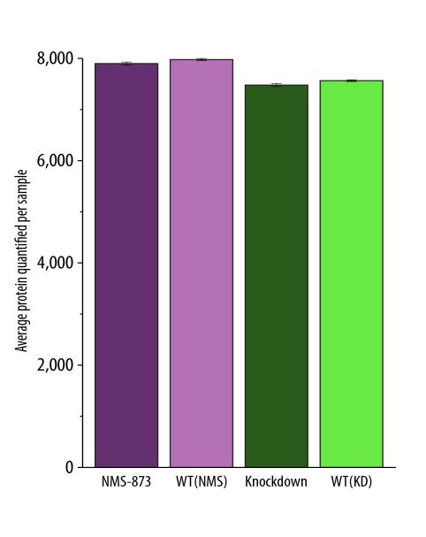
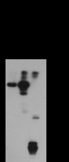
1 VCP adaptor interactions are exceptionally dynamic and subject to differential modulation by a VCP inhibitor Liang Xue 1, Emily E. Blythe 1, Elyse C. Freiberger 2, Jennifer Mamrosh 1, Alexander S. Hebert 3, Justin M. Reitsma 1, Sonja Hess 5, Joshua J. Coon 2,3,4, and Raymond Deshaies 1,6,7 SUPPLEMENTAL FIGURE LEGENDS Supplemental Fig. S1: Analysis of VCP complexes by SEC-mass spectrometry. A, Time course of efficiency of VCP knockdown induced by doxycycline (1µg/ml) using HEK293 cell line that has a stably integrated, doxycycline-inducible VCP shrna (DTC204). At the indicated time after transfection, cell lysate was prepared and evaluated by SDS-PAGE and Western blotting with anti-vcp. The 5 day point was chosen as the condition for all MS experiments, since on day 6 a large amount of cell death occurred. B, Violin plots showing the distribution of coefficients of variation (n=3) of apex measurements for proteins in each sample. C, Number of proteins quantified in each experiment (mean ± SEM, n=3). D, Total abundance change (defined as [LFQintensity] treated divided by [LFQintensity] control ) for all detected proteins in response to VCP knockdown (left panel) or NMS873 treatment (right panel). E, DAVID gene ontology keywords for proteins whose mean apex position was increased or decreased by > 2.5 fractions upon NMS873 treatment or VCP knockdown. F, Proteins from Fig. 1B with a shift in mean apex position of >2.5 fractions, along with a >4 fold increase or decrease in abundance following VCP knockdown. PTPN9 (marked with **) is the only protein that met these criteria in both the VCP knockdown and NMS873 (panel G) treatment groups. Proteins in red have been reported either in BioGRID or in the published literature to bind and/or interact with VCP in some manner. G,
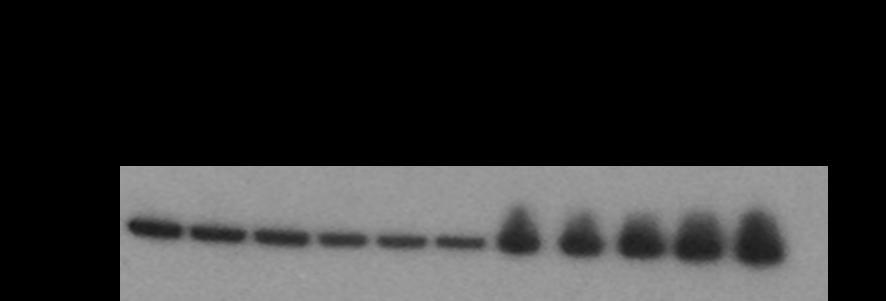
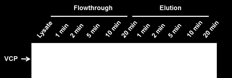
2 Same as panel E, except for the experiment in Fig. 1C in which cells were treated with 10 µm NM873 for 6h. Supplemental Fig. S2: Fractionation of VCP adaptors by SEC. SEC fractionation behavior of different classes of VCP adaptors upon perturbation of VCP activity by NMS873 or shrnamediated depletion. A, Class I adaptors co-fractionated with VCP. B, Class II adaptors were constitutively assembled in complexes of higher MW than the VCP peak. C, Class III adaptors fractionated at MWs lower than VCP and were not affected by either chemical inhibition or depletion of VCP. D, The relative LFQ intensities of VCP and adaptors in SEC fractions from HEK293 cells (bars represent standard error of the mean, n=3). E, HEK293 lysate from untreated cells was chromatographed on a Superose 6 column, and 1 ml fractions were analyzed via western blot. F, In silico-generated SEC chromatograms of representative VCP adaptors in the Kirkwood et. al. dataset. Supplemental Fig. S3: Control experiments for VCP immunoprecipitation. A, Western blotting shows the expression of endogenous VCP bearing an N-terminal FLAG tag in HEK293 cells. Lysates of HEK293 FLAGVCP cells were incubated with anti-vcp or anti-flag antibody. The input, flow through (FT) and bound fractions were separated by SDS-PAGE and blotted with anti-vcp or anti-flag antibody. Note that the FLAG tag did not cause a perceptible shift in the mobility of VCP. WB, Western blot. B, VCP was recovered in IPs as short as 6 min. The time indicated above each lane is the duration of the incubation with antibody prior to adding protein A/G beads for an additional 5 min to capture immune complexes. As the IP time increased, the amount of VCP detected by Western blot decreased in the flow through and 2
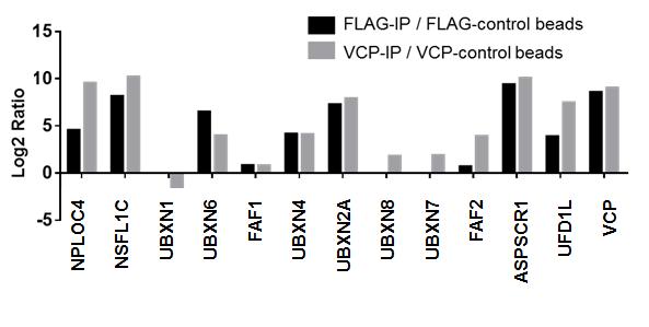
3 increased in the elution. C, Label-free quantification of VCP recovery in IPs of varying duration for the experiments in Fig. 3A and Fig. 3B. D, Venn diagram shows the overlap in protein identifications comparing FLAG IP from HEK293 FLAGVCP cells with IP of untagged endogenous VCP from wild type HEK293 cells. n=1 E, Enrichment of VCP adaptors in VCP pull-downs. Two experiments were conducted in parallel. In the first experiment (black bars), HEK293 FLAGVCP and HEK293 cells were grown in medium containing heavy or light lysine plus arginine, respectively. Cell lysates from both cultures were individually subjected to IP with anti-flag and following the IP step the samples were mixed and analyzed by mass spectrometry. In the second experiment (gray bars), Lysate from the heavy HEK293 cells was subjected to IP with anti-vcp whereas lysate from the light HEK293 cells was subjected to mock IP (no antibody was present before protein A/G beads added for capture). Following the IP step the samples were mixed and analyzed by mass spectrometry. The H/L ratios for known VCP-interacting proteins are shown. n=1. Supplemental Fig. S4: Effect of ND1L sponge on recovery of VCP binding proteins during immunoprecipitation. Titration of increasing amounts of light ND1L into heavy cell lysate progressively reduced the number of proteins recovered by IP with a VCP antibody that does not bind ND1L. Supplemental Fig. S5: Crosslinked VCP complex can be purified by immunoprecipitation. When immunoprecipitated with anti-vcp for 2 hours, most of the cross-linked VCP was recovered in the bound fraction. VCP cross-links were largely resolved upon treating the bound fraction with the reducing agent DTT. 3
4 Supplemental Fig. S6: Chemical inhibition of VCP modulates its repertoire of associated adaptor proteins in HEK293 cells and BJ fibroblasts. A, Label swap SILAC experiments were performed in which cells were either mock-treated or supplemented with 10 µm NMS873 for 6 hours. Cells were treated with 800 µm DSP for 30 minutes prior to cell lysis, mixing of cell lysates, IP with anti-vcp, and mass spectrometry analysis. The ratios for each protein identified in the replicate experiments are plotted on the x and y axes. B, same as panel A, except that cells were treated with or without 10 µm MG132 for 2 hours. C, Duplicate mass spectrometry experiments with label-free quantification were performed in which cells were either mocktreated or supplemented with 10 µm NMS873 for 6 hours. Cells were treated with 800 µm DSP for 30 minutes prior to cell lysis, mixing of cell lysates, IP with anti-vcp, and mass spectrometry analysis. The ratios for each protein identified in the replicate experiments are plotted on the x and y axes. D, same as panel C, except that cells were treated with or without 10 µm MG132 for 2 hours. Supplemental Fig. S7: Potential substrates for UFD1L NPLOC4 and UBX domain proteins as determined by covariance analyses. 4


5 A B C D Supplemental Fig. 1
6 E F G Supplemental Fig. 1
7 A B C Supplemental Fig. 2
8 D Supplemental Fig. 2

9 E Supplemental Fig. 2
10 F Supplemental Fig. 2


11 A B C D E Supplemental Fig. 3

12 Supplemental Fig. 4

13 Supplemental Fig. 5

14 A B C D Supplemental Fig. 6

15 Supplemental Fig. 7