Characterizing Particle uptake by mammalian cells. Prachi Tosniwal IIT Guwahati
|
|
|
- Merry Austin
- 5 years ago
- Views:
Transcription
1 Characterizing Particle uptake by mammalian cells Prachi Tosniwal IIT Guwahati
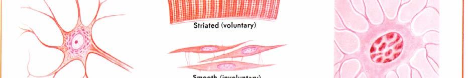

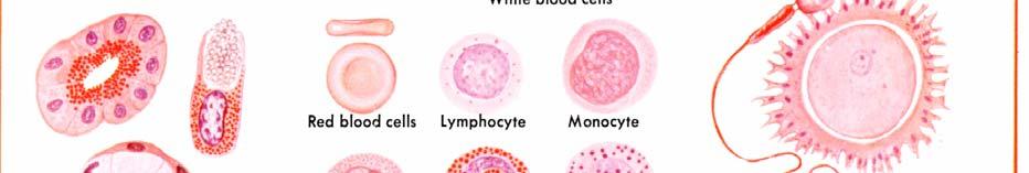
2 Mammalian Cells biology cells tissues organs systems/
3 Phagocytosis

4 Therapeutic Application and Challenges Particles used in therapeutic applications like Drug delivery Vaccination Medical diagnostics etc. These particles are eventually cleared from the body via phagocytosis, by immune cells.
5 Factors affecting phagocytic uptake? Particle parameters size, shape, surface charge and other mechanical properties influence phagocytic uptake. Studies suggest that: Smaller diameter allows the microsphere (particles) to adhere to cells faster and more strongly. gy Spheres were endocytosed more rapidly, while disks circulated in the blood longer with higher targeting specificity (spheres vs elliptical disks). Since the cell membrane is negatively charged, positively ii charged particles, strongly adhere to the cell membrane due to electrostatic attraction. AK2 AK1
6 Slide 5 AK1 AK2 Since strong adhesion between the nanoparticles and the cells is required for cells to internalize the nanoparticles, a smaller particle size would also imply that there is a higher chance that the nanoparticles can be internalized by cells Akshay Kumar, 7/6/2016 A study with various sizes ( μm) and shapes (spheres vs elliptical disks) indicates that spheres were endocytosed more rapidly, while disks circulated in the blood longer with higher targeting specificity in mice.16 Anisotropic polymeric particles, produced by the thin film stretching procedure, showed their ability to evade nonspecific cellular uptake with subsequent enhanced targeted cellular uptake and interaction Akshay Kumar, 7/6/2016
7 Factors affecting phagocytic uptake? Particle parameters size, shape, surface charge and other mechanical properties influence phagocytic uptake. AK1 AK2
8 Slide 6 AK1 AK2 Since strong adhesion between the nanoparticles and the cells is required for cells to internalize the nanoparticles, a smaller particle size would also imply that there is a higher chance that the nanoparticles can be internalized by cells Akshay Kumar, 7/6/2016 A study with various sizes ( μm) and shapes (spheres vs elliptical disks) indicates that spheres were endocytosed more rapidly, while disks circulated in the blood longer with higher targeting specificity in mice.16 Anisotropic polymeric particles, produced by the thin film stretching procedure, showed their ability to evade nonspecific cellular uptake with subsequent enhanced targeted cellular uptake and interaction Akshay Kumar, 7/6/2016
9 Objective : Study phagocytosis of particles by macrophages, with or without surface modification. Cell Culture Cells to be removed from the culture flask! What s the optimal centrifugal speed to obtain a cell pellet with highest cell viability? Phagocytosis Experimental design was modified to study uptake of particles by HeLa cells due to unavailability of RAW cell line (macrophage cell line).


10 Cell culture for mesenchymal stem cell expansion i i / h l ll i
11 Objective 1: Optimizing centrifugation speed Determine the optimal centrifugal speed to obtain a cell pellet with highest cell viability. Cell viability was estimated by determining live and dead cell count using a haemocytometer.
12 Haemocytometer PURPOSE PRINCIPLE METHOD hemocytometer counting chamber/ PURPOSE: Haemocytometer (or counting chamber) is a device used for determining the concentration of cells in cell suspension. It was originally used to determine blood cell counts. It is now used for other types of cells and microscopic particles as well. As the area bounded by the lines is known and depth of the chamber is also known, it is possible to count number of cells/particles in specific volume of liquid.
13 Haemocytometer PRINCIPLE The gridded area Nine squares. Subdivided in 3 directions Central square is further subdivided into 0.05 x 0.05 mm ( mm 2 ) squares. Distinguishing between dead dand viable cells? Sample diluted with a particular stain, such as Trypan blue(dye exclusion staining) Traverses membrane of dead cells, staining them blue. Unable to penetrate membranes of viable. cells, thus excluding them from staining. AK3 p 1 x 1mm 1mm x 0.25 mm 0.25 x 0.20 mm mm mm x 0.20 mm 0.04 mm x 0.05 mm mm 2
14 Slide 11 AK3 A number of stains are used to distinguish between viable and nonviable cells. This is based on the principle that live cells contain intact cell membranes that eliminate certain dyes, like trypan blue, Eosin, or propidium. In dead cells, the stain enters the cytoplasm and the cells take on the stain. If more than 25% of the cells are stained, the cell suspension is most likely not a viable one. Akshay Kumar, 7/7/2016
15 Haemocytometer METHOD Loading: The V notch present at the either end is the place where the sample is loaded into the haemocytometer, with the cover slip on the device. Capillary action takes place and the liquid is spread evenly inside the haemocytometer.


16 Haemocytometer METHOD Counting the cells: Count cells that are on a line? Number of squares to count? hemocytometer counting chamber/ hemocytometer counting chamber/
17 Haemocytometer Uses and Calculations: %Cell Viability = [ Total Viable cells (Unstained) / Total cells (Viable +Dead) ] X 100. Average viable cell count per square = Total #of viable cells in 4 squares / 4. Dilution Factor = Total Volume (Volume of sample + Volume of diluting liquid) / Volume of sample. AK5 Total viable cells/sample = Viable Cells/ml x The original volume of fluid from which the cell sample was removed Volume of media needed = ( #of cells needed / Total # of viable cells ) x 1000.
18 Slide 14 AK5 The primary problem faced during the first experiment was determining an accurate dilution factor. An accurate dilution factor is important because, it is difficult to count the number of cells in each square if there are too many cells. Likewise, if very few cells are present, a homogenous concentration of cells cannot be obtained. Thus, it was concluded that the dilution factor must be determined. This can be done based on initial number of cells, to make counting in each square easier. Akshay Kumar, 7/7/2016
: 0 4400 rpm We selected 5 speeds 800, 1600,")

19 EXPERIMENT Range of speeds (by centrifuge, Remi 4C): rpm We selected 5 speeds 800, 1600, 2400, 3200, 4000 rpm Into 5 tubes, centrifuged at different speeds REFERENCE POINT : INITIAL CELL COUNT
20 Objective 1: RESULTS Speed (in Live Cells Dead Cells Initial RCF) * * Cells * Number of cells ( x 105 ) ± ± ± ± ± ± ± ± ± ± Speed (in RCF) Live Cells Dead Cells Initially
21 Objective 1: INFERENCES Beyond a speed of 697 x g, the number of viable cells were seen to decrease slightly. This could be because mechanical force damaged the cells. We can conclude that the optimal range to obtain a cell pellet with highest cell viability is between x g as high number of viable cells were obtained by centrifuging gthe cells within this range is. For precise results, further experiments can be carried out within this range. We chose 700 x g for our future experiments.
22 Objective 2: Phagocytosis by HeLa cells To visualize phagocytosis and cell division in HeLa cells. Live cell imaging system was used. The experiment was carried out four times, each time carrying out improvisations to analyze cells more clearly.
23 Objective 2: Method / Experimentation ti The imaging was carried out in multiple fields using Brightfield, over a period of time. After every five minutes, an image of the field was taken. The frame images obtained were processed using IMAGEJ software.


24 AK8 Experiment 1 Visualise Individual behaviour of cells. The petri dish was almost 80% confluent. Difficult to distinguish individual cells.
25 Slide 20 AK8 The first time experiment was carried out, growth of HeLa cells was visualized. The petri dish was almost 90% confluent. Hence, growth of cells was slow and analysis was not clear. Akshay Kumar, 7/7/2016


26 Experiment 2 AK7 Particle uptake by HeLa cells and proliferation was to be analyzed. Imaging 4 hours. Higher volume of particles added. # particles >> # of cells
27 Slide 21 AK7 The second time the experiment was carried out, particle uptake by HeLa cells and proliferation was to be analyzed. Several problems were faced in analyzing the cell dynamics as the number of particles added were much more than the number of cells. The images were filled with numerous particles floating around and on the cells due to which it was very difficult to visualize the cellular dynamics. Akshay Kumar, 7/7/2016


28 Experiment 3 # of HeLa cells and particles were optimum and imaging was clearer. Imaging 4 hours. Cells did not seem to interact with the particles Short timeframes of imaging. g AK9
29 Slide 22 AK9 In the third experiment that was carried out, number of HeLa cells and particles were optimum and imaging was clearer. However, cells did not seem to interact with the particles or phagocytose them. Using fast focus for imaging of cells made them unclear, after some time, to study the effect of particles on cell proliferation and also the fields had to be selected in neighboring areas only to avoid out of focus field frames. Movement of particles around the cell was unclear, due to 2-D imaging. In order to determine accurate interaction of cells with particles, 3-D imaging is required. Akshay Kumar, 7/7/2016


30 Experiment 4 # of HeLa cells and particles were optimum and imaging was clearer. Imaging approx. 10 hours. g g pp Uptake of particles by the cell was unclear due to 2 D imaging. 3 D imaging is required to determine accurate interaction.
31 Objective 2: INFERENCES Particle to cell ratio should not be too high (ideal, 5:1), else it becomes very difficult to clearly visualize the cells. Movement of particles around the cell was unclear, due to 2 D imaging. To visualize and study the cellular dynamics precisely, 3 D frame images should be taken. Autofocus further enhances the image quality and clarity which is easier to interpret.
32 Conclusions Optimum range to spin down cells to get maximum viable cell count is between x g. Number of cells used for imaging should not be too high. Particle to cell ratio should not be higher than 5:1. Difficult to analyse uptake for short time frames. Difficult to analyse uptake for short time frames. 3D imaging and use of fluorescence could give us a better understanding of how cells are interacting with particles.
33 Acknowledgement I express my sincere and heartfelt gratitude to the Indian Institute of Science, Bioengineering Summer Training Program and Chair of Biosystems Science and Engineering(BSSE), Dr. G. K. Ananthasuresh, for giving me an opportunity to learn and gain experience. I would like to thank my guide, Dr. Siddharth Jhunjhunwala along with Dr.Vaishnavi Ananthanarayanan and Dr. Shilpee Jain for their guidance and support throughout the program. I am grateful to Ashwini Ravi, Nireekshit Tirumala and Jerrin Mathew who taught me several techniques and helped me with different aspects of my project. I would like to thank the lab members Mekhla Singhania for all the help and support.