Key components of DNA-based Biotechnology
|
|
|
- Brianne Williams
- 5 years ago
- Views:
Transcription
1 Lecture 12 DNA Recombinant Technology DNA enzymology: restriction enzymes, methylases, ligases, polynucleotide kinase, reverse transcriptases Hybridization: complementarity of DNA and RNA The DNA Carriers: Plasmid, Viral Vector Key components of DNA-based Biotechnology Restriction enzyme analysis Blotting techniques Sequencing Solid-phase synthesis PCR


2 1978 Werner Arber, Daniel Nathans, Hamilton O. Smith "for the discovery of restriction enzymes and their application to problems of molecular genetics" Werner Arber 1/3 of the prize 1978 b Switzerland Biozentrum der Universität Basel, Switzerland First discovered restriction enzyme in E. coli with Stewart Linn in the late 60s Hamilton O. Smith 1/3 of the prize b Johns Hopkins University School of Medicine Baltimore, MD Daniel Nathans 1/3 of the prize b d Johns Hopkins University School of Medicine Baltimore, MD, USA


3 The Nobel Prize in Chemistry 1980 "for their contributions concerning the determination of base sequences in nucleic acids Walter Gilbert 1/4 of the prize b Harvard University, Biological Laboratories Cambridge, MA The Nobel Prize in Chemistry 1980 "for their contributions concerning the determination of base sequences in nucleic acids Frederick Sanger 1/4 of the prize b MRC Laboratory of Molecular Biology Cambridge The Nobel Prize in Chemistry 1980 Paul Berg 1/2 of the prize 1980 b Stanford University Stanford, CA, USA "for his fundamental studies of the biochemistry of nucleic acids, with particular regard to recombinant-dna"
4 Restriction endonucleases Also called restriction enzymes: sequence-specific, target sequence mostly palindromic with 2-fold rotational symmetry, cleavage site near symmetry axis
5 Naming the restriction enzymes Restriction endonucleases was discovered as a defense mechanism in bacteria to prevent invasion of foreign DNA such as viral DNA, by cutting it up. Most of them cut at specific sites, making them an extremely useful tool in molecular biology. The name of a restriction enzyme: the first three letters derive from the Latin name of the microorganism, then strain designation, then the sequence the enzyme was discovered and isolated. EcoR 1: 1st restriction enzyme from Escherichia coli R BamH1: 1st restriction enzyme from Bacillus amyloliquefaciens H HindII : 2nd restriction enzyme from Haemophilus influenzae Rd genus species strain genus species strain
6 Restriction endonucleases Restriction enzymes cut DNA molecules at specific sequences (only 5 -->3 is shown); arrows are cutting sites Sticky end Vs. Blunt end
7 R-M system (restriction-modification system) restriction endonuclease and methylase pair confers protection of host DNA from cutting EcoRI 5 -GAATTC-3 3 -CTTAAG-5 The host DNA is protected from endonuclease digestion of EcoRI with strategic methylation at the adenine base by EcoRI methylase.
8 EcoR1 and EcoR1 methylase EcoR1 does a staggered cut at a specific 6-bp inverted repeat and gives sticky ends; the corresponding methylase also recognizes the same sequence and modifies it.
9 Restriction enzymes cut DNA into a reproducible set of restriction fragments Restriction site of Ad2 (adenovirus 2, 36 kb genome) produced by either EcoR1 (GAATTC) or HindIII (AAGCTT) from Haemophillus influenzae (labeling of A-Z according to size).
10 Restriction enzymes generate restriction maps Restriction map: mapping the restriction sites for two enzymes relative to each other in a DNA containing one copy of each site. With different pairs a detailed resitriction map can be constructed.

11 Nobel class work
12 Eco R1 Restriction enzymes, (EcoRI in this example), surrounds the DNA molecule at the point it seeks (sequence GAATTC). It cuts one strand of the DNA double helix at one point and the second strand at a different, complementary point (between the G and the A base).

13 Cut and paste cut paste Sticky ends transiently base-pair for the action of T4 DNA ligase.
14 Restriction enzymes and ligation Cut and paste form the basis of genetic engineering
15 Gel electrophoresis of nucleic acid Gel making (vertical, horizontal), agarose gel with comb inserted during solidification, sample preparation (denaturating or native), Sample loading, detection (dye staining or radioactive labeling), Quantitation and archiving
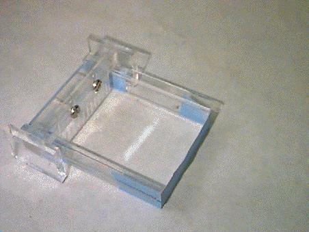
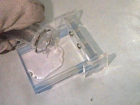
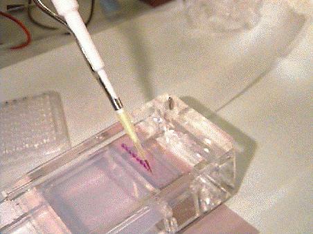
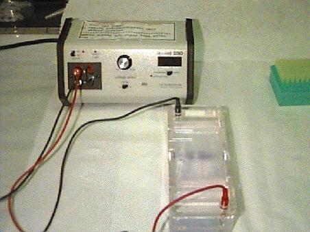
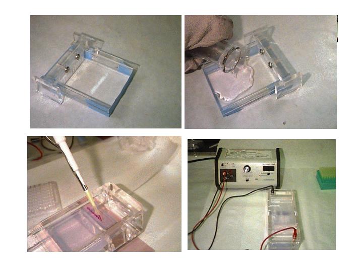
16 Gel electrophoresis of nucleic acid
17 Gel electrophoresis of nucleic acid

18 Polyacrylamide gel electrophoresis (1 nt to 2 kb) µ= Ze/f


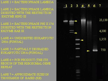
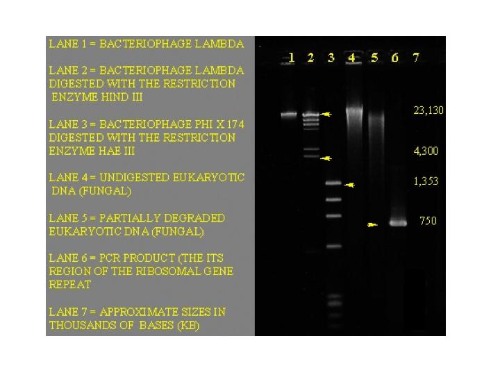
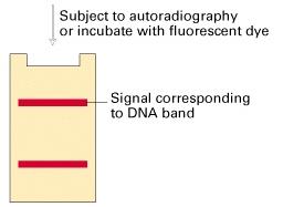
19 Agarose gel electrophoresis Agarose: repeating 1,3 linked β-d-galactopyranose and 1,4 linked 3,6 anhydroα-l-galactopyranose, pore larger for 500 bp to 20 kb DNA band detected by either autoradiography or dye staining.

20 Estimation of molecular size of DNA Electrophoretic mobility of a DNA fragment is proportional to the log of its size.
. (b) Autoradiogram of 32 P-labeled fragments separated on polyacrylamide gel.")
21 Visualization of restriction fragment (a) Different plasmid clones digested with EcoRI (lanes 1-5), HindIII of Ad2 was used as molecular size marker (M). (b) Autoradiogram of 32 P-labeled fragments separated on polyacrylamide gel.

; lane 3, NotI of E.coli chromosome. (B) Yeast chromosomes ranging from 0.2 Mb to 2.2 Mb. The gel is 13x12.")
22 Pulsed-field gel electrophoresis (A) (B) Pulsed-field gel electrophoresis can separate DNA from 20 kb to 10 Mb. Pulsed electric field forces DNA does through stretched-coiled motion, which significantly separates larger DNA of different sizes. (A) Lane 1, yeast chromosomal DNA; lane 2, concatomers of λ DNA (48.5 kb); lane 3, NotI of E.coli chromosome. (B) Yeast chromosomes ranging from 0.2 Mb to 2.2 Mb. The gel is 13x12.5 cm (reproducibility).

23 Restriction mapping A restriction mapping experiment to determine the position of a BamH1 site and the orientation of a 1.6 kb HindIII fragment.


 Two")

24 (A) Restriction mapping (B) (A) Restriction mapping of an unknown DNA. (B) Two potential maps of the unknown DNA

25 Blotting technique Analyzing specific nucleic acids in complex mixture by blotting. A specific DNA sequence isolated by cloning can serve as a probe to detect the presence and the amounts of complementary nucleic acids in complex mixtures including total cellular DNA or RNA The membrane-hybridization assay (Blotting): dsdna denature or melt and bind onto filter paper, incubate with labeled ss DNA probe, complementary DNA hybridizes, wash away non-bound labeled DNA, perform autoradiography.
: DNA separate on gel, denature by base, transfer to membrane (blot) via diffusion of electrophoresis, block with non-specific DNA or protein, hybridize with the probe and")
26 Southern blotting Southern blotting (E.M.Southern, JMB 98, 508, 1975): DNA separate on gel, denature by base, transfer to membrane (blot) via diffusion of electrophoresis, block with non-specific DNA or protein, hybridize with the probe and detect the position and amount of target DNA. Autoradiography: a technique in which a radioactive sample is exposed to a photographic emulsion, thus taking a picture of itself.


27 Another view of Southern blot analysis Southern blotting
 denature, Southern blot, (d) hybridize to labeled minisatellite DNA, detected label with")
28 DNA fingerprinting (b) (c) (d) (a) cut DNA with HaeIII into 8 fragments, only 3 contains minisatellites; (b) electrophoresis, (c) denature, Southern blot, (d) hybridize to labeled minisatellite DNA, detected label with X-ray film.


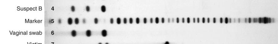
29 DNA fingerprinting and DNA typing DNA fingerprint: the use of highly variable region (e.g. hypervariable minisatellite region) of DNA to identify individuals. Lane 1-9, 9 unrelated white subjects; lanes 10 and 11 monozygotic twins.

30 DNA typing in forensics

31 DNA typing in forensics DNA typing is essentially an application of southern blotting. The frequency of a coincidental match of the DNA pattern between two individuals is one in 33 billion.
32 RNA Northern blot analysis RNA


33 Northern blotting Northern blot detects specific RNAs. It is a technique used to identify and locate mrna sequences that are complementary to a piece of DNA called a probe. Northern blot of β-globin mrna in erythroleukemia cells before and after differentiation.

34 Northern blot Northern blot: Poly(A) + RNA from isolated rat tissues, equal amounts of RNA were electrophoresed and Northern blotted. The RNAs on the blot were hybridized to a labeled probe for the rat glyceraldehyde- 3-phosphate dehydrogenase, and the blot was then exposed to X-ray film. The intensity of the band represents the relative amount of G3PDH mrna in each tissue.
35 S1 nuclease protection assay Nuclease protection method for quantitating specific RNAs in a mixture and mapping them.

36 S1 nuclease protection assay Mapping of a 1.7kb RNA on the 36-kb adenovirus genome. It is in fragments A and B
37 Mapping the start site for transcription S1 mapping vs. primer extension
38 Nulease protection assay Nulease protection assay for quantifying specific mrnas in a mixture and mapping them: (a) excess DNA probe, the protected hybrid, alcohol precipitated, electrophoresed and autoradiographed, the band intensity gives the relative amounts.


39 Northern blot Gel loading on RNA gel as determined by ethidium bromide staining of rrna Southern blot Gel loading on agarose gel as determined by ethidium bromide staining
40 DNA sequencing: Sanger s reagent

41 DNA sequencing: Sanger s method
42 DNA sequencing: Sanger s method
43 DNA sequencing: Sanger s method
44 DNA sequencing: Sanger s method 21 Primer-5 -ATGATACGGTCT-3 Template: 5 -AGACCGTATCAT-3 CAAAAAACGG...


45 DNA sequencing: the Maxam-Gilbert method Maxam-Gilbert Method: Breaking the end-labeled DNA strand at specific bases using base-specific reagents. (i) radiolabel, methylation with dimethyl sulfate, treat with piperidine, remove base, break DNA at the apurinic site, leaves 3 -phosphate on the nucleotide preceded the G. All G-removed fragments can be separated by gel.
, electrophoresed and")
46 G+A DNA sequencing: the Maxam-Gilbert method G T+C C dsdna labeled at the 5 - end, the label is removed from one end, denatured, expose four samples to different chemical reactions (controlled so that each labeled chain will break only once), electrophoresed and autoradiographed

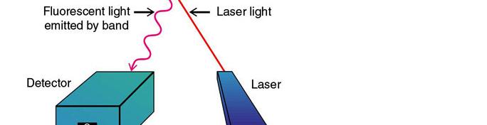

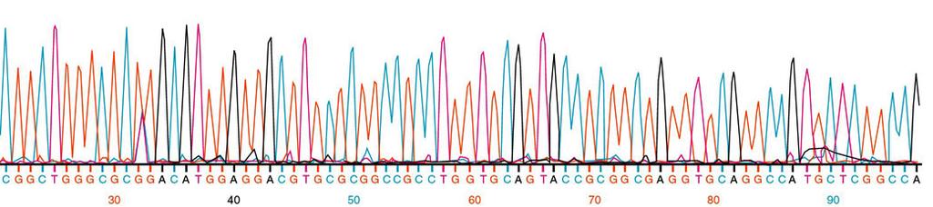
47 Automated sequencing

48 complete genome Haemophilus influenzae: complete genome Science, 269, 496, ,830,137bp encodes 1700 proteins and 70 RNAs
49 Oligonucleotide Synthesis: solid-phase synthesis of DNA by the phosphite triester method
50
51 Polymerase chain reaction: 1st cycle

52 PCR 1st cycle


53 Plasmid Plasmid: Plasmids are circular, double-stranded DNA molecules that exist in bacteria and in the nuclei of some eukaryotic cells. They can replicate independently of the host cell. The size of plasmids ranges from a few kb to near 100 kb

54 To clone blunt-ended PCR fragments amplified with Platinum Pfx, which can amplify genomic templates for up to 12 kb and plasmid template up to 20 bp. See