Prof. Steven S. Saliterman
|
|
|
- Harry Evelyn Jennings
- 5 years ago
- Views:
Transcription


1 Department of Biomedical Engineering, University of Minnesota Prof. Angela Panoskaltsis-Mortari s BMEn 5361, 3D Bioprinting
2 Tissue engineering Bioprinting Design considerations Imaging modalities Segmentation software In Part 2 we will look at some examples from the literature. Prof. Steven S. Saliterman
3 Tissue Engineering Components: The type or types of living cells being implanted (e.g. somatic, embryonic stem cells, adult stem cells, or induced-pluripotent stem cells). Type of scaffolds supporting the cells (i.e. the mechanical cues provided to the cells). Type of drugs, extra-cellular matrix (ECM), and growth factors conditioning the cells, (the additives that provide chemical cues to the cells). 3D Printing: Computer assisted process for depositing biomaterials and living cells in a determinate configuration in order to produce a defined 3D biological structure. Bioinks consist of various polymer materials, cells and additives. Prof. Steven S. Saliterman Mosadegh, B., G. L. Xiong, S. Dunham, and J. K. Min. "Current Progress in 3d Printing for Cardiovascular Tissue Engineering." Biomedical Materials 10, no. 3 (Jun 2015).

4 Our focus today Prof. Steven S. Saliterman Teodori, L., A. et al. "Three-Dimensional Imaging Technologies: A Priority for the Advancement of Tissue Engineering and a Challenge for the Imaging Community." Journal of Biophotonics 10, no. 1 (Jan 2017):
5 Foyt, D. A., M. D. A. Norman, T. T. L. Yu, and E. Gentleman. "Exploiting Advanced Hydrogel Technologies to Address Key Challenges in Regenerative Medicine." [In English]. Advanced Healthcare Materials 7, no. 8 (Apr 2018): 22.
6 Anatomically correct constructs from medical imaging data. Porous structures. Co-culturing of multiple cell types. Precise patterning of cells and ECM. Controlled deliver of growth factor and genes. Potential for high-throughput fabrication. Challenge - suitable vascularization. Diffusion length of oxygen/nutrients is µm. Prof. Steven S. Saliterman

7 Murphy, S. V., and A. Atala. "3d Bioprinting of Tissues and Organs." Nature Biotechnology 32, no. 8 (Aug 2014):
8 Today 1) Design What are your trying to achieve? What bioinks and printing method? 2) Images - Useful from the subcellular to organ level. Apply CAD tools for segmentation, freeform and other space-filling methods. 3) Slicer - G-code generation for controlling toolpath, speed, valves, droplet patterns, laser pulse, photoinitiator lights (e.g. Ingracure and other gels), temperature etc. 4) Special setups - Extruder, tips, light source (specific nm), pressure & calibration, cooling (Pluronic Gel) etc. 5) Bioprinting 6) Bioreactor - Incubation, nutrients, growth factors, oxygen supply, environment, etc. (Your 4 th dimension time!) 7) Observation - Fluorescent and transmitted light (confocal microscopy, bright-field, dark-field, confocal laser microscopy etc.). Automatic imaging with control of ambient air, humidity and temperature. 8) Characterization - Histology, growth, mechanical properties etc. Prof. Steven S. Saliterman

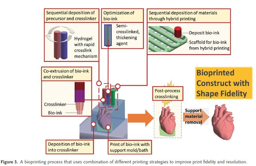
9 Lee, J. M., and W. Y. Yeong. "Design and Printing Strategies in 3d Bioprinting of Cell- Hydrogels: A Review." Advanced Healthcare Materials 5, no. 22 (Nov 2016):
10 Hospodiuk, M. et al. "The Bioink: A Comprehensive Review on Bioprintable Materials." Biotechnology Advances 35, no. 2 (Mar-Apr 2017):

11 Lee, J. M., and W. Y. Yeong. "Design and Printing Strategies in 3d Bioprinting of Cell- Hydrogels: A Review." Advanced Healthcare Materials 5, no. 22 (Nov 2016):
12 Lee, J. M., and W. Y. Yeong. "Design and Printing Strategies in 3d Bioprinting of Cell- Hydrogels: A Review." Advanced Healthcare Materials 5, no. 22 (Nov 2016):
13
14 Lee, J. M., and W. Y. Yeong. "Design and Printing Strategies in 3d Bioprinting of Cell- Hydrogels: A Review." Advanced Healthcare Materials 5, no. 22 (Nov 2016):

15 Ozbolat, I.T. 3D Bioprinting Fundamentals, Principles and Application. Elsevier, Amsterdam 2017.
16 Underlying methods in CAD systems: Constructive solid geometry (solid primitives and boolean operators) Boundary representation (vertices, edges and faces) Spacial enumeration (cubic elements) Image-based design Implicit surfaces Space-filling curves Irregular porous structures Prof. Steven S. Saliterman Giannitelli, S. M., et. al.. "Current Trends in the Design of Scaffolds for Computer-Aided Tissue Engineering." [In English]. Acta Biomaterialia 10, no. 2 (Feb 2014):

17 Nam Jet.al. Computer aided tissue engineering for modeling and design of novel tissue scaffolds. Computer-Aided Design & Applications 2004;1:

18 Honeycomb pores Hilbert recursive curves Prof. Steven S. Saliterman Giannitelli, S. M., et. al.. "Current Trends in the Design of Scaffolds for Computer-Aided Tissue Engineering." [In English]. Acta Biomaterialia 10, no. 2 (Feb 2014):
19 Giannitelli, S. M., et. al.. "Current Trends in the Design of Scaffolds for Computer-Aided Tissue Engineering." [In English]. Acta Biomaterialia 10, no. 2 (Feb 2014):


20 Our focus today Prof. Steven S. Saliterman Nam, S. Y., et. al. "Imaging Strategies for Tissue Engineering Applications." Tissue Engineering Part B-Reviews 21, no. 1 (Feb 2015):
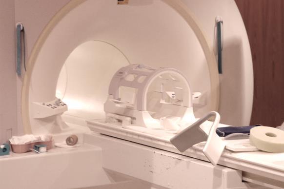
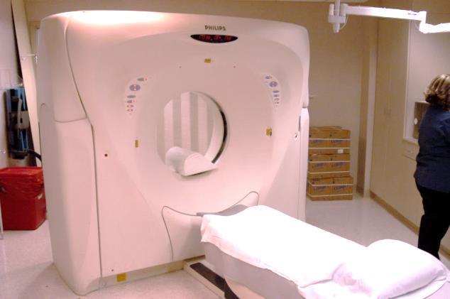
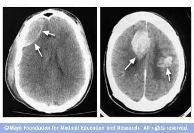
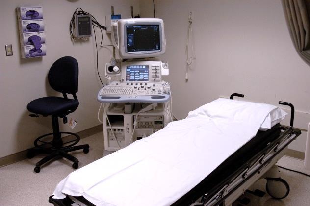
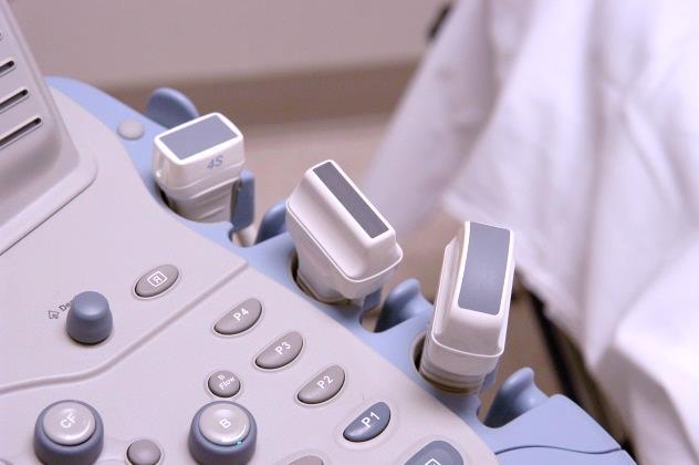
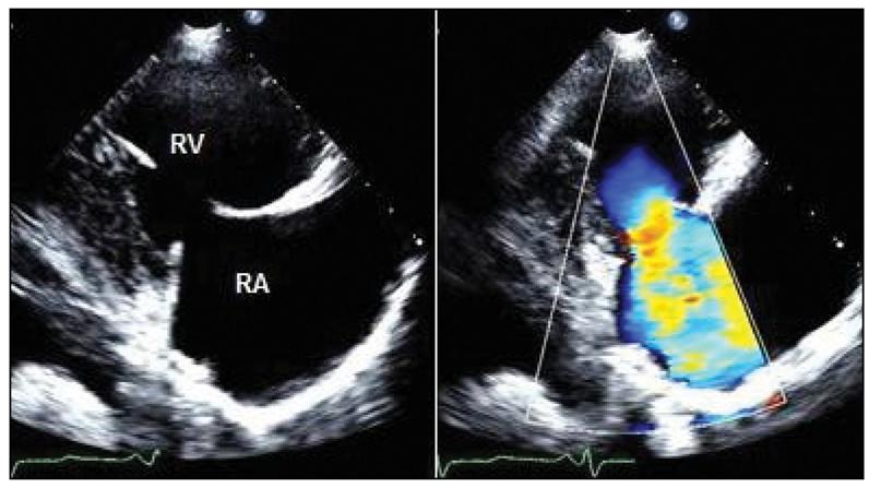
21 Magnetic Resonance Imaging (MRI) Human max. is 3T (Tesla) resolution of 250µm x 250µm 0.5mm. High spatial resolution µmri, 7-10T, 5-200µm. Magnetic nanoparticles. Computed tomography (CT) Computer Axial Tomography Typical resolution of mm. µct, resolution of 1-200µm. Ultrasound Resolution of 1mm x 1.mm x 0.2mm. PET Positron emission tomography SPECT Single photon emission computed tomography Optical Coherence Tomography (OCT) Traditional optical techniques. Prof. Steven S. Saliterman
22
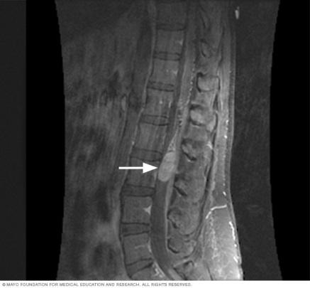
23 Mayo Foundation for Medical Education and Research


24 CT scan/pet Scan/ Combined Mayo Foundation for Medical Education and Research
 and possible deindentifying them for HIPPA regulations (DICOMCleaner).")
25 Purpose To delineate and isolate anatomical features within an imaging database- e.g. bone, cartilage, soft tissue, edema; muscle, lung, brain & other organs, and tumors. Method Extract images from DICOM files (ITK-Snap, Onis) and possible deindentifying them for HIPPA regulations (DICOMCleaner). Segmentation Software (ITK-Snap, Materialise Mimics, Materialise 3- matic). Pre-segmentation Phase - identify parts of image as foreground and background. Active Contour Phase - manual and semiautomatic methods. Editing and fixing mesh files (.STL) - Autodesk Meshmixer. Slicer software Simplify3D and Repetier. G-coding for the specific bioprinter - e.g. Slic3R (printer customized interface to control what happens in a sequence of control steps.) Prof. Steven S. Saliterman

26 Sagittal or Median Parasagittal (Yellow) Transverse or Axial Frontal or Coronal Prof. Steven S. Saliterman Image, Wikipedia

27 Manual Segmentation Prof. Steven S. Saliterman


28

29

30


31 Import the STL Mesh file generated by ITK-Snap. Edit feature here slicing in a plane, bottom view. Prof. Steven S. Saliterman
32 Tissue engineering Bioprinting Design considerations Imaging modalities Segmentation software Prof. Steven S. Saliterman