Dimeric WH2 domains in Vibrio VopF promote actin filament barbed end uncapping and assisted elongation
|
|
|
- Allan Freeman
- 5 years ago
- Views:
Transcription
1 Dimeric WH2 domains in Vibrio VopF promote actin filament barbed end uncapping and assisted elongation Julien Pernier, Jozsef Orban 1, Balendu Sankara Avvaru, Antoine Jégou, Guillaume Romet- Lemonne, Bérengère Guichard and Marie-France Carlier Cytoskeleton Dynamics and Motility group, Laboratoire d Enzymologie et Biochimie Structurale, Centre National de la Recherche Scientifique, Gif-sur-Yvette, France Contact : M.-F. Carlier, carlier@lebs.cnrs-gif.fr 1 : present address : Department of Biophysics, Medical School, University of Pecs, H-7624 Pecs, Hungary 1
2 SUPPLEMENTARY INFORMATION SUPPLEMENTARY FIGURES AND LEGENDS 2
3 3
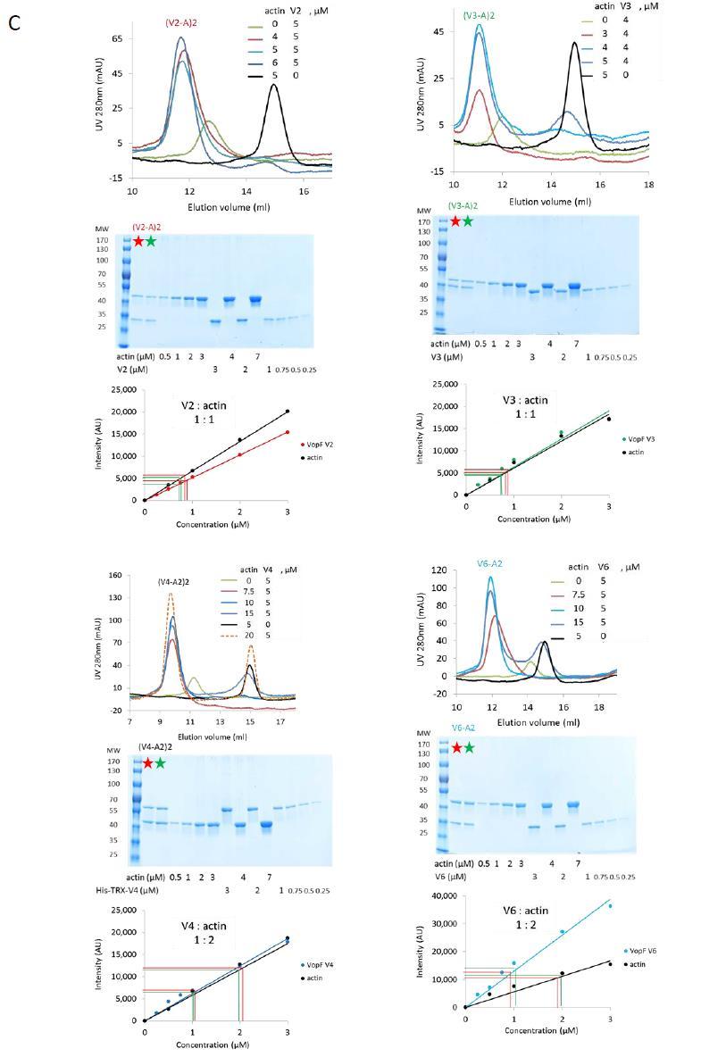
4 Supplementary Figure 1. Characterization of the various WH2 constructs and VopF complexes with actin A. Analytical size exclusion chromatography of VopF constructs. B. Fluorescence titration curves of AEDANS-actin by the isolated WH2 peptides. AEDANS- G-actin (1 μm) was titrated by W1 (blue), W2 (red) or W3 (green). The quenching of AEDANS fluorescence is normalized to represent the saturation of actin by the peptides. Binding curves are calculated as described 19. The best fit values of K D are in the inset. C. Analysis of VopFs complexes with G-actin by Size Exclusion Chromatography (Superdex /300 GL) and SDS-PAGE. Mixtures of G-actin and VopF constructs in different molar ratios (inset) were loaded on the column. Superimposed peaks for the complex ascertain that a single complex is formed. The V4-actin loaded sample represented in dashed orange line (20 µm actin, 5µM V4) contained 20 µm Latrunculin A and the elution buffer contained 2 µm G- actin and 2 µm Latrunculin A. The peak fractions (red and green stars SDS-PAGE samples) of the eluted VopF-actin complex were co-electrophoresed with actin and VopF standards. Each linear calibration curve is derived from least square analysis of the densitometric analysis of scanned gels. The VopF: actin molar ratio in the eluted complex was derived by interpolation of the amount of both proteins in the eluted fractions of the complex (red and green dashed lines). 4

5 Supplementary Figure 2. No lateral contacts of the F-actin type are formed in V2-actin and V4-actin complexes F-actin, V2, His-TRX-V4, free and in complex with actin were submitted to covalent crosslinking by ppdm, followed by SDS-PAGE and immunodetection using anti-actin or anti-histidine antibodies. F-actin lateral crosslinking yields a polypeptide of apparent MW of 120 kda. No actin-actin bonds identical to the F-actin ppdm-crosslinked dimer was seen in the V2-actin crosslinked adduct (apparent MW 80 kda) nor in V4-actin crosslinked adducts. The covalently crosslinked V4-actin adducts (3 polypeptides of apparent MW 100, 110 and 150 kda) were recognized by both anti-his and anti-actin antibodies. 5
6 Supplementary Figure 3. Actin nucleation and sequestration by VopF constructs A. Actin (2 µm,10% pyrenyl-labeled) was spontaneously assembled in the presence of 0, 1 or 5 µm of each isolated WH2 domain, color coded as indicated. B, C. Actin polymerization assays carried out with 2 μm actin in the presence of V1, V1 H, V3, V4 H, V4*, V5, V6 and V7 as indicated. 6
7 Supplementary Figure 4. V1 does not bind filament pointed ends nor gelsolin(actin) 2 complex and V4 competes with gelsolin for binding to actin A. Seeded growth (2 µm actin, 10% pyrenyl-labeled) from gelsolin actin seeds in the presence of V1 as indicated. B. Analysis of mixtures of V1 and actin, gelsolin and actin, gelsolin and V1 and actin on sizeexclusion chromatography. C. Analysis of mixtures of V4 and actin, gelsolin and actin, gelsolin and V4 and actin on sizeexclusion chromatography. V4 does not bind gelsolin(actin) 2 complex but causes its dissociation by competing with gelsolin for binding to actin. Identical results were obtained with V2 (not shown). D. Fluorescent titration curves of NBD-actin by gelsolin with or without V4. 7
8 Supplementary Figure 5. Monomeric constructs of VopF sequester G-actin A. Actin (1.8 µm, 10% pyrenyl labeled) was polymerized in the absence (closed circles) and in the presence (open circles) of 6 nm gelsolin and W1, W2 and W3 as indicated. K D were derived using equation 1 (sup. Methods) B. Actin (1.6 µm, 10% pyrenyl labeled) was polymerized in the absence (closed circles) and in the presence (open circles) of 6 nm gelsolin and V6 and V7 as indicated. K D were derived as in frame A. 8
9 Supplementary Figure 6. Dimerization of WH2 domain is required for uncapping activity and barbed end regulation by VopF A. V6 and Spire fail to uncap filaments. Barbed end growth was initiated by spectrin-actin in the presence of G-actin (2 µm), and CP (10 nm). Barbed end growth does not resume upon addition of V6 nor Spire. Profilin was present in the uncapping assay with V6, but not in the assay with Spire, because Spire blocks barbed end growth from profilin-actin (Bosch et al., 2007). B. V2 does not bind CP. CP and V2 elute in two separate species from a size exclusion column, both in absence or presence of actin. 9
 and VopF-VCD (blue).")
10 Supplementary Figure 7. Potential surface interactions between F-actin and VopF-VCD derived from docking VopF-VCD on the side of the filament Cartoon representation of surfaces of Actin (pink) and VopF-VCD (blue). Amino acids likely to form ion pairs between actin and VopF-VCD are depicted as sticks (Oxygen, red; Nitrogen, blue; Carbon, green (actin), yellow (VopF-VCD)). Bond distances less than 5 Å between ionic partners are shown as red dashed lines. 10
11 SUPPLEMENTARY TABLE Supplementary Table 1. Amino acid ionic partners derived from the model of VopF- VCD docked on the side of F-actin C-terminal dimerizationdomain (V1) amino acids Glu 456 Lys 50 Glu 461 Arg 372 Potential ionic partners on the surface of F-actin Base Glu 465 Lys 68 Lys 469 Asp 80 Lys 371 Glu 214 Arm Arg 400, Lys 417 Glu 311 Arg 402 Glu 334 Lys 410 Asp 25 11
12 SUPPLEMENTARY VIDEO LEGENDS Supplementary Movies 1 and 2. These movies (taken at 1 frame every 30s) represent the evolution with time of the two filaments shown in Figure 5C. Movies are accelerated 210-fold. Supplementary Movie 1. Filament remains capped In this movie (field width= 8 µm) the filament remains capped and does not grow upon flowing in 2 nm CP. Supplementary Movie 2. VopF uncaps barbed ends from capping protein In this movie (field width= 17 µm), the filament is uncapped and grows at the same rate as when free upon flowing in 2 nm CP and 20 nm V2. 12