DTI of White Matter in Brain Diseases; Potentials and Pitfalls
|
|
|
- Lillian Thompson
- 5 years ago
- Views:
Transcription
1 DTI of White Matter in Brain Diseases; Potentials and Pitfalls Rick M. Dijkhuizen, PhD Biomedical MR Imaging and Spectroscopy Group Center for Image Sciences University Medical Center Utrecht Utrecht, The Netherlands


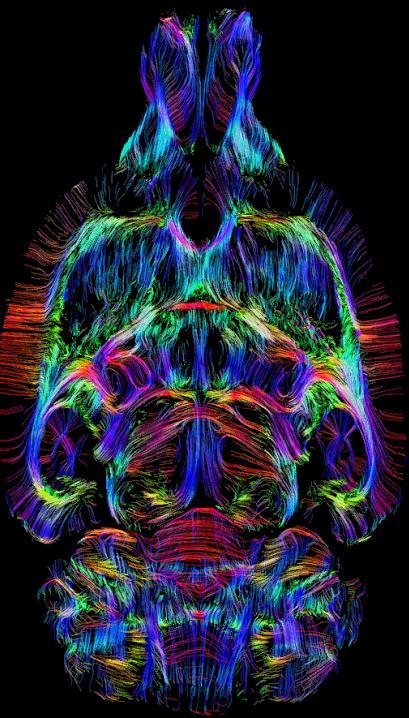
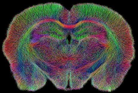
2 Diffusion tensor imaging of white matter
 and shape (anisotropy) of water displacement Mean")

3 Diffusion tensor imaging Diffusion Tensor Imaging: Mapping of degree (diffusivity) and shape (anisotropy) of water displacement Mean diffusivity MD Fractional anisotropy FA Cerebrospinal fluid High, free, isotropic diffusion White matter fibers Low, restricted, anisotropic diffusion


 Mean")
")
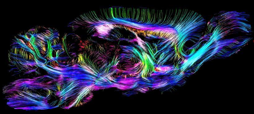
4 Diffusion tensor imaging of tissue architecture Diffusion tensor decomposition: The three diffusion eigenvectors Axial (D//) and radial diffusivities (D ) Mean diffusivity (MD) Fractional anisotropy (FA) Orientation-based FA map Fiber tractography


 map")

5 DTI of white matter changes after stroke Middle cerebral artery occlusion 1 week post-stroke 10 weeks post-stroke Fractional anisotropy (FA) map Fiber reconstructions Schaechter et al., Hum Brain Mapp 2009
 vs. Controls (n=10) Reduced FA Increased FA Schaechter et al.")
6 Altered fractional anisotropy in white matter tracts after stroke Patients (>6 months post-stroke; n=10) vs. Controls (n=10) Reduced FA Increased FA Schaechter et al., Hum Brain Mapp 2009
 posterior Stroke > Controls FA reduction: white matter degeneration FA increase: white matter remodeling? anterior 0.95 1-P 1.")
7 Degeneration and remodeling of perilesional white matter after stroke Tract-based spatial statistics of FA maps: Stroke (day 70; n=5) vs. Controls (n=10) posterior Stroke > Controls FA reduction: white matter degeneration FA increase: white matter remodeling? anterior P 1.00 Stroke < Controls H&E staining FA map LFB staining (myelin) Van Meer et al., J Neurosci 2012


8 Functional connectivity (z ) Sensorimotor performance (points) Correlation between changes in white matter structure, gray matter activity and behavior after stroke White matter structure Functional connectivity Behavioral function Medium stroke Van Meer et al., J Neurosci 2012


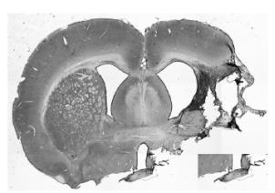

9 Increased FA may be related to various microstructural changes Fractional anisotropy (FA) 8 weeks after 120-min MCA occlusion LFB (myelin) GFAP (glia) APP (amyloid precursor protein) Rudrapatna et al., Neuroimage 2014

10 Degree of staining Correlations between diffusion parameters and histological stainings Kendall's-τ test Ipsi- and contralesional Ipsilesional Contralesional Rudrapatna et al., Neuroimage 2014


11 Neuroanatomical connectivity in perilesional white matter In vivo neuronal tract tracing with manganese-enhanced MRI Tracer injection Neuronal uptake Axonal transport a r Mn 2+ - Ca 2+ analogue - Paramagnetic Mn 2+ SMCX Th/IC Accumulation T 1 W MRI 2 days post-mn 2+ DTI and MEMRI of white matter connectivity at 10 weeks after stroke Areas with progressive FA increase IC FA map Post-manganese R 1 map Van der Zijden et al., Exp Neurol 2008


 Large")


12 DTI-based tractography after experimental stroke Control Medium stroke (10 weeks) Large stroke (10 weeks) Po et al., PLoS One 2012 Sinke et al., unpublished


13 Accuracy of DTI-based fiber reconstructions?


14 DTI-based tractography A Diffusion elipsoids Streamlines C Free, isotropic diffusion Restricted, anisotropic diffusion B A A C B Connectivity matrix A B C x B x C x x


15 Structural cortical connectome in rat brain 60 DW directions 150 µm resolution 50,000 streamlines 3D high-resolution post mortem DTI Cortical regions based on rat brain atlas from Paxinos & Watson


16 Rat cortical connectivity matrix


17 Rat connectome NeuroVIISAS Online database with connectivity information from >5,000 histological tract tracing studies Schmitt and Eipert, Neuroinformatics 2012

18 Rat cortical connectivity matrix
] x 100% Specificity: [TN/(TN+FN)] x")
19 Rat cortical connectivity matrix Sensitivity: [TP/(TP+FP)] x 100% Specificity: [TN/(TN+FN)] x 100%


20 DTI-based cortical connectome reconstructions match incompletely with true axonal projections DTI-based tractography is hampered by considerable amount of false positives and false negatives
21 DTI of White Matter in Brain Diseases Potentials: - In vivo whole-brain assessment of white matter - Identification of white matter degeneration and remodeling Pitfalls: - Complex relationship between FA and underlying tissue microstructure - Inaccuracy of fiber reconstructions Solutions: - Assessment of additional parameters (e.g., diffusion kurtosis) - Improved tractography strategies (e.g., anatomically constrained)


22 Acknowledgements Biomedical MR Imaging & Spectroscopy Group, UMC Utrecht Erwin Blezer Julia Boonzaier Caroline van Heijningen Eline van Lieshout Kajo van der Marel Maurits van Meer Wim Otte Tessa Roelofs Umesh Rudrapatna Michel Sinke Milou Straathof Geralda van Tilborg Annette van der Toorn Gerard van Vliet Pavel Yanev Jet van der Zijden Dept. Neurology, UMC Utrecht Kees Braun Jaap Kappelle Casper van Oers Bart van der Worp Lab. Neuroimmunology and Origin of Diseases, UMC Utrecht Cobi Heijen Cora Nijboer Lab. Experimental Brain Research, Lund University Tadeusz Wieloch Dept. Anatomy, University of Rostock Oliver Schmitt Martinos Center for Biomedical Imaging, Harvard Medical School Judith Schaechter Ona Wu Netherlands Organisation for Scientific Resarch European Stroke Network Utrecht University High Potential Program Qatar National Research Fund Dutch Heart Foundation TACTICS