General Guidelines for Handling Feeder Dependent Human ipscs
|
|
|
- Edwin Barnett
- 5 years ago
- Views:
Transcription
1 General Guidelines for Handling Feeder Dependent Human ipscs This document provides guidance on how to resuscitate, culture and cryopreserve human induced pluripotent stem cells (ipscs) supplied by the Human Induced Pluripotent Stem Cells Initiative (HipSci). All recommendations refer to the culture of ipscs in 6- well plates. All cell manipulations, tissue culture vessel preparations and medium preparations should be performed under aseptic conditions within a Class II Microbiology Safety Cabinet. The cabinet should be cleaned thoroughly before use and after processing each cell line by wiping all surfaces with Trigene/Distel or equivalent disinfectant and 70% ethanol. Each cell line should be handled separately to avoid mislabelling or cross- contamination between cell lines. It is advisable that a small number of vials are cryopreserved as a master stock, as soon as possible. Cells provided were cultured in the presence of Penicillin and Streptomycin. Contents Materials... 2 Equipment... 2 Reagent Preparation... 2 Medium ME Working stock... 2 Collagenase IV... 3 Dispase % Gelatin preparation... 3 MEF Medium... 4 Reconstitution of FGF... 4 Advanced DMEM F12 for Feeder Dependant cultures... 4 Freeze Medium... 5 ROCK inhibitor mm EDTA Solution... 5 Complete Essential 8 Medium (E8) well culture plates... 6 MEF feeder layer... 6 Vitronectin coating (Feeder Free use only)... 6 Culture Methods... 7 Thawing Human ips Cells... 7 Culturing of Human ips cells... 8 Passaging Human ips cells... 8 Cryopreservation of Human ips cells... 9 Transfer to Feeder Free Troubleshooting APPENDIX 1 FEEDER DEPENDANT GRADING SYSTEM
2 Materials 6 well tissue culture treated plate Corning ml 0.2 µm filter unit Thermo Scientific µm filter Thermo Scientific Syringe VWR International Mouse Embryonic Fibroblasts (MEFs) cells AMSBIO GSC- 6001G Gelatin powder (Porcine derived) Sigma G1890 Vitronectin Stemcell Technologies Essential 8 medium Life Technologies (50 X 10 ml) A Advanced DMEM F12 Invitrogen Knockout Serum Replacer (KSR) Invitrogen (100 ml) L- Glutamine Invitrogen (5 ml) Mercaptoethanol (2- ME) Sigma (25 ml) M3148 Zebrafish Fibroblast Growth Factor (FGF) University of Cambridge Protein Production Facility - Undiluted stock 0.5 mg Dispase Gibco Collagenase IV Invitrogen ml Cryovials Scientific Laboratory Supplies (x450) K DPBS (no calcium, no magnesium) Life Technologies Dimethyl sulfoxide (DMSO) Sigma- Aldrich (50 ml) D2438 Foetal Bovine Serum (FBS) Sigma- Aldrich (500 ml) F2442 ROCK inhibitor (ROCK) Sigma- Aldrich (1 mg) Y0503 Sterile filtered water Sigma Aldrich (500 ml) W ml / 50 ml Falcon Tubes Falcon / Penicillin Streptomycin (10,000 U/ml) Invitrogen (100 ml) Equipment Class II Microbiology Safety Cabinet Incubator set at 37 o C / 5% CO 2 Water bath set at 37 o C - 80 o C Storage Liquid Nitrogen or appropriate Cryo storage unit Cell freezing containers (also known as Mr Frosty ) Phase contrast microscope (4x, 10x, 40x magnification) Pipette boy and selection of stripettes (5 ml / 10 ml) Pipettes (P1000 / P200 / P100 / P20) and corresponding sterile tips ParafilmTM Reagent Preparation Medium 2- ME Working stock It is essential the preparation of 2- ME is performed within a fume hood. Calculate how much working stock of 2- ME is required (each bottle requires 350 µl working solution). Perform a 1:100 dilution of the original stock solution (14.3 M) using filter sterile water. Filter sterilise working solution using a 0.22 µm filter and syringe. 2
3 Working solution should be used on day of production and any excess should be disposed of according to manufacturer s chemical safety procedures. Collagenase IV Thaw aliquots of frozen KSR and L- Glutamine at 4 o C overnight or on day of use at 37 o C (do not overheat the components). Weigh 500 mg of Collagenase IV powder. Dissolve collagenase IV in 50 ml of Advance DMEM at 37 o C (for approximately 30 minutes). Add the following to a 500 ml 0.2 µm filter unit: 350 ml of Advanced DMEM, 50 ml of dissolved collagenase solution, 100 ml of thawed KSR, 5 ml of L- Glutamine, 350 µl of 2- ME working solution, Label with preparation date, and store at 4 o C for up to 14 days. Perform a sterility check before use (optional). Allow to warm to room temperature before use, cold aliquots can be placed in a 37 o C water bath to warm. Dispase Weigh 0.5g of dispase powder. Dissolve dispase in 50 ml of Advance DMEM at 37 o C for approximately 30 minutes. Add the following to a 500 ml 0.2 µm filter unit: 450 ml of Advanced DMEM, 50 ml of dissolved dispase solution, Label with preparation date, and store at 4 o C for up to 14 days. Perform a sterility check before use (optional). Allow to warm to room temperature before use, cold aliquots can be placed in a 37 o C water bath to warm. 0.1% Gelatin preparation Weigh 1 g of gelatin powder and add to 500 ml sterile filtered water. Heat in a water bath to 56 o C until gelatin is fully dissolved (approximately 30 minutes). Pour 500 ml of sterile filtered water into a 1000 ml 0.2 µm filter unit and filter through. To the same filter unit add the dissolved gelatin solution and filter through. 3
4 Perform a sterility check before use (optional). MEF Medium Thaw aliquots of frozen reagents (FBS and L- Glutamine) overnight at 4 o C or on day of use in a water bath at 37 C. Remove 50 ml of Advanced DMEM F12 from stock bottle. To the remaining 450 ml of Advanced DMEM F12 add: 50 ml of FBS 5 ml of L- Glutamine 350 µl of 2- ME working solution (x100 stock dilution) 5 ml of Penicillin Streptomycin (10,000 U/ml stock solution) (optional) Label with preparation date, and store at 4 o C for up to 14 days. Perform a sterility check on complete medium before use (optional) Allow complete medium to warm to room temperature before use, cold bottles can be placed in a 37 o C water bath to warm. Reconstitution of FGF To obtain as working solution of 4 µg/ml: Prepare a tube with 125 ml 0.1% BSA (BSA reconstituted in DPBS). Take ~1 ml of the 0.1% BSA to dissolve the 0.5 mg of FGF before transferring it back to the pre- prepared tube of 0.1% BSA. If needs be take another 1 ml of the dissolved FGF to rinse out any remaining FGF powder in the original tube. Mix well to ensure all the FGF is dissolved before making aliquots and store at - 80 o C. Once reconstituted FGF can be stored at - 80 o C for 10 months. If transferred to - 20 o C it should be used within 2 months. Once thawed, aliquots should be discarded after use. Advanced DMEM F12 for Feeder Dependant cultures Thaw aliquots of frozen reagents (KSR and L- Glutamine) overnight at 4 o C or on day of use in a water bath at 37 C. Remove 100 ml of Advanced DMEM F12. To the remaining 400 ml of Advanced DMEM F12: Add 100 ml of KSR. Add 5 ml of L- Glutamine. Add 350 µl of 2- ME working solution 5 ml of Penicillin Streptomycin (optional) 4
5 Label with preparation date and store at 4 o C for up to 14 days. Perform a sterility check on complete medium before use (optional). Allow complete medium to warm to room temperature before use, new, cold bottles, without FGF, can be placed in a 37 o C water bath to warm. Before use in IPS derivation, add 1 ml FGF (4 µg/ml) to 500 ml of medium for a final concentration of 8 ng/ml. Label bottle with date the FGF was added. Storage: +4ᵒC (once FGF is added do not warm in water bath) Expires: 3 days after addition of FGF. Freeze Medium Prepare a 10% DMSO in Knock- out Serum Replacement (KSR) solution (for example 1 ml DMSO to 9 ml KSR). Store at 4 o C until use. Use on day of preparation only. ROCK inhibitor Reconstitute ROCK inhibitor by diluting 5 mg in 1.5 ml of sterile filtered water to make 10 mm stock solution. Aliquot and store at - 20 o C for up to 6 months; aliquots can be thawed once and should then be discarded. 0.5 mm EDTA Solution Prepare fresh 0.5 mm EDTA by diluting Ultrapure 0.5 M EDTA, ph 8.0 with DPBS using a 1:1000 dilution (for example, 10μl Ultrapure 0.5M EDTA in 10 ml DPBS). Store at room temperature. Use on day of preparation only. Complete Essential 8 Medium (E8) Thaw aliquots of frozen E8 Supplement at 4 o C overnight (do not thaw at 37 o C as this will degrade the FGF) Add 10 ml of thawed E8 supplement to 500 ml of E8 basal medium. Add 5 ml of Penicillin Streptomycin (optional) Swirl bottle to mix (avoid creating air bubbles). Label with preparation date and store at 4 o C for up to 14 days. Perform a sterility check on complete medium before use (optional). Allow complete medium to warm to room temperature before use, new, cold bottles can be placed in a 37 o C water bath to warm. Take care to not leave in the water bath or at room temperature for too long as this will result in degradation of the FGF. 5
6 6 well culture plates MEF feeder layer Upon receipt, store MEF cells cryogenically. Warm MEF medium to room temperature. Coat the required number of plates in 0.1% gelatin. Dispense 1.5 ml of the 0.1% gelatin solution to as many wells of a 6 well plate as required. Gently rock the 6 well plate back and forth to spread the solution across the whole surface of the well. Incubate at room temperature for a minimum of 20 minutes in the flow hood. Calculate the number of MEF cells needed: 6 well plate = 1.14x10 6 per plate. Calculate the number of vials to be thawed depending on the cell density per vial (provided by the manufacturer). Aliquot 5 ml of warm MEF medium for each vial to be thawed into a falcon tube. Thaw cells in the water bath or dry bath at 37 C until a small ice crystal remains. Add cells drop wise using a 1 ml pipette to the medium. Centrifuge at 200 x g for 3 minutes and aspirate supernatant. Re- suspend in 1 ml MEF medium using a 1 ml pipette. Top up cell suspension with 5 ml of MEF medium for each vial used. Perform two viability cell counts using Trypan blue. Calculate the viable cell density and calculate the total volume of medium required to dilute the cells to a density of 7.6x10 4 per ml. Aspirate gelatin from plates. Swirl MEF cell suspension regularly to homogenise cells and aliquot: 6 well plate = 2.5 ml per well i.e. 15 ml per plate. Transfer plates to incubator and agitate in stacks of four plates or less (do not swirl). Incubate overnight at 37 o C and 5% CO 2. Store at 37 C, 5% CO 2 for up to 3 days. Plates are best used the next day. Vitronectin coating (Feeder Free use only) Upon receipt, store vitronectin at - 80 C. Prior to use, thaw the stock vial of vitronectin at room temperature or overnight at 4 C. Dilute the vitronectin in DPBS to a final concentration of 10 μg/ml (example: 2 ml vitronectin to 48 ml DPBS). Gently mix the solution by inverting or swirling the container. Do not vortex Immediately dispense 1 ml of the vitronectin solution to as many wells of a 6 well pate as required. 6
7 Gently rock the 6 well plate back and forth to spread the matrix across the whole surface of the well. Incubate at room temperature for one hour before use. Prepared plates can be sealed with ParafilmTM then stored at 4 C for up to 7 days. Allow the vessel to equilibrate to room temperature for 1 hour prior to use. Culture Methods We strongly recommend that you thaw HipSci vials in accordance with this protocol, using the reagents stated above. We also recommend you make a master bank of the lines as soon as possible to avoid any risk of losing the lines. Once the lines have thawed and are established, after 1-2 passages you can consider transitioning them to a different culture system if you wish. Bear in mind the cells may not respond well to this change and a maintenance plate using the original culture system is recommended until the new method stabilises. Thawing Human ips Cells Prior to starting, prepare a stock of Feeder Dependant medium + ROCK by adding 10 mm ROCK inhibitor to an aliquot of Feeder Dependant medium (12 ml per line being thawed) to a final concentration of 10 µm (1:1000 dilution) and allow to warm to room temperature. Partially thaw the frozen vial of ips cells at 37 o C, using a water bath, until there is a small ice crystal remaining. Dry and spray the vial with 70% ethanol before placing in the culture hood. Add 1 ml of the Feeder Dependant medium + ROCK solution drop- wise to the cryovial with a 5 ml strippete, then gently collect and transfer the entire cell suspension to a 15 ml Falcon tube. Add 8 ml of the Feeder Dependant medium + ROCK solution to tube and let the visible colonies sediment. While waiting for colonies to sediment, aspirate medium from 1 well of the pre- prepared feeder plate and wash with 2 ml of DPBS. Aspirate DPBS and add 1 ml Feeder Dependant medium + ROCK solution to the well. Aspirate supernatant from the cell pellet and gently re- suspend the pellet in 1 ml of Feeder Dependant medium + ROCK solution using a 5 ml stripette (pipette slowly once or twice maximum, taking care not to break colonies too much) and transfer to one well of a 6 well plate. Agitate plate gently (do not swirl) within a tissue culture incubator set at 37 o C and 5% CO 2 to ensure even distribution of cells across well. Top up with 1 ml of fresh Feeder Dependant medium + ROCK solution after 24 hours. 48 hours post thawing medium should be changed daily without ROCK inhibitor, except for on the day of passaging. 7
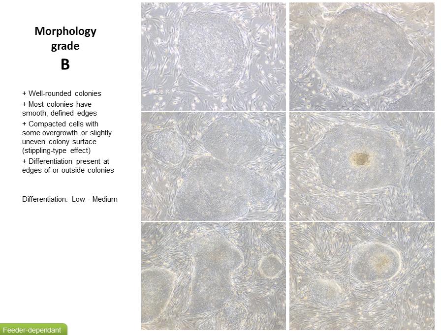
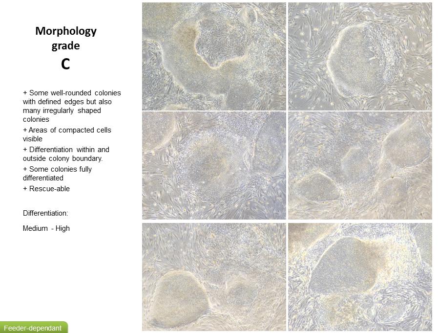
8 Culturing of Human ips cells It is good practice to observe ipsc lines daily under phase contrast microscope (4x, 10x, 40x magnification) for ipsc- like morphology, the presence of differentiated cells and confluence (see appendix 1 for a grading system) Cells are fed by removing 95% of the medium from the wells using an aspirator pipette. Aseptically add 2 ml of fresh medium per 1 well of a 6 well plate by gently adding to the side of the well. Incubate cells at 37 C, 5% CO 2. Medium should be changed daily except for on the day of passaging. Passaging Human ips cells Feeder Dependant ipsc should be observed on Day 4-7 after previous passage; refer to grading system to assess morphology and confluency (see appendix 1 for Feeder Dependant grading system). Passaging should be performed as below on Day 5-7 after previous passage when colonies are compacted and have well defined edges (grade A- B). Cells may also require passaging if levels of differentiation start to exceed that of ipsc or colonies start to look overgrown or unhealthy (grade C- D). Aspirate the spent medium from the cells and wash with 2 ml of DPBS per well. Aspirate DPBS and add 1 ml each of both collagenase and dispase per well. Rock the dish to cover the surface of the cells and set a timer for 45 minutes. Incubate in a tissue culture incubator set at 37 o C, 5% CO 2 for 45 minutes. If after 45 minutes no colonies appear to be lifting, incubate for a further minutes. Do not exceed 60 minutes. Differentiated cells should remain attached. Collect suspension of floating colonies and gently dispense into a falcon tube, taking care not to break up the colonies. Leave the colonies to sediment out of suspension and form a pellet. Do not centrifuge. Aspirate the supernatant from the pellet and re- suspend in 5-10 ml of medium (depending on pellet size). Leave the colonies to sediment out of suspension again and repeat the previous step. Remove MEF medium from feeder plate(s), wash with 2 ml DPBS per well. Add 1.5 ml of warm Feeder Dependant medium (containing FGF) per well. Aspirate as much spent medium as possible from the falcon tube so only the cell pellet is left. Re- suspend cell pellet in 1 ml of fresh Feeder Dependant medium (containing FGF). Using a 1 ml pipette, collect and dispense the cell suspension up to 3 times to gently break up the colonies to medium/small fragments. Be careful not to over pipette, or pipette too harshly as this will result in single cells. Add the appropriate volume of Feeder Dependant medium (containing FGF) to the cell suspension (between 3 10 ml depending on original pellet size). 8
9 Make sure the suspension is fairly homogenous (by gentle agitation); assess by eye the gradient of the fragment sizes. Take 1 ml of cell suspension from around the middle of the tube (to collect only medium sized fragments). Dispense drop- wise into each well, distributing evenly across the well. Assess confluence of fragments (by phase contrast microscopy). Add more suspension if required (remember to agitate suspension again before re- plating). Feeder Dependant cultures can sustain a maximum split ratio of approximately 1:3. Agitate plate gently (do not swirl) within a tissue culture incubator set at 37 o C and 5% CO 2, to spread the fragments equally around the well, leave to settle overnight before moving. Cryopreservation of Human ips cells Colonies should be large enough to freeze 5-7 days after passaging when cells are approximately 70-80% confluent. A confluent well of a 6 well plate will have enough cells to generate 1 frozen vial. Cells are usually frozen when their morphology lies between grade A- B (refer to the Feeder Dependant grading system to assess morphology in appendix 1). Aspirate the spent medium from the cells and wash with 2 ml of DPBS per well. Aspirate DPBS and add 1 ml of both collagenase and dispase per well. Rock the dish to cover the surface of the cells and set a timer for 45 minutes. Incubate in a tissue culture incubator set at 37 o C, 5% CO 2 for 45 minutes. Prepare appropriate volume of freeze medium to freeze 1 ml cell suspension per vial. Prepare a cell freezing container (if required make sure to use appropriate volume of replenished Iso- propanol) and store at 4 o C until use. If after 45 minutes no colonies appear to be lifting, incubate for a further minutes. Do not exceed 60 minutes. Differentiated cells should remain attached. Collect suspension of floating colonies and gently dispense into a falcon tube, taking care not to break up the colonies. Leave the colonies to sediment out of suspension and form a pellet. Do not centrifuge. Aspirate the supernatant from the pellet and re- suspend in 5-10 ml of medium (depending on pellet size). Leave the colonies to sediment out of suspension again and repeat the previous step. Leave the colonies to sediment out of suspension once more. Aspirate as much medium as possible so only the cell pellet is left. Re- suspend in appropriate volume of freezing medium, giving a total volume of 1 ml per cryovial. Do not over pipette the cells as this will result in single cells rather than cell clusters. Dispense 1 ml of cell colony suspension into each cryovial and seal tightly. Immediately place the cryovials into a pre- chilled cell freezing container (4 C) then immediately transfer the container to a - 80 C freezer. Allow the cells to remain at - 80 C overnight (16-36 hours). 9
10 Once frozen transfer the cells, on dry ice, to an Ultra- low temperature storage vessel (LN2 or C freezer). Transfer to Feeder Free Prior to attempting to transfer a Feeder Dependant culture to Feeder Free ensure the quality of the line is of grade A- B (refer to the Feeder Dependant grading system to assess morphology in appendix 1) and that confluence is around 70%. 2-3 days post passaging the Feeder Dependant culture replace the Feeder Dependant medium with E8 and continue to media change using E8 until the culture is ready to passage. If you have not had a chance to change medium prior to passage, but feel your culture is of good enough quality to transfer, you can still attempt transfer but maybe seed with a Feeder Dependant, E8 medium 50:50 mix. On day of passage aspirate the spent medium from ½ a 6 well plate and wash with 2 ml of DPBS per well. Aspirate DPBS and add 1 ml each of both collagenase and dispase per well. Rock the dish to cover the surface of the cells and set a timer for 45 minutes. Incubate in a tissue culture incubator set at 37 o C, 5% CO 2 for 45 minutes. If after 45 minutes no colonies appear to be lifting, incubate for a further minutes. Do not exceed 60 minutes. Differentiated cells should remain attached. Collect suspension of floating colonies using E8 medium and gently dispense into a falcon tube, taking care not to break up the colonies. Leave the colonies to sediment out of suspension and form a pellet. Do not centrifuge. Aspirate the supernatant from the pellet and re- suspend in 5-10 ml of medium (depending on pellet size). Leave the colonies to sediment out of suspension again and repeat the previous step. Aspirate Vitronectin solution from pre- prepared coated ½ 6 well plate. Add 1 ml of E8 medium to each well. At this point you may also want to prepare a MEF feeder plate and create a Feeder Dependant maintenance plate in case the transfer does not work. Aspirate as much spent medium as possible from the falcon tube so only the cell pellet is left. Re- suspend cell pellet in 1 ml of fresh E8 medium. Using a 1 ml pipette, collect and dispense the cell suspension up to 3 times to gently break up the colonies to medium/small fragments. Be careful not to over pipette, or pipette too harshly as this will result in single cells. Add the appropriate volume of E8 medium to the cell suspension (between 3 6 ml depending on original pellet size). 10
11 Make sure the suspension is fairly homogenous (by gentle agitation); assess by eye the gradient of the fragment sizes. Take 1 ml of cell suspension from around the middle of the tube (to collect only medium sized fragments). Dispense drop- wise into each well, distributing evenly across the well. Assess confluence of fragments (by phase contrast microscopy), seed the vitronectin plate at a higher density to a normal passage. Add more suspension if required (remember to agitate suspension again before re- plating). Agitate plate gently within a tissue culture incubator set at 37 o C and 5% CO 2 to ensure even distribution of cells across the well. Transferring ipscs into a new culture system such as transferring to feeder free is a stress to the cells and high levels of spontaneous differentiation can occur. It can take up to 4 passages before the cells adapt, it is advised to not try anything other than basic passaging until this point. If it becomes apparent that the transfer has not been successful the easiest solution is to try again from the feeder dependant culture. Troubleshooting Problem Low viability after thaw. Little to no colonies visible within 2 weeks after recovery Low viability after passage Cells do not attach properly Non- typical morphology High levels of cell death Cells do not proliferate Non- uniform distribution of colonies within plate Possible Solutions/Precautions Ensure you screen round the edges of the well, cells have a tendency to congregate against the sides. Feeder dependants cultures can take up to 2 weeks to recover, you could potentially leave them for longer than this if nothing is identified on the dish but this is not ideal. To ensure quality of future vials: Ensure that cryovials are thawed quickly and that medium is added to the cells very slowly (drop- wise while gently swirling the tube). Ensure that cells were banked at log phase of growth. Try thawing cells into a smaller tissue culture vessel. Ensure 10 µm ROCK inhibitor is added at thaw and 24 hours post thaw top up with 1 ml of medium plus 10 µm ROCK inhibitor, but after this point cells should be media changed daily and without ROCK inhibitor. Try and avoid dissociating thawed colonies to single cells when plating. Ensure the confluence and health of the MEFs is adequate. If necessary top up the MEFs, this can occur even with ipscs on the dish but take care to not over seed the MEFs. Ensure cells are in log phase of growth at passaging. Avoid leaving collagenase and dispase on for too long (don t exceed 1 hour), as cells will dissociate to single cells which won t survive the passage. Ensure the confluence and health of the MEFs is adequate. Make sure that MEFs are evenly distributed across the dish 11
12 Areas with too high density of ips cells In addition some areas may have fewer colonies at sufficient confluence. Ensure that the cell aggregates are evenly distributed by gently rocking the plate back and forth and side to side. Not in a circular motion. Take care when placing plate into the incubator and leave undisturbed for 24h. Ensure incubators are level and free from vibrations. Colonies are not coming off the plate post 45 minute incubation in collagenase and dispase Centre of colonies become over grown Darker discolouration in the centre Centres potentially detach, leaving donut colonies This can lead to differentiation in the centre of the colonies Spontaneous differentiation Colonies do not have defined edges Cells within the colonies are less compact Cells appear flattened and bigger Morphology as depicted in C and D of the grading system (appendix 1) Do not allow colonies to become too large and dense. Do not let cells become more than 80 % confluent. Extend incubation of collagenase and dispase from 45 minutes to 1 hour (do not exceed 1 hour). After a 1 hour incubation take what you can, but remember to scale back the surface area you seed over in accordance with what you obtained from the original dish. Ensure the collagenase and dispase is between room temperature and 37 o C before use. Ensure the collagenase and dispase has not become inactive over time due to thermal cycling. Passage when the individual colonies are of a reasonable size and clearly compacted, don t wait for the whole surface to be of a reasonable confluence. In a bid to avoid in the first instance: Ensure the plates used have healthy MEFs. Avoid leaving plates outside the incubator for more than 15 minutes. Ensure media is within date and has not been left in temperatures of room temperature or higher for extended periods (1-2 hours). ipscs are extremely sensitive and any changes in reagent type or user- to- use technique variability can cause an increase in spontaneous differentiation. Fragment size at seeding can have an impact, too small or single cells, can spontaneous differentiate, too large and differentiation can form in the centre of the colony. Don t allow individual colonies to over grow or become too compact in the centre. To tackle differentiation once it sets in: Standard passaging is often the safest first option for 3-4 passages, especially if you are transitioning culture systems, as the cells need time to adapt. Passaging early i.e. before the dish has reach it usual confluence, as long as the ipsc colonies are compacted and of a reasonable size. 12

13 Be more gentle with the dish post collagenase and dispase incubation in a bid to just take healthy colonies and leave the single cells APPENDIX 1 FEEDER DEPENDANT GRADING SYSTEM 13
14 14

15 Many thanks to Lucy Weston- Stiff for producing this document. 15