MEDICAL LASER SYSTEMS
|
|
|
- Pierce Arnold
- 5 years ago
- Views:
Transcription

1 MEDICAL LASER SYSTEMS
 is an international developer, manufacturer and supplier of medical laser systems. MELSYTECH Ltd.")


2 MELSYTECH Ltd. MELSYTECH Ltd. (Nizhny Novgorod Region, Russia) is an international developer, manufacturer and supplier of medical laser systems. MELSYTECH Ltd. offers all well-known laser types - Nd:Yag, diode, KTP, Erbium, Holmium, Thulium, Q-switched and picosecond lasers - for such applications as cosmetology, dermatology, gynecology, phlebology, ENT, aesthetic medicine and more. The company is the first and only supplier of medical laser systems, whose whole product range is based on diode pumping technology. The enterprise has in-house production of laser and optical components, modules, and accessories that provide for full cycle manufacturing of laser systems. MELSYTECH Ltd. today: A leading company on the Russian and CIS market for innovations and production research in laser technologies for medical application. A large network of distribution in Russia and partnerships overseas. A holder of patents for unique proprietary technologies. A team of qualified specialists, including medical advisers, and engineers doctorate and highest academic degree. Clinical trials of technologies and new equipment in close cooperation with leading dermatologists and plastic surgeons. Quality, reliability, efficiency of laser systems. Full production cycle - design and development, manufacturing of components, assembly year of establishment of the company as a medical laser manufacturer 70 over 70 laser devices in sales per year 9 nine different laser wavelengths 1 number one in medical laser sales in Russia 2


3 MEDICAL LASER SYSTEMS LASER TECHNOLOGY On a daily basis doctors all over the world undertake thousands of laser treatments to efficiently deal with medical, cosmetology, and aesthetic challenges. Since 1960, when the laser phenomenon was discovered, the technology has constantly advanced to transform it into routine and safe application. The efficiency and high technology nature of lasers drive their presence in our life on a bigger and bigger scale. In particular, aesthetic medicine, dermatology and cosmetology have become those areas which cannot be imagined without laser technologies anymore. A laser with certain parameters is capable to incise and prosect tissues, as well as remove and coagulate malformations, and treat developments and anomalies of intradermal vessel. At the same time application of laser provides for relative ease of use, low traumas and high selectivity of effect. Laser emission features a number of specific characteristics that define how and at what depth biological tissue is affected. Depending on a wavelength the laser targets various substances within human skin, that can either reflect or absorb laser emission, such as water, hemoglobin, melanin, oxyhemoglobin. The latter ones, according to a known theory of selective photothermolysis, are chromophores, that is components that selectively absorb laser energy. For instance, since green KTP laser (532nm) penetrates through upper skin layers and is well absorbed by hemoglobin, it is widely used for vascular lesion removal. As a result, the affected vessel undergoes intense emission of heat, the blood clots, and the internal vascular wall deteriorates. The vicious vessel then heals with connective tissue, and the skin color returns back to natural. You can check how well each chromophore can absorb laser emission depending on a wavelength on a graph below. ABSORPTION DEPTH Diode 808nm red Pacinian corpuscles Capillaries Nd:YAG 1064nm Er:YAG infra 2940nm red Epidermis Nerves Dermis Sudoriparous gland Subcutaneous tissue Arteries Er :YA G :YA G Ho MELANIN 103 Hair Sebaceous gland ND 105 Hair follicle ABSORPTION COEFFICIENT (1/cm) КТР 532nm green Di KT P od e :YA G ABSORPTION AND TISSUE PENETRATION DEPTH HEMOGLOBIN WATER 101 PROTEIN 10-1 SCATTER Wave length (nm) 3


4 MAGIC MAX A three-wave medical laser for gynecology and aesthetic medicine. 532 nm 808 nm 1064 nm Nd: YAG+KTP Diode Nd: YAG The MAGIC MAX provides for application of various modes within a single procedure by switching between the wavelengths in seconds, which makes it universal for a wide range of medical applications. The diode-pumping technology eliminates any flash lamps in the laser systems, and therefore is not subject to cumbersome and expensive aftersales service. Application: OTORHINOLARYNGOLOGY removal of polyps, coagulation of Kiesselbach s area vessels, OSAHS (obstructive sleep apnea hypopnea syndrome), rhonchopathy, tonsillectomy, adenoidectomy, nasal septum correction. Laser surgery in ENT secures best attenuated treatments of mucous coat of pharyngeal and nasal cavities, and thus saves extremely valuable functions of epithelial and adenoid tissues. REMOVAL OF VASCULAR LESIONS (including child malformations) at various depths (telangiectasia, Rosacea, hemangiomas, wine stains). The mechanism of action is based on selective absorption of laser energy by oxy- and ferrihemoglobin, as well as by protein structures responsible for blood clotting process. Through the absorption of laser energy the protein undergoes direct coagulation while the vessels intima undergoes indirect thermal destruction, so that blood clotting is initiated. GYNECOLOGY removal of polyps, endometriosis treatment, vaginal rejuvenation, stress urinary incontinence treatment, laser coagulation of cervical erosion. Besides reactive collagen reduction in the tissue, reconstruction and neocollagenesis processes are initiated. The heated tissue grows in collagen, the fibers become shorter and thicker, which makes the affected tissue contract and strain. PHLEBOLOGY endovenous laser ablation, reticular varicosity. The destroying effect on the vessels endothelium comes through a thermal impact by laser emission. A laser carrying fiber gets introduced into a sore part of the vein through a centesis in the skin. The laser energy heats up the blood inside the vessel and makes it bubble-boil. These are the steam bubbles that bring the heat effect to the wall of the vein which directly damages the endothelium and coagulates the protein in it. ONYCHOMYCOSIS TREATMENT (nail fungus). The laser passes the nail plate, penetrates into the nail bed and thus heats up the fungus matter. The fungus clumps undergo high temperature impact and their growth stops. LASER LIPOLYSIS The treatment is ideal for effective reduction of pockets of fat such as love handles, upper back, double chins, and saddlebags. The laser probe breaks fat cells with thermal energy and provides for better separation of the fat from the neighboring tissue. 4


5 One for all. Three different wavelengths in one optical output. Wave length, nm Operation mode Free running Q-switched Free running Q-switched Pulse energy, mj 3,000 1,500 9,000 1,500 Max pulse length 10ms - 2s 30ns 10ms - 2s 30ns Energy density, J/cm Max pulse repetition rate, Hz Max average output, W Pulse power, W 33,000 33, ,000 Cooling system Air-liquid, integrated Spot size, mm mm Scanner Scanning area Ø30mm Through our proprietary developments and latest research about laser effect on biological tissue at Melsytech we generate the laser beam so that to keep energy distribution uniform all through the profile and avoid hot spots or fall-offs in the working spot. FLAT-TOP BEAM PROFILE No hot spots in the range No peripheral fall off Diode-pumping is a state-of-the-art technology in medical application. The design of devices offers a huge potential, as both software and hardware can be easily upgraded. You may rest assured that Melsytech laser systems will stay in high demand and up-to-date equipment for years. Not only will this secure a fast payback, but it will be bringing you profits long after. 5
6 6 MEDICAL LASER SYSTEMS

7 MAGIC FR The Magic FR is a long-pulsed Nd:YAG laser system. The 1064nm wavelength, high laser emission energy and extended spot size of the device provide for treatment of malformations deep in the skin beyond reach by any other wavelength. This is the best tool to remove vascular lesions of medium size and bigger. The laser system comes in a standard Magic FR version and an advanced Magic FR Full version with higher energy settings. Application: REMOVAL OF VASCULAR LESIONS over body and face; Nonablative skin rejuvenation; ACNE TREATMENT Acne inflammations treatment by Nd:YAG laser is pathogenetic due to direct sterilization of inflammations. REMOVAL OF PIGMENTED LESIONS. Magic FR Wave length, nm 1064 Operation mode Free running Max pulse energy 50J Max energy density 6,300J/cm 2 Pulse length ms Max pulse repetition rate, Hz 100 Cooling system Air-liquid, integrated Spot size, mm 1-6mm, or 3-15mm Magic FR Full Wave length, nm 1064 Operation mode Free running Max pulse energy 70J Max energy density 8,900J/cm 2 Pulse length ms Max pulse repetition rate, Hz 100 Cooling system Air-liquid, integrated Spot size, mm 1-6mm, or 3-15mm 7


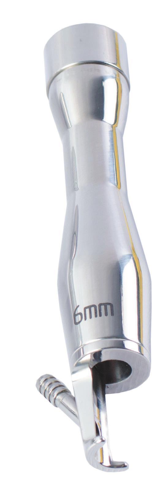
8 Magic max handpieces and adaptors Scanner 532, 808, 1064 nm Enhances performance and ease of application. User can adjust the shape, size and density of scanning. Maximum scanning area makes up 30mm in diameter. 1mm 2mm 4mm Adjustable scanning pitch Adjustable shape and size of scanning area Zoom handpiece mm. Shot-by-shot treatment, convenient switching of spot size without changing handpieces; effective for treatment of vascular lesions, wine stains. Gynecological handpiece This tip locks up onto a standard scanner and is designed for gynecology treatments such as vaginal rejuvenation, laser coagulation of cervical erosion, polypectomy, stress and postpartum urinary incontinence treatment. Lipolysis adapter Laser lipolysis for subsequent liposuction ENT handpiece The handpiece is designed for out-patient treatment of various medical conditions of oral and nasal cavities. Phlebological fiber The fiber tip is designed for EVLA procedures - treatment of varicose veins and vein ablation, the procedure being an alternative to conventional surgery. Fiber core diameter 400 or 600micron. Single spot handpieces 1.2, 2, and 6mm Shot-by-shot treatment of spider veins, wine stains etc. With integrated air skin cooling. The handpieces can be used with any of the wave lengths. 8


9 BEFORE and AFTER Hemangioma Immediately after In 4 weeks Facial vascular lesions Immediately after Wine stain Immediately after Leg vascular lesions Immediately after 9
 is a life-time method for biological tissues")
 and being an")

.")


10 OCT is a unique device designed for noninvasive biological tissue diagnosis Sudoriparous gland Hair and sebaceous gland Optical coherence tomography (OCT) is a life-time method for biological tissues structure imaging. The method is referred to as optical biopsy owing to high image resolution (20-25 micron) and being an alternative to traditional excisional biopsy when the latter is not possible or undesirable in cases when no cell identification is required. It is best used for investing tissues study. The method is registered to be used in Russia (For life-time examination of skin morphology in health and disease by the optical coherence tomography method: Novel medical technology, 2008). Visualization occurs as a result of registration of probing emission (low intensity light, near infrared band, up to 1.5mW) radiating back from tissue elements, that come to feature different refractive index and re-radiation characteristics. OCT image is a 2D and 3D image of optical irregularities of the tissue at 1.5mm depth, in pseudocolor brown palette. It is aligned with the cross microscopic section. Principal OCT applications: ophthalmology, gynecology, surgery, carcinology, dentistry, angiology, urology, dermatology, and cosmetology. 10

, right - thick skin (planta).")


11 Dermatology application: The method allows to acquire data related to morphological condition of healthy and pathologically modified skin in real time. OCT allows for visual differentiation of the epidermis and upper part of the dermis. The method is a tool to evaluate the condition of the dermo-epidermal junction area, dermis vessels, sebaceous and sudoriparous glands, hair follicle, all the ungual elements. OCT image of healthy human skin: Left - thin skin (forearm), right - thick skin (planta). 1 - superficial part of the horny layer with poorly cohesive scales, 2 - middle and lower part of the horny layer with adherent scales, 3 - superpapillary region of cell layers of the epidermis, 4 - interpenetrating area of epidermal excrescences and dermal papillae, 5 - upper part of the reticular layer of the dermis. The OCT visualizes major pathologic processes in dermis, and can be used for life-time diagnostic of dermatoses, inlcuding basal cell carcinoma, melanoma, differential diagnostic of melanomas and nevil, onychopathy. Spongiosis in eczema - vesicles Healthy skin OCT provides for multifocal and manifold examination which allows to monitor abnormal changes during cosmetic procedures and treatments to determine effect and find early signs of side effects (for instance, preclinical development of atrophia cutis during corticosteroid therapy). OCT images are interpreted via the following concepts: structural properties, layering, height, homogeneity, signal intensity within a layer, contrast of layers and areas with different signal intensity, boundary characteristics of layers and areas. ADVANTAGES OF APPLICATION: - noninvasive - imaging on-the-fly - multifocal and manifold examination, over-time examination - high spatial resolution and high image contrast - portable size, user friendly control, flexible probe, to be compatible with endoscopic gear. Technical specification Central wave length A-Scan Line Rate Scanning depth Longitudinal resolution Lateral resolution Sensitivity Optical power at object 1300nm 92 khz 1,5 mm 20 micron 25μm 90 db 0.75mW, complies with ANSI standard 11

12 CONTACT ООО MeLSyTech address: , Nizhny Novgorod Region, Dzerzhinsk, 1, Igumnovskoe Shosse, Russia tel , fax web melsytech.com