Cross-Linker Modulation to Maintain Phenotype of RGD-Alginate-Embedded Mesenchymal Stem Cells
|
|
|
- Percival Small
- 5 years ago
- Views:
Transcription
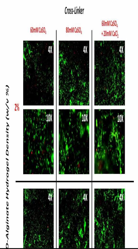
1 Cross-Linker Modulation to Maintain Phenotype of RGD-Alginate-Embedded Mesenchymal Stem Cells Ashley B. Allen, Hazel Y. Stevens, Robert E. Guldberg. Georgia Institute of Technology, Atlanta, GA, USA. Disclosures: A.B. Allen: None. H.Y. Stevens: None. R.E. Guldberg: None. Introduction: Alginate hydrogels have been used in a wide range of tissue engineering applications to deliver protein and biologics due to favorable properties such as biocompatibility and ease of chemical modification. Functionalizing the polysaccharide backbone, through covalent addition of the adhesive peptide RGD, has been shown to enhance the attachment, spreading, and proliferation of surface-seeded cells. However, despite desirable cell-matrix interactions in 2D, mesenchymal stem cells (MSCs) mostly exhibit a rounded morphology when embedded within a 3D RGD-alginate hydrogel environment and it is known that the morphology of the stem cells can impact both cell viability and differentiation [1-2]. The type and concentration of cross-linker are known modulators of alginate gelling kinetics and consequent mechanical and biochemical properties of the hydrogel. The extent of cross-linker solubility in aqueous medium (CaCl2 > CaSO4 > CaCO3) has been shown to correlate positively with gelling speed and negatively to resulting hydrogel stiffness [3]. The concentration of calcium ion, or cross-linker, and density of the hydrogel also influence these parameters. The objective of this preliminary study was to build on a previously established RGD-alginate carrier system for large bone defect repair [4] to better maintain embedded stem cell phenotype and viability. To this end, hydrogel matrix properties were manipulated through modulation of cross-linker molecule and concentration as well as hydrogel density. The resultant effects on stem cell morphology, phenotype, and viability were evaluated in vitro. Methods: Rat Mesenchymal Stem Cell (rmsc) Isolation: Bone marrow was harvested from Lewis Rat femurs and cultured until passage 2. Construct Preparation and Culture: Constructs containing 0.25x10⁶ rmscs in 1% or 2% RGD-alginate were crosslinked using one of four methods - (1) 60 mm CaSO4, (2) 60 mm CaCl2, (3) 80 mm CaSO4, or (4) 60 mm CaSO4 with concomitant 20 mm CaCl2. Hydrogels were mixed using a dual-syringe method, injected into perforated PCL nanofiber mesh tubes (150 µl gel/tube), and incubated in PBS for 20 minutes to remove excess soluble calcium. Constructs were cultured vertically in custom made holders on a rocker plate with media changes twice weekly. Live/Dead Stain: Embedded rmsc morphology was evaluated at 48 hours using a live (calcein) / dead (ethidium homodimer) stain (Invitrogen) and fluorescent microscopy (n=2). Flow Cytometry: Constructs prepared using 60 mm CaSO4 +/- 20 mm CaCl2 were digested using an EDTA solution on day 7 (n=1). Recovered cells were labeled with fluorescently-tagged antibodies for CD49 (FL2) and CD90 (FL1) and analyzed using flow cytometry. DNA Quantification: Constructs prepared using 60 mm CaSO4 +/- 20 mm CaCl2 were digested on day 14 and DNA content was measured using a PicoGreen assay (n=4). Statistics: DNA content values were analyzed using a t-test. Error bars represent standard error of the mean. Results: RGD-alginate cross-linked using 60 mm CaSO4, 80 mm CaSO4, or 60 mm CaSO4 and 20 mm CaCl2 (groups #1, 3, and 4) resulted in an injectable rmsc-seeded hydrogel, suitable to culturing in vitro. However, substitution of 60 mm CaSO4 with 60 mm CaCl2 (group #2) resulted in a non-injectable hydrogel and no morphological data could be collected from this group. Live/dead staining revealed that the addition of 20 mm CaCl2 to the previously established carrier system gave the best improvement in embedded rmsc spreading (Figure 1). Use of a reduced hydrogel density (1%) showed no qualitative impact on cell morphology and resulted in a gel of insufficient mechanical stability. Thus, 2% hydrogels from groups #1 and 4 were selected for analysis of rmsc phenotype and DNA content at later time-points. A higher proportion of rmscs recovered from CaCl2-supplemented constructs (group #4) expressed MSC cell surface markers following 7 days of culture, as quantified using mean fluorescence intensity (Figure 2). DNA content measured at 2 weeks indicated that both alginate gels supported rmsc viability to a similar extent (Figure 3). Discussion: CaCl2 supplementation during the incorporation of rmscs into 2% RGD-alginate hydrogels in this preliminary study resulted in enhanced cell spreading and improved retention of stem cell surface markers. These results indicate that CaCl2- supplemented alginate gels better maintain embedded stem cell phenotype, and thus may provide a more favorable delivery approach for cell-based regenerative therapy techniques. However, the improved rmsc properties observed within hydrogels cross-linked using CaCl2 did not translate to higher cell content after 2 weeks in culture. Further investigation into the relationship between phenotypic and characteristics of rmscs within this system is warranted. Future work will assess these delivery systems in vivo for their ability to support embedded cell viability and to foster construct mineralization. Significance: Cell-based strategies for volumetric tissue regeneration have promise, but are challenged by poor tissue integration and early death of delivered cells upon implantation. Manipulation of a hydrogel vehicle to better preserve the
2 phenotype and proliferative capacity of embedded stem cells in vitro may translate to improved cell viability and therapeutic efficacy upon delivery in vivo. Acknowledgments: This work was supported by the United States Department of Defense through the Center for Advanced Bioengineering for Soldier Survivability (CABSS) grants W81XWH and W81XWH References: 1.Pek, Y.S. et al Biomaterials, Engler, A.J. et al., J Musculoskelet Neuronal Interact, Kuo, C.K. et al Biomaterials, Boerckel, J.D. et al., Biomaterials,
3
4
5 ORS 2014 Annual Meeting Poster No: 1170
6