AIP1 functions as an endogenous inhibitor of VEGFR2-mediated signaling and inflammatory angiogenesis
|
|
|
- Sharon Burns
- 5 years ago
- Views:
Transcription
1 SUPPLEMENTAL MATERIALS functions as an endogenous inhibitor of VEGFR2-mediated signaling and inflammatory angiogenesis Haifeng Zhang 1*, Yun He 1*, Shengchuan Dai 1*, Zhe Xu 2*, Yan Luo 2, Ting Wan 2, Dianhong Luo 1, Dennis Jones 1, Shibo Tang 2, Hong Chen 1, William C. Sessa 1, and Wang Min 1,3 1 Interdepartmental Program in Vascular Biology and Therapeutics, Department of Pathology, Yale University School of Medicine, 10 Amistad St., New Haven, CT Zhongshan Ophthalmic Center, Sun Yat-Sen University, Guangzhou, P.R. China. 3 Corresponding author: Dr. Wang Min, Interdepartmental Program in Vascular Biology and Transplantation and Department of Pathology, Yale University School of Medicine, 10 Amistad St., New Haven, CT Tel: ; Fax: ; wang.min@yale.edu * These authors contributed equally to this work. Supplemental Zhang, H. et al., inhibits VEGFR2 signaling 1
2 Fig.S1. deletion in tissues. a. expression in paraffin section of lung (a) and brain (b) was determined by immunohistochemistry with anti-. Arrow indicates vascular endothelium and arrowhead for lung bronchial epithelium. b: Arrow indicates axon and arrowhead for cell body of a purkinjie cell. Supplemental Zhang, H. et al., inhibits VEGFR2 signaling 2
3 Supplemental Zhang, H. et al., inhibits VEGFR2 signaling 3
4 Fig.S2. expression in vasculature during embryogenesis and effects of deletion on vascular development. a-b. Effects of deletion on vascular development. -WT and -KO embryos were obtained by mating +/- mice and embryos were harvested at indicated times (E9.5-E16.5). For E embryos, embryo vasculature was visualized by whole mount-staining with anti-cd31 antibody. Shown images in a are E10.5 embryos. For E embryos, freshly dissected embryos without staining were photographed. Shown images in b are E13.5 embryo, york sac and placenta. c. expression in vasculature during development. E13.5 embryos were co-stained by immunostaining with anti- (rabbit) and anti-cd31 (an EC marker, goat) antibodies followed FITC-conjugated secondary antibody against rabbit IgG and phycoerythrin (PE)-conjugated secondary antibody against goat IgG. Images were taken under fluorescence microscope. Co-localization of with CD31 is detected in -WT but not -KO mice (indicated by arrows). Fig.S3 α-sma-positive cells/mm2 b. * 400 Non-ischemic Ischemic WT KO Fig.S3. -KO mice showed enhanced vessel maturation. Pericyte/smooth muscle cells were immunostained with smooth muscle α-actin (SMA, a smooth muscle/pericyte marker). Representative sections from non-ischemic and ischemic groups of -WT and -KO mice on day 28 post-ischemia are shown in a. Quantification of SMA-positive 2 capillaries (number/mm muscle area) are shown. Data are mean ± SEM from 10 fields per section (3 sections/mouse and n=4 for each strain). *, p<0.05. Supplemental Zhang, H. et al., inhibits VEGFR2 signaling 4


5 Supplemental Zhang, H. et al., inhibits VEGFR2 signaling 5
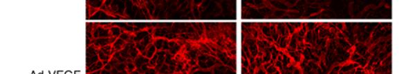
 (Ad-VEGF) or β-galactosidase (Ad-LacZ) was intradermally injected into the mice right an")
6 Fig.S4. VEGF-induced ear neovascularization was greatly augmented in -KO mice. VEGF-induced ear angiogenesis. Adenovirus encoding VEGF 164 (1x10 9 pfu) (Ad-VEGF) or β-galactosidase (Ad-LacZ) was intradermally injected into the mice right and left ear skin, respectively. a. VEGF-induced angiogenesis in -WT and -KO mice was accessed by a direct microscopy. b. Ear permeability was measured by Evan s blue dye (EBD) assay. Ear skin containing the extravasated protein-bound dye was excised and the dye was extracted from the tissue. Dye concentrations were measured at 630 nm using a spectrophotometer. The values obtained are expressed as total nanograms of EBD extracted, and are a measure of the total amount of protein-bound dye that extravasated in response to adenoviral-expressed VEGF or LacZ. Data are expressed as mean ± SEM from n=3 for each strain. *, p<0.05. c. Ear vasculature was visualized by a whole-mount staining with PE-conjugated anti-cd31. d. Quantification of vessel density from c. Data are mean ± SEM from 10 fields per ear (n=5 for each group). *, p<0.05. b. Ad- LacZ Ad- β-tubulin d. 100 * Retina vascular area (%) Ad-LacZ Ad- Ad-LacZ Ad-VEGF Fig.S5. Overexpression of inhibits VEGF-induced in vivo angiogenesis. a-b. Transgene expression of LacZ reporter and. Ad-LacZ or Ad- (2x10 8 pfu) was injected intravetrously into -KO mice. LacZ expression in retina was visualized by β-galactosidase staining (a) and expression was detected by Western blot with anti- (b). c-d. expression inhibits VEGF-induced retina angiogenesis. Ad- or Ad-LacZ was co-ministrated intravetrously into -KO mice. Retina vasculature was visualized by isolectin staining (c) with quantification of vessel density in d. Data are mean ± SEM from 10 fields per retina (n=4 for each group). *, p<0.05. Supplemental Zhang, H. et al., inhibits VEGFR2 signaling 6

7 Fig.S6 b. a. Ctrl sirna sirna VEGF p-vegfr2 VEGFR2 p-plc-γ PLC-γ p-akt Akt Fig.S6. VEGF-induced EC migration and signaling are augmented by knockdown. HUVEC were transfected with control sirna or sirna oligonucleotides. 36 h post-transfection, cells were starved for 12 h. a. knockdown increases VEGF-induced EC migration. Cells were subjected to a monolayer wound injury assay as described in Fig.5 in the presence of VEGF (10 ng/ml) for indicated times. Data presented are means (±SEM) of the triplicates from three independent experiments. b. knockdown increases VEGF-induced VEGFR2 activation. Cells were treated with VEGF (10 ng/ml) for 5 min. Phosphorylation of VEGFR2, total VEGFR2, and β-tubulin were determined by Western blot with respective antibodies. Fig.S7 MLEC Fig.S8 Muscle WT KO WT KO bfgf Ischemia p-plc-γ p-ask1 ASK1 PLC-γ (p-ask1/ask1) p-akt β-tubulin Akt Fig.S7. deletion has no effect on bfgf signaling in cultured EC. -WT and -KO MLEC were cultured overnight in 0.5% FBS followed by bfgf treatment (10 ng/ml) for indicated times (0-60 min). Phosphorylation of PLC-γ and Akt were determined by Western blot with phospho-specific antibodies. Total PLC-γ and Akt as well as were determined by Western blot with respective antibodies. Fig.S8. deletion has no effect on ischemia-induced ASK1-JNK signaling in tissue. -WT and -KO were subjected to ischemic ligation, and tissues were harvested on day 7. Activation of ASK1 was determined by Western blot with a phospho-specific antibody (pthr845). Relative ratios of p-ask1/ask1 are shown, with untreated WT as 1.0. As controls, and β-tubulin proteins were also determined. Supplemental Zhang, H. et al., inhibits VEGFR2 signaling 7