Amino Acids and Proteins
|
|
|
- Doris Chapman
- 5 years ago
- Views:
Transcription
1 Various Functions of Proteins SB203 Amino Acids and Proteins Jirundon Yuvaniyama, Ph.D. Department of Biochemistry Faculty of Science Mahidol University Enzymes Transport proteins utrient and storage proteins ontractile or motile proteins Structural proteins Defense proteins Regulatory proteins Etc... Figure taken from Fibrous and Globular Proteins Interior of protein is generally hydrophobic ydrophilic ollagen Parvalbumin ydrophobic


2 Protein: Polymer of Amino Acids Val Ala Amino Acids Leu Gln Glu R Asp Asn Arg -terminus -terminus Gly Phe Ile + 23 α Pro ys Tyr Trp Met is Thr Ser Lys Amino Acids Amino Acids Figure taken from Figure taken from


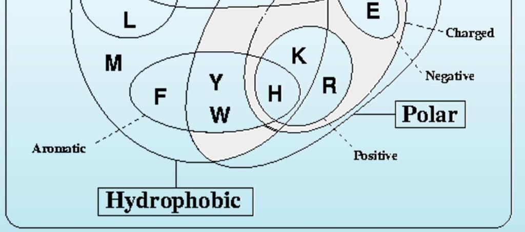
3 Amino Acids Amino Acids A B D E F G I Ala ys 8.3 Asp Glu Phe Gly is Ile J K L M P Q R Lys Leu Met Asn Pro Gln Arg S T U V W X Y Ser Thr Val Trp Tyr 10.1 Z Figure taken from Various Functions of Proteins Figure taken from Figure taken from


 R i Psi")


4 Peptide Bond Formation 2 -terminus R R 2 R 1 2 R 3 R 4 R 2 Primary Structure R 5 -terminus R 6 Peptide unit is planar and rigid Phi (φ) R i Psi (ψ) + R j Along the polypeptide backbone, the peptide units are planar and rigid, leaving only two rotatable bonds (phi and psi) per amino-acid unit to define folding of the protein chain. 180 Psi (ψ) Ramachandran Plot β Examples of 3D Structures All α All β 0 α Secondary Structure Tertiary Structure α/β α+β Phi (φ)
 Enzymes with onprotein omponents ofactors May")


5 Assembly of Sub-units Quarternary Structure R Structure ierarchy R 2 R 3 R 4 R 5 R 6 GroEL (sp60) Enzymes with onprotein omponents ofactors May dissociate from enzyme May be further classified as: Metal ions, e.g. a 2+, Mg 2+ oenzymes: small organic compounds some vitamins Prosthesticgroup Tightly bound to enzyme, e.g. heme in hemoglobin Apoenzyme vs oloenzyme Apoenzyme: enzyme lacking cofactor oloenzyme: complete enzyme Protein Modifications Disulfide bond formation Acetylation of -terminus increase protein stability ydroxylation at Pro collagen fibers Glycosylation at Asn(-linked) or Ser/Thr(-linked) Phosphorylation at Ser/Thr or Tyr Sorting and Maturation: Preproenzyme Myristoylationat -terminal Gly membrane anchoring Etc...
6 Disulfide Linkage Protein Ligand Interaction ovalent bond between 2 nearby ys aused by oxidation of thiol groups S Provide additional stabilization of 3D structure Thermophilic proteins Pepsin in stomach Recognition of a ligand by a receptor is provided by combination of several types of interactions at specific geometry. The term lock and key emphasizes complementarity in shapes due to VDW interactions. Protein Ligand Interaction Structural Flexibility In fact, combination of complementarities in shape (VDW) and chemical interactions, e.g. vs. + charges in ionic interactions and -bond donor and acceptor pairs in -bond formation all contribute to recognition. As 3D structures of proteins are stabilized by weak interactions, they are flexible and dynamically changing conformation. Some parts of the structure may be more flexible than others and may provide functions.


7 Structural Flexibility DF TF ADP eme in emoglobin ADP 2 transfer 2 Flexibility of DFR structure allows for binding of DF and ADP as well as its catalysis. emoglobin structure interacts with heme as a prosthetic group which can bind 2. Sensing of 2 Binding to b Allosteric hange upon 2 Binding


8 Sickle-ell Anemia aused by Single Mutation on β-globin Sickle-ell Anemia aused by Single Mutation on β-globin Mutations affect protein structures, and thus their properties. Summary Proteins are flexible polymer of amino acids Various properties of amino acid side chains lead to variety of structures 3D structure of protein is formed in hierarchy and stabilized mainly by weak-force interactions 3D structure of protein determines its function through Shape of molecule and active site hemical interactions with substrate, cofactor, coenzyme, or prosthetic group The End