Tissue Culture Sterilization and Contamination
|
|
|
- Anastasia Pierce
- 5 years ago
- Views:
Transcription
1 Tissue Culture lab #4 Tissue Culture Sterilization and Contamination Nowf Aldouweghri
2 Introduction Successful cell culture depends heavily on keeping the cells free from contamination by microorganisms. Non-sterile supplies, media, and reagents, airborne particles laden with microorganisms, unclean incubators, and dirty work surfaces are all sources of biological contamination. Aseptic technique, designed to provide a barrier between the microrganisms in the environment and the sterile cell culture, depends upon a set of procedures to reduce the probability of contamination from these sources.
3 Introduction The elements of aseptic technique are: A sterile work area Good personal hygiene Sterile reagents and media, and sterile handling.
4 Sterile Work Area The simplest and most economical way to reduce contamination from airborne particles and aerosols (e.g., dust, spores, shed skin, sneezing) is to use a cell culture hood. The cell culture hood should be properly set up and be located in an area that is restricted to cell culture that is free from drafts from doors, windows, and other equipment, and with no through traffic.
5 Sterile Work Area The work surface should be organized and contain only items required for a particular procedure; it should not be used as a storage area.
6 Sterile Work Area Before and after use, the work surface should be disinfected thoroughly, and the surrounding areas and equipment should be cleaned routinely. For routine cleaning, wipe the work surface with 70% ethanol before and during work, especially after any spillage. You may use ultraviolet light to sterilize the air and exposed work surfaces in the cell culture hood between uses. Leave the cell culture hood running at all times, turning them off only when they will not be used for extended periods of time.

7 Why is 70% ethanol used for wiping microbiological working areas? 70% percent of alcohol is ideal to a stronger solution. Pure alcohol coagulates protein in contact. Suppose the pure alcohol is poured over a single celled organism. The alcohol will go through the cell wall of the organism in all direction, coagulating the protein just inside the cell wall. The ring of the coagulated protein would then stop the alcohol from penetrating farther from the cell, and no more coagulation would take place. At this time the cell would become inactive but not dead. Under the favorable conditions the cell would then begin to function.
8 Why is 70% ethanol used for wiping microbiological working areas? If 70 percent of alcohol is poured to a single celled organism, the diluted alcohol also coagulates the protein, but at a slower rate, so that it penetrates all the way through the cell before coagulation can block it. Then the entire cell is coagulated and the organism dies.

9 Good Personal Hygiene Wash your hands before and after working with cell cultures.

10 Good Personal Hygiene In addition to protecting you from hazardous materials, wearing personal protective equipment also reduces the probability of contamination from shed skin as well as dirt and dust from your clothes.


11 Sterile Reagents and Media Commercial reagents and media undergo strict quality control to ensure their sterility, but they can become contaminated while handling. Follow the guidelines below for sterile handling to avoid contaminating them. Always sterilize any reagents, media, or solutions prepared in the laboratory using the appropriate sterilization procedure (e.g., autoclave, sterile filter).
12 Sterile Handling Always wipe your hands and your work area with 70% ethanol. Wipe the outside of the containers, flasks, plates, and dishes with 70% ethanol before placing them in the cell culture hood. Avoid pouring media and reagents directly from bottles or flasks. Use sterile glass or disposable plastic pipettes and a pipettor to work with liquids, and use each pipette only once to avoid cross contamination.
13 Sterile Handling Always cap the bottles and flasks after use and seal multi-well plates with tape to prevent microorganisms and airborne contaminants from gaining entry. Never uncover a sterile flask, bottle, petri dish, etc. until the instant you are ready to use it and never leave it open to the environment. Return the cover as soon as you are finished.
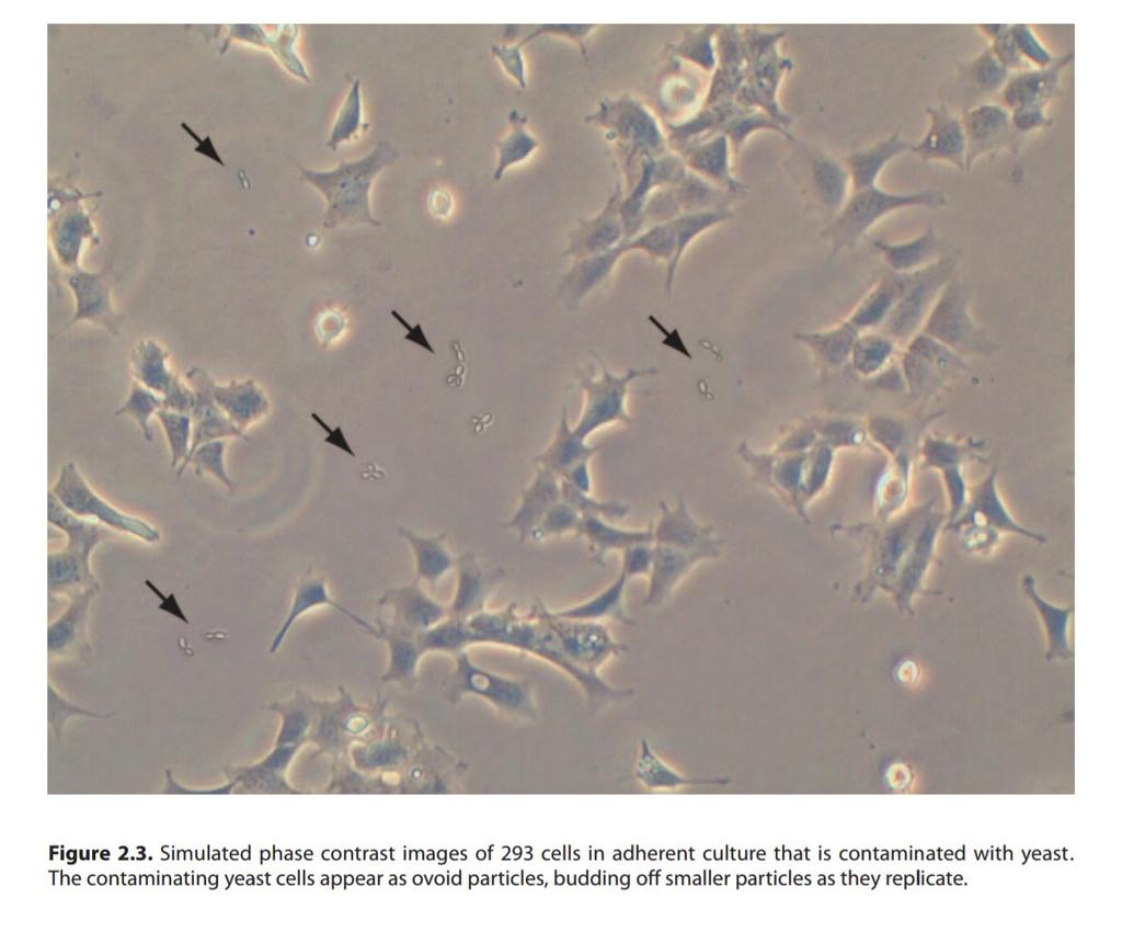
14 Sterile Handling If you remove a cap or cover, and have to put it down on the work surface, place the cap with opening facing down. Use only sterile glassware and other equipment. Be careful not to talk, sing, or whistle when you are performing sterile procedures. Perform your experiments as rapidly as possible to minimize contamination.
15 Part 2 Tissue Culture Contamination
16 Introduction A cell culture contaminant can be defined as some element in the culture system that is undesirable because of its possible adverse effects on either the system or its use. These elements can be divided into two main categories: 1. Chemical contaminants: the presence of nonliving substances that affect the culture. Usually chemical contaminants come from reagents or the water used to make the media. Such as impurities in media, sera, and water, endotoxins, plasticizers, and detergents. N. A
17 Introduction 2. Biological contaminants: by viable organisms or viruses Some easy to detect such as bacteria, molds, yeasts. Some are more difficult to detect such as viruses, mycoplasma, protozoa, as well as cross contamination by other cell lines.

18 Bacteria Bacteria are a large unicellular microorganisms. They are the most commonly encountered biological contaminants in cell culture. The media becomes yellow, because the bacteria deplete the O2 from the culture and produce CO2 and acid, the acidification of the medium, turning it yellow. Media may have distinct odor.
19 Bacteria Bacterial contamination is easily detected by visual inspection of the culture within a few days of it becoming infected. Infected cultures usually appear cloudy (turbid), sometimes with a thin film on the surface. Sudden drops in the ph of the culture medium frequently occur. Under a low- power microscope, the bacteria appear as tiny, moving granules between the cells, and observation under a high-power microscope can resolve the shapes of individual bacteria.

20 Bacteria The simulated images below show an adherent 293 cell culture contaminated with E. coli.
21 Yeast Yeasts are unicellular eukaryotic microorganisms in the kingdom of Fungi. Like bacterial contamination, cultures contaminated with yeasts become turbid, especially if the contamination is in an advanced stage. There is very little change in the ph of the culture contaminated by yeasts until the contamination becomes heavy, at which stage the ph usually increases.
22 Yeast Under microscopy, yeast appear as individual ovoid or spherical particles, that may bud off smaller particles. The simulated image below shows adherent 293 cell culture 24 hours after plating that is infected with yeast.
23 Yeast
24 Molds Molds are eukaryotic microorganisms in the kingdom of Fungi that grow as multicellular filaments called hyphae. A connected network of these multicellular filaments contain genetically identical nuclei, and are referred to as a colony or mycelium. Similar to yeast contamination, the ph of the culture remains stable in the initial stages of contamination, then rapidly increases as the culture become more heavily infected and becomes turbid.

25 Molds Under microscopy, the mycelia usually appear as thin, thread-like filaments.
26 Viruses Viruses are microscopic infectious agents that take over the host cells machinery to reproduce. Their extremely small size makes them very difficult to detect in culture, and to remove them from reagents used in cell culture laboratories.
27 Viruses Using virally infected cell cultures can present a serious health hazard to the laboratory personnel, especially if human or primate cells are cultured in the laboratory. Viral infection of cell cultures can be detected by electron microscopy, immunostaining with a panel of antibodies, PCR with appropriate viral primers.
28 Mycoplasma Mycoplasma are simple bacteria that lack a cell wall, and they are considered the smallest self-replicating organism. Because of their extremely small size (typically less than one micrometer).
29 Mycoplasma Mycoplasma are very difficult to detect until and grow slowly Signs you may have a mycoplasma contamination: o Maximum confluent cell density is reduced o Morphological deterioration of the cells o Cell doubling time is increased o Some slow growing mycoplasma may persists in culture without causing cell death, but they can alter the behavior and metabolism of the host cells in the culture.
30 Mycoplasma Chronic mycoplasma infections might manifest themselves with decreased rate of cell proliferation, reduced saturation density, and agglutination in suspension cultures; however, the only assured way of detecting mycoplasma contamination is by testing the cultures periodically using fluorescent staining, PCR, immunostaining.

31 Mycoplasma Uninfected infected infected Animal cell stained for mycoplasma using MycoFluor TM reagent

32 Precaution the waste products produced by the mammalian cells themselves will slowly decrease the ph, gradually turning the solution orange and then yellow. This color change is an indication that even in the absence of contamination, the medium needs to be replaced (generally, this should be done before the medium has turned completely orange).
33 Cross contamination While not as common as microbial contamination, extensive cross-contamination of many cell lines with contaminated and overgrown (fast growing cell lines) by other, more aggressive cells is a clearlyestablished problem with serious consequences. Obtaining cell lines from trustworthy cell banks, periodically checking the characteristics of the cell lines, and practicing good aseptic technique are practices that will help you avoid crosscontamination.
34 Cross- contamination Contaminated cell lines should never be used for research demanding the specific type of cell line they are assumed to be, and most of them should ideally be discarded or at least not used in research at all, except when the contaminant cells have acquired novel characteristics. Many techniques are used to detect cross contamination of cells such as Giemsa stain karyotyping under a light microscope.
35 Using antibiotics Antibiotics should not be used routinely in cell culture, because their continuous use encourages the development of antibiotic resistant strains and allows low-level contamination to persist, which can develop into full-scale contamination once the antibiotic is removed from media, and may hide mycoplasma infections and other cryptic contaminants.
36 Using antibiotics Antibiotics should only be used as a last option and only for short term applications, and they should be removed from the culture as soon as possible. If they are used in the long term, antibioticfree cultures should be maintained in parallel as a control for cryptic infections.

37
38 Reference EU.pdf N. A