Custom 3D Scaffolds for Regenerative Medicine Applications
|
|
|
- Shawn Parsons
- 5 years ago
- Views:
Transcription
1 Custom 3D Scaffolds for Regenerative Medicine Applications Eric M. Brey, Ph.D. South Texas Veterans Health Care System University of Texas at San Antonio

2 Craniofacial Defects Craniomaxillofacial reconstruction often requires complex, multistage surgical procedures. There is a critical need for improved methods for reconstruction of complex skeletal defects. Sutradahar et al., PNAS, 2010 Craniomaxillofacial injury is the primary unfitting condition in many soldiers evacuated from current conflicts.

3 Tissue Engineering Tissue engineering is the application of the principles and methods of engineering and the life sciences toward the fundamental understanding of structureformation relationships in normal and pathological mammalian tissues and the development of biological substrates to restore, maintain or improve functions. Sutradahar et al., PNAS, 2010 Skalak and Fox (eds.), Tissue Engineering, Alan Liss 1, 1, 1995

4 Approach Image the defect Identify the structure required Generate PMMA chambers Load tissue engineering strategy Implant against the periosteum Harvest Transfer to the defect Akar et al., Tissue Engineering Part B, 2018
 in an uncompromised location Brey et al.")
5 in vivo bioreactor growth of vascularized bone within the body by implantation of a molded chamber against the periosteum (vasculogenic, osteogenic) in an uncompromised location Brey et al., Plast Rec Surg, 2007; Cheng et al, Tissue Eng, 2005, Plast Rec Surg, 2006, 2009.


6 Periosteum guided prefabrication using morcellilzed bone graft can lead to the growth of vascularized bone of clinical size and volume Brey et al., Plast Rec Surg, 2007; Cheng et al, Tissue Eng, 2005, Plast Rec Surg, 2006, 2009.
7 Design a tissue engineering scaffold that stimulates directed vascularization into the material while maintaining tissue volume. z


 units generate copolymers that")
8 Poly(ethylene glycol) hydrogels Biocompatible, resistant to protein and cell adhesion Incorporation of biofunctionality into hydrogels by immobilization of bioactive derivatives of photopolymerizable monomers Introduction of poly(l-lactic acid) units generate copolymers that are degradable by hydrolysis


9 Pore Size Porosity allowed invasion in the absence of degradation. Vessel invasion varies with pore size in vitro and in vivo Chiu et al., Tissue Eng Part C Methods Very little vessel invasion observed in pore size < 50 µm Vessel invasion depth Chiu et al., Biomaterials. 2011


10 Fibrin loaded pores Fibrin loaded in the pores throughout the scaffold volume Fibrin stimulated vascular ingrowth in a dose dependent manner Jiang et al., Tissue Eng Part A Vessel density


11 Degradation Introduction of poly(l-lactic acid) units into PEG hydrogels to generate copolymers that are degradable by hydrolysis By varying the ratio of PEG-DA to PEG-PLLA-DA we can generate porous hydrogels with varying degradation times Degradation time The hydrogels maintain their pore size and structure during degradation Pore size Audie L. Murphy Memorial Chiu et VA al. Hospital PLoS One, 2013; Chiu et al., J Fluoresc 2012
 Fibrinogen within")


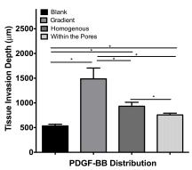
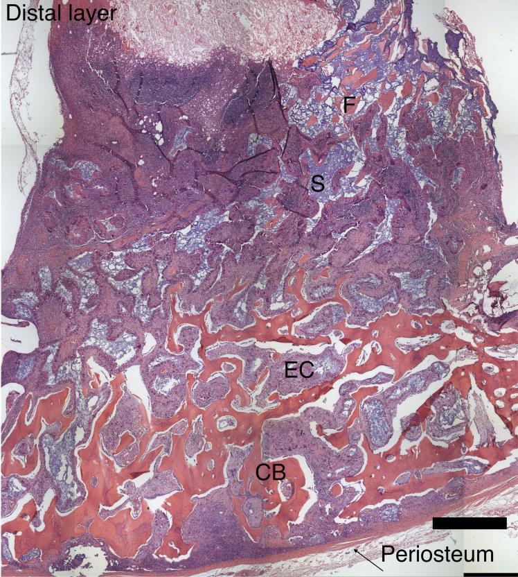
12 Gradient Scaffold Preparation Distal layer A B C Porous Hydrogel 4 mm 10 mm 25% PEG-DA (Mw 8000) Fibrinogen within the pores µm pores PLGA microspheres in 10 % PEG-PLLA-DA PEG-DA: Polyethylene glycol diacrylate PLGA : Poly(lactic-co-glycolic acid) PDGF-BB: Platelet-derived growth factor PEG-PLLA-DA: Poly (ethylene glycol)-co-(l-lactic acid) diacrylate


13 NIR Imaging Akar et al., Biomaterials 2015

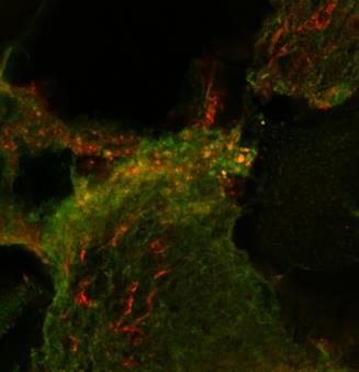
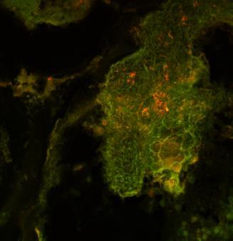
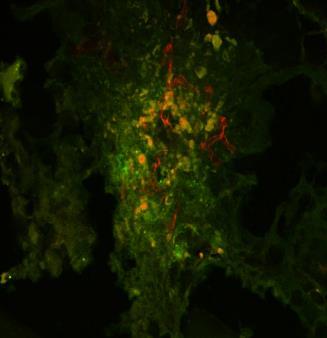
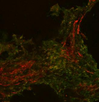
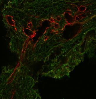
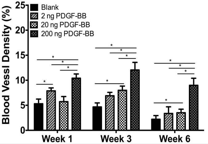
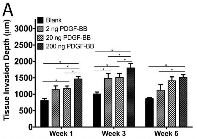
14 Invasion and Vascularization Week 1 Week 3 Week 6 Blank 100 µm 100 µm 100 µm Invasion Depth 200 ng PDGF- BB 100 µm 100 µm 100 µm Green: Tissue Red: Blood vessels Vessel density Akar et al., Biomaterials 2015
 ceramic particles were incorporated into")
.")

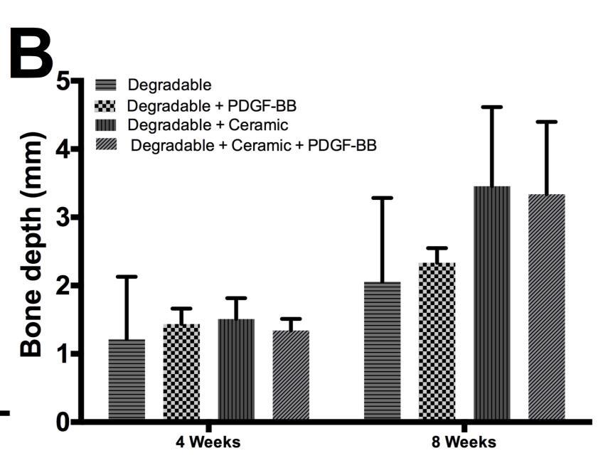
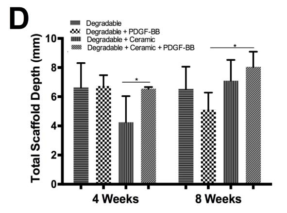
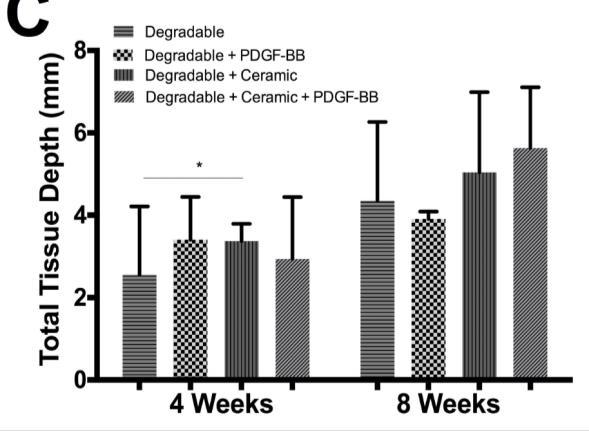
15 Ceramic Composites Hydroxyapatite (HA) and β-tricalcium phosphate (β-tcp) ceramic particles were incorporated into the composite scaffolds Varying weight ratios (70:30, 50:50, 30:70% HA: β-tcp). The presence of ceramic particles within scaffolds significantly extended the degradation time. HA and TCP ceramics to this scaffold system enhanced bone formation.
 17.5:7.")
 70:30%: HA:TCP 200")
16 10 mm Porcine Model PMMA chamber Gradient scaffold Cuff (adhesion) 17.5:7.5 % PEG-PLLA-DA:PEG-DA/Fibrin Polymer: Ceramic = 2:1 (w/w) 70:30%: HA:TCP 200 ng PDGF-BB
17 Evaluation

18 Modeling Agent based model of angiogenesis in porous scaffolds Results predicted experimental results for the influence of pore architecture Invasion depends Pore size Porosity Interconnectivity Size distribution Mehdizadeh et al., Biomaterials 2013


19 Precise control of internal and external architecture 3D Printing Wang et al., Advanced Materials. 2015

20 Large Animal Defect Model Porcine mandibular defect model Evaluating surgical transfer of functional engineered bone formed in the in vivo bioreactor Imaging courtesy of John Decker
21 Conclusions The in vivo bioreactor can result in vascularized bone of clinical volume and customized shape. A cell free approach requires optimization of structure, degradation kinetics and spatio-temporal release of growth factors. We are currently applying our approaches for engineering vascularized bone in a large animal, clinically-translatable model of an in vivo bioreactor.

;")


22 Acknowledgements - Funding Veterans Administration (5 I01 BX ); NIH (5R01EB , 1R01AR ); AHA (Innovators Research Grant); NSF (IIS , CBET , EEC , DSES )
23 Acknowledgements - Collaborators Army Institute of Surgical Research Washington University in St. Louis John Decker, D.M.D., M.S. Mark Anastasio, Ph.D. Erik Weitzel, M.D. University of Maryland College Park Chang Gung Memorial Hospital John Fisher, Ph.D. Ming-Huei Cheng, M.D. Hui-Yi Hsiao, Ph.D. University of Chicago Ronald Cohen, M.D. Illinois Institute of Technology Ali Cinar, Ph.D. Elisabeth Hildt, Ph.D. Georgia Papavasiliou, Ph.D. Rice University Tony Mikos, Ph.D. Wake Forest Institute for Regenerative Medicine Emanuel C. Opara, Ph.D. University of Belgrade Lada Zivcovic, Ph.D. University of Los Andes Juan Carlos Briceno, Ph.D. University of Texas at San Antonio Gabriela Romero, Ph.D. Amina Qutub, Ph.D. Chris Rathbone, Ph.D.


24 Acknowledgements - Lab Jacob Brown Brenda Carrillo Madeleine Farrer Maria Gonzalez Porras, Ph.D. Christina Jones Kelly Langert, Ph.D. Paola Lerma Samantha Mann Meritxell Martinez Favour Obuseh Binita Shrestha, Ph.D. Katerina Stojkova Feipeng Yang