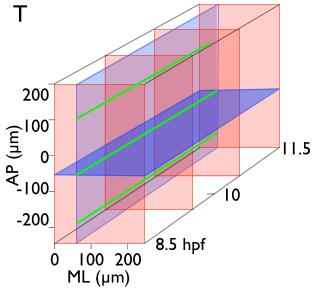
Keller et al., 2008, Science. Guy Blanchard. Movie. Movie. Tomer et al., 2012 Nat. Methods. Stephen Young. Movie. Movie.
|
|
|
- Katherine Nelson
- 5 years ago
- Views:
Transcription


1 Big data solutions to dynamic imaging Keller et al., 2008, Science Guy Blanchard Stephen Young Tomer et al., 2012 Nat. Methods Zallen lab

2 Mechanics Morphogenetic genotype Mechanics Morphogenetic phenotype Gene expression Protein composition of cell Cell organisation & connectivity? Elastic & plastic changes in cell shape, arrangement & cell number From in situ/live protein imaging From live imaging of tissues & strain rate analyses Normal development Cancer! Biomaterials etc.

3 Actomyosin cortex Apical Junctional Basal Medial
4 Dynamic imaging analysis pipeline Imaging Image analysis Tagged cells Tracked cells
 Anterior Cell-cell interface properties & dynamics (length, orientation, vertex angles, neighbour connectivity.")
5 Automated cell shape tracking Raw movies (ventral view of Drosophila germ-band, with Lucy Butler & Benedicte Sanson) Cadherin-GFP Cell properties & dynamics (location, movement, area/volume, best-fit ellipsoid, elongation ratio...) Anterior Cell-cell interface properties & dynamics (length, orientation, vertex angles, neighbour connectivity...) Cell speed (µm / min) Apical cell area Interface length...to calculating small multi-cellular domain deformation rates neighbour gain neighbour loss
6 Dynamic imaging analysis pipeline Imaging Image analysis Tagged cells Tracked cells Strain rates

")
7 Tissue tectonics Centroid trajectories Remove mean domain translation Relative cell movements Orientation and strength of greatest deformation Best-fit cell shape ellipses Domain average Differentiate w.r.t. time Individual cell shape strain rates Short Medium Long Size change expansion contraction 4% / min 2% 4% / min 2% - = 4% / min 2% Tissue (total) strain rate Cell shape strain rate - = Blanchard, Kabla, Mahadevan & Adams (2009) Nat. Methods Intercalation strain rate
 Ventral view Cell shape change Cell intercalation Blanchard, Kabla, Mahadevan & Adams, 2009,")
8 Small domain strain rates ant cf lat med post Extension rate Convergence rate lat 0.04 (pp/min) (pp/min) Ventral view Cell shape change Cell intercalation Blanchard, Kabla, Mahadevan & Adams, 2009, Nat. Methods


9 3D morphogenetic maps (GAMMs) Blanchard*, Schultz*, Kabla & Adams
10 Dynamic imaging analysis pipeline Imaging Image analysis Tagged cells Tracked cells Strain rates Tagged Myosin Quantified cell Myosin
 Myosin")


11 Dual-labelled Drosophila embryos Cell membranes (Gap43-Cherry) Myosin (Sqh-GFP) Ventral(view( A( P( A Cephalic(furrow( Mesoderm(invagina:on( Image ventral embryo view Ventral( ( Use curved plane through Adherens Junctions Tetley*, Blanchard*, Fletcher, Adams & Sanson P Apical cell shape tracking (random cell colours) x6 WT embryos Cell-cell interface myosin fluorescence intensity Fluorescence legend

 60")

 Proportion -0.2 0 0.")
 Project")


12 Myosin polarity patterns across AP axis Bidirectional polarity Unidirectional polarity Time (minutes from start of GBE) Proportion AP coordinate (µm) Proportion Collapse DV 135axis 180 AP coordinate (µm) Project polarity onto AP Single embryos. To find stereotypical behaviour want standardised AP axis coordinate system. Time (minutes from start of GBE)


13 Within-parasegment patterns 6 embryos, 3-4 parasegments each Stereotypical within-parasegmental cable locations Tetley*, Blanchard*, Fletcher, Adams & Sanson
14 Dynamic imaging analysis pipeline Imaging Image analysis Mechanical inference Tagged cells Tracked cells Strain rates Tissue stress & mechanical properties Myosin force Tagged Myosin Quantified cell Myosin
 Stress ( ) 100 120 140 160 180 200 220-20")
 Myosin (MHC-YFP) Machado et al., 2015, BMC Biol.")
15 Myosin motors drive morphogenetic contraction stage 13 Membrane (ECad-GFP) Dorsal view of amnioserosa Cell Area (µm^2) Stress ( ) Time (Minutes from start of DC slow phase) Cell myosin fluorescence (0-255) Myosin (MHC-YFP) Machado et al., 2015, BMC Biol. Strain ( )


16 Emergent physical properties Machado et al., 2015, BMC Biol.

17 Dynamic imaging analysis pipeline Imaging Image analysis Mechanical inference Modelling Tagged cells Tracked cells Strain rates Tissue stress & mechanical properties Model stress & mechanical properties Myosin force Tagged Myosin Quantified cell Myosin Model forces


18 Vertex-based (mechanical?) simulation A D V P Chaste modelling platform Alex Fletcher (University of Sheffield)


19 3D and in toto tracking & modelling Claire Lye Interface surface area (µm^2) Jocelyn Etienne, Grenoble


20 Thanks to Amnioserosa Pedro Machado Alfonso Martinez Arias Nicole Gorfinkiel Julia Duque Jocelyn Etienne Salivary placode Katja Roeper Alex Booth Germ-band extension Bénédicte Sanson Claire Lye Rob Tetley Huw Naylor Zebrafish Richard Adams Nora Schultz Stephen Young Alexandre Kabla Joel Jennings Vertex-based modelling & Chaste Alex Fletcher