FFGWAS. Fast Functional Genome Wide Association AnalysiS of Surface-based Imaging Genetic Data
|
|
|
- Emmeline Newton
- 5 years ago
- Views:
Transcription


1 FFGWAS Fast Functional Genome Wide Association AnalysiS of Surface-based Imaging Genetic Data Chao Huang Department of Biostatistics Biomedical Research Imaging Center The University of North Carolina at Chapel Hill, Chapel Hill, NC 27599, USA Joint work with Hongtu Zhu The TheUNIVERSITY of of NORTH CAROLINA at at CHAPEL HILL
2 Outline Motivation for Imaging Genetics Statistical Methods for Imaging Genetics Fast Functional Genome-wise Association Analysis Conclusion

3 Motivation for Imaging Genetics


4 Imaging Genetics E B E: environmental factors G D Selection G: genetic markers D: disease
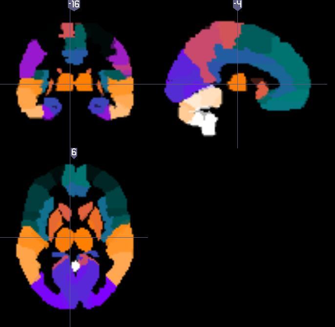
5 Structural MRI Diffusion MRI Imaging Data - Variety of acquisitions - Measurement basics - Limitations & artefacts - Analysis principles - Acquisition tips Functional MRI (task) Functional MRI (resting) PET EEG/MEG CT Calcium

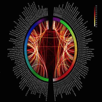

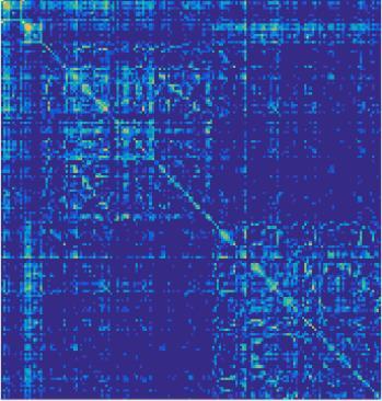
6 Neuroimaging Phenotype! Goal 1: From raw data to conn! Goal 2: From connectomics to Multivariate, smoothed functions, and piecewisely smoothed functions Dimension varies from 100~500,000.
. Nature Review Genetics")
7 Multi-Omic Data Ritchie et al. (2015). Nature Review Genetics
 related to disease risk in response to environmental adversity. (Hariri AR, Holmes A. Genetics of emotional regulation: the role of the serotonin transporter in neural function.")
8 Motivation Imaging genetics allows for the identification of how common/rare genetic polymorphisms influencing molecular processes (e.g., serotonin signaling), bias neural pathways (e.g., amygdala reactivity), mediating individual differences in complex behavioral processes (e.g., trait anxiety) related to disease risk in response to environmental adversity. (Hariri AR, Holmes A. Genetics of emotional regulation: the role of the serotonin transporter in neural function. Trends Cogn Sci. [10: ])
9 Statistical Methods for Imaging Genetics

10 Statistical Methods Hibar, et al. HBM 2012
:g ÎG 0 } Phenotype Genotype Error Y X B E n p y n p x p x p y n")
11 High Dimensional Regression Model Data Y i = {y i (v):v ÎV} {X i (g):g ÎG 0 } Phenotype Genotype Error Y X B E n p y n p x p x p y n p y

12 Challenges n p y p 10 7 x Hibar, et al. HBM 2012
13 Fast Voxel-wise Genome-wise Analysis (I) Spatially Heteroscedastic Linear Model (II) Global Sure Independence Screening Procedure (III) Detection Procedure Huang, et al. Neuroimage 2015 Issues to be addressed: -- Spatially correlated functional data -- Multivariate imaging phenotypes
14 Fast Functional Genome-wise AnalysiS

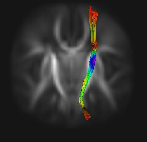
 Fiber bundles Data")


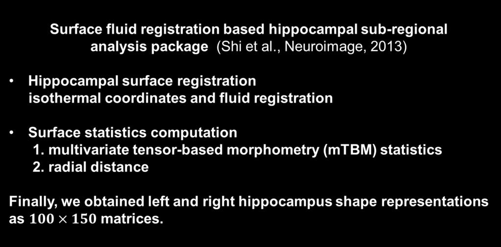
15 A low dimensional representation is necessary for statistical inference (1) Connectivity matrix (2) Fiber bundles Data Structure Sub-Cortical Structure Models Incorporate prior anatomical information via explicit shape models Have 15 different sub-cortical structures (left/right separately) 20 Thalamus Caudate 40 Accumbens 60 Pallidum 80 Brainstem 100 Putamen 120 Amygdala Hippocampus courtesy of P. Aljabar Jin et. al

16 Data Structure Person No.1. Person No.100 Hippocampal Surface. SNP1 SNP2. SNP2000 Person No. 1 Genetic Variation... Person No. 100

17 FFGWAS Multivariate Varying Coefficient Model Global Sure Independence Screening Procedure Significant Voxel-locus Cluster-locus Detection SNP 1 SNP N
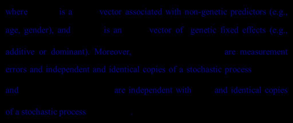
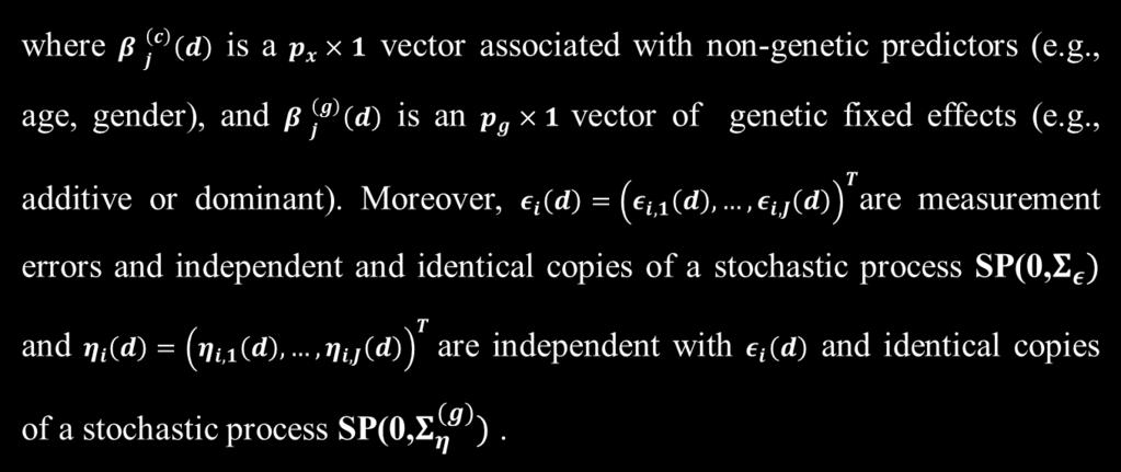
18 Multivariate Varying Coefficient Model
19 Multivariate Varying Coefficient Model We need to test: We first consider a local Wald-type statistic as: where
20 Big-data Challenges Several big-data challenges arise from the calculation of as follows. Calculating across all loci and vertices can be computationally. Bandwidth selection in across all loci can be also computationally. Holding all in the computer hard drive requires substantial computer resources. Speeding up the calculation of.
21 FFGWAS To solve these computational bottlenecks, we propose three solutions as follows. Calculate under the null hypothesis for all loci. Divide all loci into K groups based on their minor allele frequency (MAF), and select a common optimal bandwidth for each group. Develop a GSIS procedure to eliminate many noisy loci based on a global Wald-type statistic.
22 A Global Sure Independence Screening (1) The global Wald-type statistic at locus g is defined as (2) Calculate the p-values of for all loci (3) Sort the -log 10(p)-values of and select the top N0 loci The candidate significant locus set
23 Detection Procedure (1) The first one is to detect significant voxel-locus pairs (2) The second one is to detect significant cluster-locus pairs. Wild Bootstrap method

24 Simulation Studies and Real Data Analysis
25 Simulation Studies: Data Generation Covariate Data (non-genetic data) Generated from either U(0,1) or the Bernoulli distribution with success probability 0.5. Genetic Data Linkage Disequilibrium (LD) blocks ( Haploview & PLINK ) 1. Generate 2,000 blocks; 2. Randomly select 10 SNPs in each block; 3. Chose the first 100 SNPs as the causal SNPs

26 Imaging Data Simulation Studies: Data Generation Step 1: Fitting the model without genetic predictors Estimates of True values Step 2: Specifying effected Regions Of Interest associated with causal SNPs Step 3: Generating imaging data with prespecified parameters and ROIs
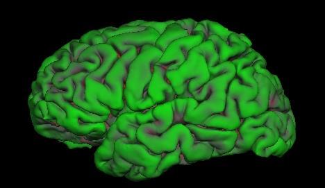
27 Simulation Studies Simulation settings: the green and red regions in the figure, respectively, represent Hippocampal surface, and the effected ROI associated with the causal SNPs among first SNPs. Simulation results for comparisons between FFGWAS and FVGWAS in identifying significant voxel-snp pairs.


28 Imaging Genetics for ADNI PI: Dr. Michael W. Weiner detecting AD at the earliest stage and marking its progress through biomarkers; developing new diagnostic methods for AD intervention, prevention, and treatment. A longitudinal prospective study with 1700 aged between 55 to 90 years Clinical Data including Clinical and Cognitive Assessments Genetic Data including Ilumina SNP genotyping and WGS MRI (fmri, DTI, T1, T2) PET (PIB, Florbetapir PET and FDG-PET) Chemical Biomarker
29 ADNI Data Analysis: Dataset Description 708 MRI scans of AD (186), MCI (388), and healthy controls (224) from ADNI-1. These scans on 462 males and 336 females are performed on a 1.5 T MRI scanners. The typical protocol includes the following parameters: (i) repetition time (TR) = 2400 ms; (ii) inversion time (TI) = 1000 ms; (iii) flip angle = 8 o ; (iv) field of view (FOV) = 24 cm with a 256 x 256 x 170 acquisition matrix in the x, y, and z dimensions, (v) voxel size: 1.25 x 1.26 x 1.2 mm 3. Covariates: gender, age, APOE ε4, and the top 5 PC scores in SNPs

30 Imaging Data Preprocessing
31 ADNI Data Analysis Top 10 SNPs (Left Hippocampus) SNP CHR BP -LOG 10(p) rs e rs e rs e rs e rs e rs e rs e rs e rs e rs e Top 10 SNPs (Right Hippocampus) SNP CHR BP -LOG 10(p) rs e rs e rs e rs e rs e rs e rs e rs e rs e rs e
 (Right")
32 ADNI Data Analysis (Left Hippocampus) (Right Hippocampus)
33 ADNI Data Analysis: Left Hippocampus Significant Loci Zoom (Left Hippocampus) (Right Hippocampus)
34 ADNI Data Analysis: Left Hippocampus (Left Hippocampus) Top 1 SNP: rs Closed Gene: HRH4 HRH4 (Histamine Receptor H4) is a Protein Coding gene. Diseases associated with HRH4: cerebellar degeneration An important paralog of this gene: CHRM4 (Right Hippocampus) Top 1 SNP: rs Closed Gene: C3orf58 Mirshafiey & Naddafi, Am J Alzheimers Dis Other Demen C3orf58 (Chromosome 3 Open Reading Frame 58) is a Protein Coding gene. Diseases associated with C3orf58: hypoxia
35 ADNI Data Analysis Left 12 rs rs Right 0 rs rs log 10 (p) values on Hippocampus (L & R) corresponding to Top 2 SNPs
 values of significant")
36 ADNI Data Analysis Left 1.5 rs rs Right rs rs log 10 (p) values of significant clusters on Hippocampus (L & R) corresponding to Top 2 SNPs 0
37 Conclusion We have developed a FFGWAS pipeline for efficiently carrying out whole-genome analyses of multimodal imaging data. Our FFGWAS consists of a multivariate varying coefficient model, a global sure independence screening (GSIS) procedure, and a detection procedure based on wild bootstrap methods. Two key advantages of using FFGWAS include (i) Much smaller computational complexity; (ii) GSIS for screening many noisy SNPs. We have successfully applied FFGWAS to hippocampal surface data & genetic data of ADNI study.

38 A Software for FFGWAS