Platelet Factor IV- Heparin Antibodies. Presenter: Michael J. Warhol, M.D.
|
|
|
- Scot Boone
- 5 years ago
- Views:
Transcription

1 Platelet Factor IV- Heparin Antibodies Presenter: Michael J. Warhol, M.D.
2 Learning Objectives Describe the mechanism of interaction between Heparin and Platelet Factor 4 Review the chemistry of Heparin Identify the consequences of antibodies to the Heparin Platelet Factor 4 Examine the testing methodology for the anti-platelet Factor 4 Heparin anti-body Enhance the clinical awareness of Platelet Factor IV Antibodies Population at risk Clinical signs Diagnosis and treatment Importance of protocol Medical Consequences of Poor Quality Patient Satisfaction
3

4
5
6
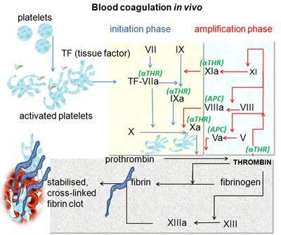
7 Extrinsic system Intrinsic system Surface Contact Tissue Damage XII XIIa Tissue Factor XI XI IX XIa (Xa VIIa VII VII X II V VIIa V a Xa Major Site Major Site Iia (Thrombin) Fibrinogen Fibrin
8
9
10
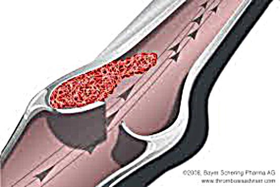
11 CASE STUDY 57 year old female admitted with pneumonia and respiratory failure Admission platelet count was 230,000 Prophylactic heparin administered On the 7 th ICU day, the patient arrested Platelet count 110,000 Result Patient expired Diagnosis-Heparin Induced Thrombocytopenia HIT
12 Heparin Induced Thrombocytopenia Most common adverse event with heparin use is bleeding. Some patients develop a pro-thrombotic state known as heparin induced Thrombocytopenia (HIT) HIT Type I: Mild asymptomatic decrease in platelet count HIT Type II: Severe, potentially devastating thromboembolic complication; life and limb threatening
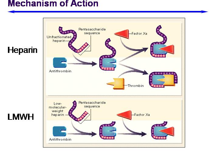
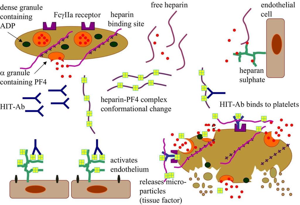
13 Heparin Induced Thrombocytopenia Type II An immune complex can form between heparin and platelet Factor 4(PF4) released by platelets. This complex becomes an antigen and elicits an antibody response. The antibody response destroys the platelets Observed in 2-5% of patients treated with heparin The risk of thrombosis is 33-50%
14
15 Clinical Signs of HIT Deep venous thrombosis (50%) Pulmonary Embolism (25%) Skin lesions at injection site (10-20%) Acute limb ischemia (5-10%) Warfarin associated limb gangrene (5-10%) Acute CVA or myocardial infarction (3-5%)
16 Patient Population Cardiopulmonary Bypass Surgery and Orthopedic Surgery are greatest risks HIT may also occur through: -Heparin flushes or subcutaneous administration -Heparin-coated catheters and prosthesis -Chronic dialysis patients
17 Factors Influencing the Frequency of HIT Type of Heparin and route of administration Bovine UFH>Porcine UFH>LMWH Intravenous>subcutaneous Patient Population Duration of heparin therapy-use beyond day 5 increases the risk of HIT Sex: Female>Male
18 Probability of HIT 50% fall in platelet count Onset between 5 and 10 days after therapy or <1 day if heparin administered within 100 days New thrombosis or thrombotic signs
19 The Diagnosis of HIT-The four Ts 1. Thrombocytopenia 2. Timing of Platelet count 3. Thrombosis 4. Other causes of thrombocytopenia
20 HIT Type II-Clinico-Pathologic Diagnosis >50% platelet fall from Baseline or <100,000/ml. Onset varies-typical 5-10 days after heparin exposure; rapid < 1 day of UFH re-exposure (prior exposure within 100 days); delayed-up to 40 days after UFH exposure New thrombosis, skin necrosis No other causes Antibodies to complexes of HPF4
21 Laboratory Diagnosis of HIT Platelet Count H-PF4 antibody check Platelet Functional Analysis
22 Antigen-Base Tests Standardized Reagents Not dependent on platelet donors Direct testing for Anti-Platelet Factor IV antibody is available as a stat test with results in 10 minutes
23 Treatment of HIT Discontinue heparin Delay Warfarin until platelet count recovers Avoid platelet transfusion Treat with direct thrombin inhibitors, e.g. argantroban(acova), bivalirudin
24 Conclusions HIT is a clinical and laboratory Diagnosis Patients with HIT are at risk for life and limb threatening thrombotic disease In critically ill patients, a negative antigen test paired with the 4T s can exclude the presence of anti-pf4 antibodies
25 Elisa Vs Immuno Precipitation Elisa is a two step method versus a one step immuno precipitation method. Immuno precipitation can be performed in less than one hour.