Ilya Turchin. Institute of Applied Physics of the RAS, Nizhny Novgorod, Russia.
|
|
|
- Asher Gilmore
- 5 years ago
- Views:
Transcription
1 Fluorescence 3D imaging of small animals Ilya Turchin Institute of Applied Physics of the RAS, Nizhny Novgorod, Russia German-Russian Forum Biotechnology Munich 2010
.")
 in experimental animals. Hoffman R.M.")
2 Fluorescence whole-body maging applications: Preclinical study of new photosensitizers for fluorescent diagnostics and photodynamic therapy. Visualization of the drug delivery process using fluorescent labeles (quantum dots or fluorescent proteins). Investigation of tumor growth, metastasis and retardation under therapy using tumor cells transfected with fluorescent proteins. Detection of molecular processes, in particular, fluorescence resonance energy transfer (FRET) in experimental animals. Hoffman R.M. Lab Animal 31, 2002
 illumination) - 2D images Good")
")


3 Whole-body fluorescence imaging techniques Reflectance (epi( epi-illumination) illumination) - 2D images Good transverse resolution for subcutaneous tumors Very fast, no reconstruction Transillumination (or Projection) method Visualisation of deep and subcutaneous tumors Fast, no reconstruction Fluorescent diffuse tomography (FDT) allows for 3D reconstruction of the fluorophore concentration Complicated, need for reconstruction. Inverting matrix is big size, not sparse (unlike in CT), ill-conditioned Diffuse pattern V. Ntziachristos, et al., Nature Biotechnology 23, 2005


4 FDT setup with a single source-detector pair Projection method 2D images, no reconstruction fluorescent proteins, photosensitizers, quantum dots Fluorescent diffuse tomography 3D reconstruction if the fluorophore is well localized fluorescent proteins IV Turchin, AP Savitsky, et al. // JBO 13, (2008)
5 Algorithm of scanning an experimantal animal projection method obtaining a general view of the animal (source and detector are moving synchronously) 3D reconstruction scanning of the selected region with small step sizes and different shifts between source and detector. d x = -5 mm d x =0 d x =5 mm d y =-5 mm d y =0 d y =5 mm Reconstructed distribution of fluorophore concentration z = 0mm z = 1mm z = 2mm z = 3mm z = 4mm
 Accumulation of Photosens (1")
 in mouse cervical carcinoma Biodistribution Elimination Tumor selectivity")
6 Preclinical study of new photosensitizers for fluorescent diagnostics and photodynamic therapy (non-tomographic imaging) Accumulation of Photosens (1 mg/kg, i.v.) in mouse cervical carcinoma Biodistribution Elimination Tumor selectivity Dosage studies M.Shirmanova, IV Turchin, et al. // JBO, accepted for publication in 2010
 and I.V.")
, conjug")
7 Whole-body imaging of delivering fluorescent agents to the tumor (non-tomographic imaging) In collaboration with S.M. Deyev (IBCH RAS) and I.V. Balalaeva (NNSU) Human breast carcinoma SKBR-3 in nude mice Intravenous injection of Qdot 705 ITK (Invitrogen Inc.), conjugated with anti-her2/neu 4D5 scfv-antibody by barnase-barstar protein module Ex 635 nm, Em max 705 nm A.u Tumor nodes before 1.5 hours after injection
8 Investigation of tumor growth, metastasis and retardation under therapy using tumor cells transfected with fluorescent proteins DsRed2 TurboFP635 (scientific name Katushka) nm нм Wavelength, nm Wavelength, nm Larger wavelengths gives higher penetration depths. Penetration depth [mm]
 and I.V.")

9 Intravital monitoring of tumor growth In collaboration with S.M. Deyev (IBCH RAS) and I.V. Balalaeva (NNSU) Nude mice carrying human ovarian carcinoma SKOV-3 expressing Katushka, provided by Scanning area Control before tumor cells injection 700 a.u days 19 days 22 days 200 a.u. 700 a.u a.u
10 Algorithm of scanning an experimantal animal projection method obtaining a general view of the animal (source and detector are moving synchronously) 3D reconstruction scanning of the selected region with small step sizes and different shifts between source and detector. d x = -5 mm d x =0 d x =5 mm d y =-5 mm d y =0 d y =5 mm Reconstructed distribution of fluorophore concentration z = 0mm z = 1mm z = 2mm z = 3mm z = 4mm

11 3D reconstruction of the fluorophore distribution (at depth Z, object width is 1.2 cm)


12 CCD-based FDT experimental setup Fluorescent diffuse tomography 3D reconstruction if the fluorophore is well localized fluorescent proteins Backreflection for subcutaneous tumors 2D images, no reconstruction fluorescent proteins, photosensitizers, quantum dots


13 3D reconstruction of the DsRed2-expressing expressing tumor in-vivo In collaboration with A.P. Savitsky (INBI RAS) Back Reflection
14 Model experiment: reconstruction accuracy Source Detectors X Z Y Reconstruction accuracy - position of the object Δx, Δy = 1 mm Δz =1.5 mm - object shape ΔR=0.5 mm - separate reconstruction of 2 nearest objects Δx, Δy = 3 mm
15 Is it possible to have better resolution in 3D imaging? In vivo -Photoacoustic tomography Ex vivo - Transparent tissue - Ultramicroscopy


16 Ultramicroscopy In collaboration with K.V. Anokhin (INP RAMS) Coherent Sapphire 500 HP 488 nm SM Fiber 3dB Fiber optic beam splitter CCD Camera Imaging optics Spectral filter Aperture slit Cylinder lens Objective Cylinder lens Aperture slit Optical clearing Collimator Collimator Translation stage

17 Ultramicroscopy Confocal microscopy Excitation light z, mm Detector sensitivity area Point spread function х, mm


18 Experimental setup for ultramicroscopy In collaboration with K.V. Anokhin (INP RAMS) 5 А Б В б 1 а г д в


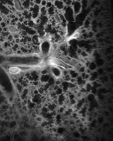
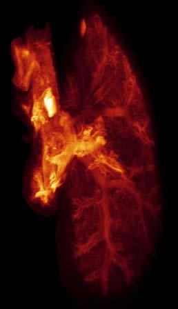
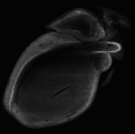
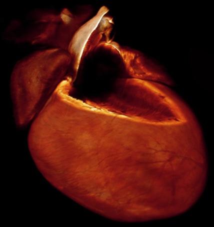
19 Autofluorescence visualization for experimental medicine (3D and virtual section) In collaboration with K.V. Anokhin (INP RAMS) А B А B lung kidney heart
 Mouse embryo,")
20 In collaboration with K.V. Anokhin (INP RAMS) Mouse embryo, autofluorescence
 Mouse hippocampus,")
21 In collaboration with K.V. Anokhin (INP RAMS) Mouse hippocampus, labeled with GFP


.")
22 IAP, RAS I.V. Turchin V.A. Kamensky A.G. Orlova N.M. Shakhova M.Yu. Kirillin M.S. Kleshnin I.I. Fiks V.I. Plehanov V.A. Vorob ev M.B. Prudnikov Acknowledgements Our collaborators A.P. Savitsky, INBI RAS I.G. Meerovich, INBI RAS A.L. Rusanov, INBI RAS K.V. Anokhin, INP RAMS I.V. Balalaeva, NNSU A.A. Brilkina, NNSU E.V. Zagaynova, NNSMA RAMS M.V. Shirmanova, NNSMA RAMS S.M. Deyev, IBCH RAS T.A. Zdobnova, IBCH RAS This work was partly supported by the Russian Foundation for Basic Research (project # ,# ) and the Science and Innovations Federal Russian Agency (project # ).