W E R E A L L A B O U T P E O P L E. $ORNIER
|
|
|
- Benjamin King
- 5 years ago
- Views:
Transcription

1 Modular lithotripter W E R E A L L A B O U T P E O P L E.
2


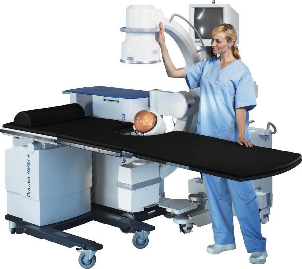
3 The fully motorized, four-axis table is ideal for both Extracorporeal Shock Wave Lithotripsy (ESWL) and endourology. It incorporates an exchangeable radiolucent stretcher that offers a wide range of movement as well as Trendelenburg tilting capability. Extensive urological accessory options are also available. Select one of the advanced scanners from the ultrasound leader in urology, BK Medical. Both the Merlin and ProFocus provide special transducers for shockwave applications, prostate scanning, and abdominal diagnosis. Selected surgical C-arms, from a variety of manufacturers, can be mechanically linked to the Compact Sigma to create a single stable unit. This provides high quality imaging for lithotripsy as well as other applications. 2


4 The Dornier is the result of vast experience and expertise in shock wave technology. Real world therapeutic effectiveness and minimization of side effects were the design objectives. Today hundreds of customers worldwide trust the Dornier technology. With high penetration depth and a wide dynamic range, the is ideal for a spectrum of applications from low-energy analgesic therapy to low-anesthesia stone treatment to high-energy pseudarthrosis application. 3


5 The Dornier Compact Sigma s isocentric design allows both the shock wave and the imaging systems to revolve around a single focal point. In practical terms, this translates into the precise alignment of the targeting system with the shock wave focus. The projection angles of the X-ray localization and the ultrasound windows can be varied over a wide range for optimal imaging, without losing the relationship to the therapy focus. Thanks to this isocentric design, elaborate and costly navigation systems for stone localization are unnecessary with the Compact Sigma. Depending on the indication, shock waves can be introduced from different orientations. This flexibility allows all treatments to be conducted with patients positioned comfortably on their backs, a unique feature of Dornier s Compact line of lithotripters. 4


6 Simply slide the X-ray C-arm under the connecting plate and turn the lever. That s it. The mechanical coupling is fast to set up, easy to operate and extremely stable. A variety of C-arms from well-known manufacturers, are supported. 5


7 QuickLinx, Dornier s optical position control, ensures proper alignment between the C-arm and Compact Sigma. The laser aiming device is mounted to the top of the therapy arm and is equipped with three manually adjustable beams, which can be turned on or off with the touch of a button. 6


8 The use of the isocentric localization arm allows the shock wave and ultrasound to remain independent of each other. This is advantageous in situations where interaction between the shock waves and ultrasound picture can create issues. The results are improvements in the effectiveness of the shock wave as well as the lifetime of the electronic probe. During therapy, the ultrasound transducer is always in direct contact with the skin to provide optimal image quality. The localization arm provides the flexibility needed for excellent image quality and accurate targeting. 7


9 Whether kidney, bladder or prostate ultrasound has the potential to be a major diagnostic aid. BK-Medical has long specialized in urology and understands clinical needs. This is reflected in the choice of urological options: rectal transducers, intraoperative probes, urological software, puncture attachments and more. B-K Medical s advanced models have extended capabilities to include 3D ultrasound, color Doppler and brachytherapy. 8


10 Proximal ureteral stones can be localized by both vertical and oblique X-ray projections providing posterior lateral coupling for treatment. This leaves the X-ray image clear and unobstructed. Avoidance of bone or intestinal gas is simple thanks to the flexible imaging design. Lithotripsy in the medial and distal ureter is typically performed ventrally due to the osseous pelvis. The overtable position of the therapeutic unit provides excellent access while patients remain comfortably on their backs. 9


11 The simultaneous use of X-ray and ultrasound can be used for stones that are not clearly seen with ultrasound alone. X-ray localization allows instant positioning while ultrasound provides real time information about respiration and disintegration. 10


12 UIMS is configured to accept a wide range of imaging devices. For easy set-up, a wide variety of imaging device templates is included in the system. UIMS Dornier s comprehensive Urology Information Management System is optionally available on the Compact Sigma for viewing, processing, and archiving of images and for patient data management. Furthermore, it offers DICOM capabilities and can be connected to a PACS network to maximize patient data collection and workflow. The UIMS Viewing Station can operate as a stand alone viewer, when attached to a hospital network or as a secondary capture system for a variety of imaging devices including C-arms, ultrasound systems and endoscopes. Based upon the viewing station model selected, the secondary capture feature can directly acquire live images and store single images or sequences from a standard line or highline image device. The acquired images can be transmitted to the facility s PACS system using the DICOM interface or viewed on the LCD monitors. 11


13 UIMS features a user-friendly, intuitive graphical interface allowing patient data and treatment information to be entered quickly. Using a simple search function or DICOM Worklist, patient information can be retrieved easily enhancing workflow. Additionally, UIMS is available on Dornier s urology imaging table and most Dornier lithotripters providing inter-operability on Dornier UIMS systems. ESWL treatment information can be documented in the Advanced Patient Data Management System for a complete patient treatment history. Pre-treatment, ESWL data and post-treatment information can be included and select data presented in a graph or histogram. User-defined treatments reports can be created to meet a variety of needs. Images can be stored to an assortment of media storage devices including PACS, DVD, CD, memory stick in a variety of formats. When exporting the images to a local or network printer, the operator can use the Print Preview function to select the desired images. 12


14 Settings are fast and reliable. In a matter of seconds the Compact Sigma can be adjusted for various therapeutic positions. The foot-activated safety brake ensures the Compact Sigma stays parked. Large and stable wheels provide easy transportation. Door thresholds, uneven pavement and slight inclines don t cause transportation challenges for the Compact Sigma. 13


15 The Compact Sigma s cables are easily recoiled and hidden in a specially designed side-compartment. The connecting plate for the mechanical linkage of the C-arm, tucks away neatly. Just fold it out when needed. The Compact Sigma s sleek ergonomic design includes a convenient pull-out storage drawer for a variety of accessories. 0 14
16 Dornier MedTech GmbH Argelsrieder Feld 7 D Wessling Germany Dornier MedTech America, Inc Roberts Boulevard Suite 100 Kennesaw, GA USA Phone: Fax: info@dornier.com Dornier MedTech Asia Pte. Ltd. 2 Corporation Road #05-11/12 Corporation Place Singapore Phone: Fax: infoasia@dornier.com Dornier MedTech Japan Meguro Estate Building Kami-Ohsaki Shinagawa-ku, Tokyo Japan Phone: Fax: info@domedtech.co.jp Dornier MedTech Europe GmbH Argelsrieder Feld 7 D Wessling Germany Phone: Fax: infoeurope@dornier.com Dornier MedTech Italia s.r.l. Via Arrigo Cavaglieri N Rome Italy Phone: Fax: Dornier MedTech Espana Pedro Y Pons 9-11 Edificio Masters E Barcelona Spain Phone: Fax: Dornier MedTech France SARL 156 rue des Castors Epagny France Phone: Fax: Dornier MedTech Europe GmbH Moscow Representative Office Uliza Mytnaya 3 Office Moskau Russia Phone: Fax: info@dornier.com DMTA20-07/08