Human IFN-α (Interferon-Alpha) Pre-Coated ELISA Kit
|
|
|
- Morgan Dorsey
- 5 years ago
- Views:
Transcription
1
2
3 Human IFN-α (Interferon-Alpha) Pre-Coated ELISA Kit Catalog No: well Format (96 tests) Detection Range: pg/ml Sensitivity: <9.4 pg/ml This immunoassay kit allows for the in vitro quantitative determination of Human IFN-α concentrations in serum, plasma and other biological fluids. This kit is for Research Use Only. Not for use in diagnostic/therapeutics procedures. ABEOMICS, Inc Pacific Heights Blvd, STE D. San Diego, CA info@abeomics.com Website:
4 TABLE OF CONTENTS I. Background II. Overview III. Advantages IV. Storage V. Precautions for Use VI. Standard Curve VII. Reagent Preparation and Storage VIII. Assay Procedure IX. References X. Troubleshooting ABEOMICS, Inc. info@abeomics.com Website: 2
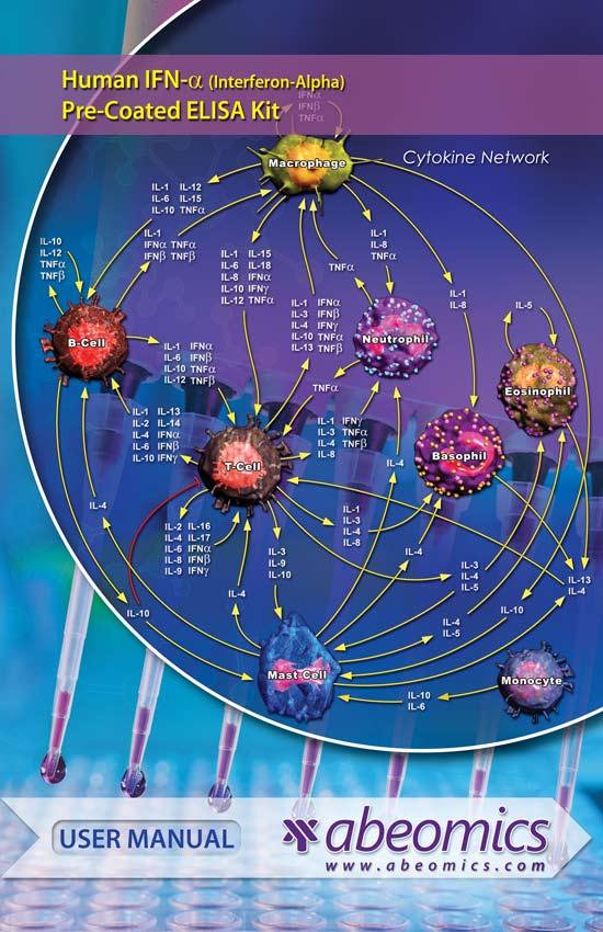
5 I. BACKGROUND IFN-α (Interferon Alpha) is an anti-viral cytokine of type I IFN family. There are 13 functional IFNα genes, one IFNβ gene, one IFNκ gene encoding a protein that is preferentially expressed in skin. IFN-α is classically induced following activation of viral pattern recognition receptors such as the endosomal Toll-like receptors and cytosolic nucleic acid sensors. IFN-α signaling results in wide range of effects upon the immune system, including upregulation of MHC molecules and activation of antigen presenting cells. These functions place IFN-α in a critical position bridging the innate and adaptive immune responses, and suggest that IFN-α could be important in setting thresholds for autoimmunity. A significant role for IFN-α in the pathogenesis of systemic lupus erythematosus is well supported, and clinical trials of anti-ifn-α monoclonal antibodies are in progress in this disease. In other autoimmune diseases characterized by substantial inflammation and tissue destruction, the role of type I interferons is less clear. II. OVERVIEW This assay employs an antibody specific for Anti-human IFN-α coated on a 96-well plate. Standards and samples are pipetted into the wells and IFN-α present in a sample is bound to the wells by the immobilized antibody. The wells are washed and biotinylated anti-human IFN-α antibody is added. After washing away unbound biotinylated antibody, HRPconjugated streptavidin is pipetted to the wells. The wells are again washed, a TMB substrate solution is added to the wells and color develops in proportion to the amount of IFN-α bound. The Stop Solution changes the color from blue to yellow, and the intensity of the color is measured at 450 nm. III. ADVANTAGES Multiple samples can be analyzed in a low volume, highthroughput format. Full analysis can be complete in 2 hours. ABEOMICS, Inc. info@abeomics.com Website: 3
6 IV. STORAGE Kit can be stored in 4 C, if you are using within a week. If you are using within 6 months, lyophilized standard can be stored in -20 C and other components at 4 C. Kit Components Item Specifications Storage 96 well Strip ELISA Plate 8 X 12 well 4 C Lyophilized Standard 2 vials -20 C Sample and Standard Dilution Buffer 20 ml 4 C Biotinylated Detection Antibody for 120 µl 4 C h IFN-α Antibody Dilution Buffer 10 ml 4 C HRP Conjugated Streptavidin 120 µl 4 C Streptavidin Dilution Buffer 10 ml 4 C TMB Substrate 10 ml 4 C Stop Solution 10 ml 4 C 25X Wash Buffer 30 ml 4 C Plate Sealer Product Manual 1 5 pieces Material Required, (Not Supplied) Microplate Reader 37 C Incubator Plate Reader Multi Chanel Pipette and disposable tips Eppendorf Tubes Deionized Water V. PRECAUTIONS FOR USE 1. To inspect the validity of experiment operation and the appropriateness of sample dilution proportion, pilot experiment using standards and a small number of samples is recommended. ABEOMICS, Inc. info@abeomics.com Website: 4
7 OD 450nm MANUAL Human IFN-α (Interferon-Alpha) Pre-Coated ELISA Kit 2. After opening and before using, keep plate dry. 3. Before using the Kit, spin tubes and bring down all components to the bottom of tubes. 4. Storage TMB reagents avoid light. 5. Washing process is very important, not fully wash easily cause a false positive. 6. Duplicate well assay is recommended for both standard and sample testing. 7. Don t let Micro plate dry at the assay, for dry plate will inactivate active components on plate. 8. Don t reuse tips and tubes to avoid cross contamination. 9. Avoid using the reagents from different batches together. VI. STANDARD CURVE Human IFN-α Standard Curve is shown below. Human IFN-a y = 0.017x R² = Human IFN-a Conc (pg/ml) X pg/ml Y O.D ABEOMICS, Inc. info@abeomics.com Website: 5

8 ABEOMICS, Inc. Website: 6
.")
9 VII. REAGENT PREPARATION AND STORAGE Included buffers and reagents are optimized for use with this kit. Substitution with other reagents is not recommended and may not give optimal results. 1. Prepare Standard Curve: One hour before the experiment. a. Quick spin down one vial of lyophilized standard. (DO NOT dilute standard directly on the plate). Add 1ml of sample/standard dilution buffer into one of the standard tube. Incubate at room temperature for 10 min. Mix thoroughly by vortex. Stock Standard concentration is 1000 pg/ml. b. Label 6 eppendorf tubes with 500pg/ml, 250 pg /ml, 125 pg/ml, 62.5 pg/ml, 31.2 pg/ml, 15.6 pg/ml respectively. Add 0.3 ml of sample/ standard dilution buffer into each tube. Add 0.3 ml of stock standard (1000pg/ml) into 1 st tube and mix thoroughly. Transfer 0.3 ml from 1 st tube to 2 nd tube and mix thoroughly. Transfer 0.3 ml from 2 nd tube to 3 rd tube mix thoroughly, and so on. Fig-1: Dilution tubes Note: Standard Solutions are best used within 2 hrs. Standard solution should be stored at 4 C for up to 12 hrs. or store at -20 C for up to 48 hrs. Avoid repeated freeze-thaw. 2. Sample Preparation and storage: Test samples should be collected, analyze immediately (within 2 hrs.) or aliquot and store at -20 C for long term. Avoid multiple freezethaw cycles. ABEOMICS, Inc. info@abeomics.com Website: 7
10 a. Cell culture supernatants: Centrifuge to remove precipitate, analyze immediately or aliquot and store at -20 C. b. Serum: Coagulate the serum at room temp about 1 hr. Centrifuge approximately 1000 g for 15 min. Analyze serum immediately or aliquot and store at -20 C. c. Plasma: Collect plasma with heparin or EDTA as the anti-coagulant. Centrifuge for 15 min at 2-8 C at 1500 g within 30 min of collection. For eliminating the platelet effect, suggesting that further centrifugation for 10 min at 2-8 C at 10,000 g. Analyze immediately or aliquot and store frozen at -20 C. d. Tissue Homogenates: For general information, hemolytic blood may affect the results, you should rinse the tissues with ice cold PBS (0.01M, ph 7.4) to remove excess blood thoroughly. Tissue pieces should be weighed and then minced to small pieces. This will be homogenized in PBS in a cold glass homogenizer. (Volume depends on the weight of the tissue, 1gram of tissue requires 9 ml of ice cold PBS with protease inhibitor). To further break the cells, you can sonicate the suspension with an ultrasonic cell disrupter or subject it to freeze- thaw cycle. Homogenates are then centrifuged for 5 min. at 5000 g to get the supernatant. Note: Samples to be used within 5 days may be store at 4 C, otherwise sample should be stored at -20 C (< 1 month) or-80 C (< 2 months) to avoid loss of bioactivity and contamination. Hemolyzed samples are not suitable for use in this Assay. e. End user should estimate the concentration of the target protein in the test samples first, then select proper dilution factor to make the diluted target protein concentration falls the optimal detection range of the kit. Dilute the samples with the provided dilution buffer. Several trials may be necessary in practice. The test sample should be well mixed with the dilution buffer. ABEOMICS, Inc. info@abeomics.com Website: 8
11 Standard curve and sample should be made before the experiment. High target protein concentration ng/ml: Dilute 1:100 (add 1 µl of sample into 99 µl of sample/ standard dilution buffer). Medium target protein concentration 1-10 ng/ml: Dilute 1:10 (add 10 µl of sample into 90 µl of sample/ standard dilution buffer). Low target protein concentration pg/ml: Dilute 1:2 (add 50 µl of sample into 50 µl of sample/ standard dilution buffer). Very low target protein concentration < 15.6 pg/ml: Do not dilute, use 100 µl of sample. 3. Preparation of Biotin detection antibody working solution: Prepare within one hour before the experiment. Calculate total volume working solution required. (0.1 ml/ well number of wells. Add µl extra). Dilute Biotin detection antibody with antibody dilution buffer at 1:100 and mix thoroughly. (i.e. add 1 µl of Biotin conjugated detection antibody into 99 µl of antibody dilution buffer). 4. Preparation of HRP-Streptavidin Conjugate (SABC) working solution: Prepare within 30 min before the experiment. Calculate total volume working solution required. (0.1 ml/well number of wells. Add µl extra). Dilute SABC with SABC dilution buffer at 1:100 and mix thoroughly. (i.e. add 1 µl of SABC into 99 µl of SABC dilution buffer). 5. Preparation of 1 X Wash buffer: Prepare 1 X Wash buffer by diluting 25X Wash buffer in sterile water. Diluted Wash buffer may be stored at 4 C, however we recommend preparing fresh 1X wash buffer for each experiment. For example: 10 ml of 25X Wash buffer in 240 ml of sterile water. ABEOMICS, Inc. info@abeomics.com Website: 9
12 VIII. ASSAY PROCEDURE Before starting the experiment, equilibrate the SABC working solution and TMB substrate for at least 30 min at room temp. When diluting samples and reagents, they should be mixed completely and evenly. It is recommended to plot a standard curve for each test. * If not all microplate strips will be used, remove the excess strips by pressing up from underneath each strip. Place excess strips back in the foil pouch with the included desiccant pack and reseal. 1. Set standard, test sample and blank (control zero) wells on the pre-coated plate and then record their position. It is recommended to measure each standard and sample in duplicate. Note: Wash plate twice before adding standard, sample and blank into the well. 2. Add 0.1 ml of standard 1000pg/ml, 500 pg/ml, 250 pg/ml. 125 pg/ml, 62.5 pg/ml, 31.2 pg/ml, 15.6 pg/ml, and Blank (control zero dilution buffer) into standard well. 3. Add 0.1 ml of diluted samples into test sample wells. 4. Seal plate with a cover and incubate at 37 C for 90 min. 5. Remove the cover and discard samples and standard solution by tapping plate on an absorbent paper. Note: DO NOT let the wells completely dry any time. DO NOT wash plate. 6. Add 0.1 ml of Biotin-detection antibody working solution into the above wells (Standards, control zero and samples). 7. Seal plate with cover and incubate at 37 C for 60 min. 8. Remove the cover, and wash plate 3 times with 1X wash buffer. 9. Add 0.1 ml of SABC working solution into each well. Cover the plate and incubate at 37 C for 30 min. 10. Remove the cover and wash plate 5 times with 1X wash buffer. Each time let the wash buffer stay in the well for 1-2 min. ABEOMICS, Inc. info@abeomics.com Website: 10
13 11. Add 90 µl of TMB substrate into each well, cover the plate and incubate at 37 C in dark within min. (Note: This incubation time is for reference use only. The optimal time should be determined by end user). The shades of blue can be seen in the first 3-4 wells, only on most concentrated standards. Other wells show no obvious color. 12. Add 50 µl of stop solution into each well and mix thoroughly. Color will change into yellow immediately. 13. Read O.D. absorbance at 450 nm in a micro-plate reader immediately after adding the stop solution. 14. Calculation: Relative O.D. 450 = O.D. for each well O.D. 450 control zero well. The Standard curve can be plotted as the relative O.D. 450 of each standard solution in Y axis vs. the respective concentration of the standard in X axis. Concentration of the samples can be incorporated from the standard curve. If the samples were diluted, multiply the dilution factor to the concentration. Table-1 Standard1 Standard A 1000pg/ml 1000pg/ml B 500pg/ml 500pg/ml C 250pg/ml 250pg/ml D 125pg/ml 125pg/ml E 62.5pg/ml 62.5pg/ml F 31.2pg/ml 31.2pg/ml G 15.6pg/ml 15.6pg/ml H 0 0 ABEOMICS, Inc. info@abeomics.com Website: 11
14 IX. REFERENCES 1. Interferon alpha as a primary pathogenic factor in human lupus. PMID: Interfering with immunity: detrimental role of type I IFNs during infection. PMID: Critical role of interferon-α constitutively produced in human hepatocytes in response to RNA virus infection. PMID: Type I interferon in organ-targeted autoimmune and inflammatory diseases. PMID: X. TROUBLE SHOOTING Problem Probable Cause Suggestion No signal Forgot to add all components. Prepare check list and add the components in the correct order. Low signal Not enough lysates per well. Check the protein concentration. Add more lysates. High background Washing is not sufficient. Wash plates thoroughly after incubation with Streptavidin-HRP secondary ABEOMICS, Inc. info@abeomics.com Website: 12
15

16
Human TIMP-2. Pre-Coated ELISA Kit
 Human TIMP-2 (Metalloproteinase Inhibitor-2) Pre-Coated ELISA Kit Catalog No: 90-2125 1 96 well Format (96 tests) Detection Range: 0.156 10 ng/ml Sensitivity: < 0.094 ng/ml This immunoassay kit allows
Human IFN-γ. Pre-Coated ELISA Kit
 Human IFN-γ (Interferon Gamma) Pre-Coated ELISA Kit Catalog No: 90-2067 1 96 well Format (96 tests) Detection Range: 15.6 1000 pg/ml Sensitivity: < 9.4 pg/ml This immunoassay kit allows for the in vitro
HumanIL-11(Interleukin11) Pre-CoatedELISAKit
 Pre-CoatedELISAKit") HumanIL-11(Interleukin11) Pre-CoatedELISAKit Human IL-11 (Interleukin 11) Pre-Coated ELISA Kit Catalog No: 90-2077 1 96 well Format (96 tests) Detection Range: 31.2 2000 pg/ml Sensitivity: 18.7 pg/ml
HumanIL-11(Interleukin11) Pre-CoatedELISAKit Human IL-11 (Interleukin 11) Pre-Coated ELISA Kit Catalog No: 90-2077 1 96 well Format (96 tests) Detection Range: 31.2 2000 pg/ml Sensitivity: 18.7 pg/ml
Chicken Calprotectin ELISA Kit. User Manual
 Nordic BioSite AB Propellervägen 4A, 183 62 Täby, Sweden T +46 (0)8 544 433 40, F +46 (0)8 756 94 90 info@nordicbiosite.com, www.nordicbiosite.com Org. No: 556539-9374, Residence: Täby Chicken Calprotectin
Porcine IgM (Immunoglobulin M) ELISA Kit
 ELISA Kit") Porcine IgM (Immunoglobulin M) ELISA Kit Catalogue No: EP0085 Size: 48T/96T Reactivity: Porcine Detection Range: 0.156-10ng/ml Sensitivity:
Porcine IgM (Immunoglobulin M) ELISA Kit Catalogue No: EP0085 Size: 48T/96T Reactivity: Porcine Detection Range: 0.156-10ng/ml Sensitivity: