System of protein filaments in the cytoplasm of. eukaryotic cell that gives the cell shape and capacity for
|
|
|
- Jasmin Wilkins
- 5 years ago
- Views:
Transcription
1 Cytoskeleton System of protein filaments in the cytoplasm of eukaryotic cell that gives the cell shape and capacity for directed movement. The protein filaments are responsible for the shaping, moving, dividing and interaction of cells.
2 Function 1) Give the cell shape. 2) Give the organization to the cytoplasm Anchor the cell organelles. Provide attachment points for organelles. Make communication between parts of the cell possible. 3) Provide the machinery for intracellular movement e.g., The transport of organelles from one place to another. Segregation of chromosomes into the two daughter cells at mitosis.
3 There are three general types of cytoplasmic filaments: 1) Microfilaments (Actin filaments) 2) Microtubules 3) Intermediate filaments These types differ in width, composition, and specific function but all provide structure and organization to the cytoplasm and shape to the cell. Each of the cytoskeletal component is composed of simple protein subunits that polymerize to form filaments of uniform thickness.

4
.")

5 1) Microfilaments (Actin filaments) Found in virtually all eukaryotic cells and 7 nm diameter The protein subunit is actin (globular protein). In the presence of ATP, the monomeric protein (actin) spontaneously associates into linear, helical polymer to form actin filament. Actin filament is two stranded helical polymer of the protein actin. They are present in linear bundles or networks rather than a single filaments.
6 Cells contain proteins that bind to actin monomers or filaments and infleunce a state of actin aggregation. e.g., filamin, fodrin (cross-link actin filaments to each other) Although actin filaments are dispersed through the cell, but large numbers of actin filaments bound to specific plasma membrane proteins lie just beneath, parallel to the plasma membrane, conferring shape and rigidity on the cell surface. Actin-myosin complexes form contractile ring that squeeze the cytoplasm in two during cytokinesis in all eukaryotes.


7
8 Function 1) In generation of tension. 2) In the contraction of muscle. 3) Play a role in cell division and motility. 4) In folding or extension of the cell membrane. 5) In movement of structures within the cell.
9

10 Microvilli Fingerlike extensions of plasma membrane. Increase surface area for absorption. Core of actin filaments for stiffening. The shape of the microvilli in this intestinal cell are supported by microfilaments, anchored to a network of intermediate filaments.
.")

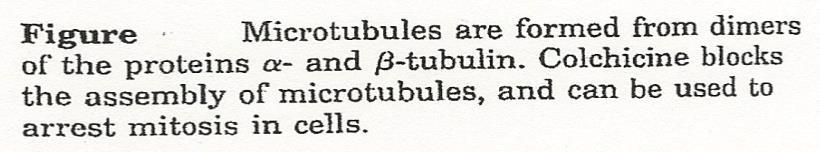
11 2) Microtubules Diameter 22 nm. The protein subunit is tubulin(dimmer composed of α- tubulin and β-tubulin). Dimmers of α- and β-tubulin polymerize to form linear polymers (protofilaments). 13 protofilaments associate side by side to form the hollow microtubules. Microtubules grow out from a small structure near the center of the cell, called the centrosome (organizing structure) and the other free in the cytoplasm.

12
13 Function 1) In nerve cells, bundles of microtubules participate in the movement of materials from the cell body towards the end of the cell (exon terminal). 2) Required in the formation of mitotic spindle during cell division. 3) Are the motile units in eukaryotic cilia and flagella.
14
 Microtubule of cytoskeleton (a) Motor molecules can attach to receptors on vesicles or organelles, and walk the")
15 Vesicle ATP Receptor for motor molecule Motor molecule (ATP powered) Microtubule of cytoskeleton (a) Motor molecules can attach to receptors on vesicles or organelles, and walk the organelles along the microtubules of the cytoskeleton. ATP Motor molecule (ATP powered) Cytoskeletal elements (microtubules or microfilaments) (b) In some types of cell motility, motor molecules attached to one element of the cytoskeleton can cause it to slide over another element, as in muscle contraction and cilia movement. Figure 3.24
 intermediate between actin filaments and microtubules.")


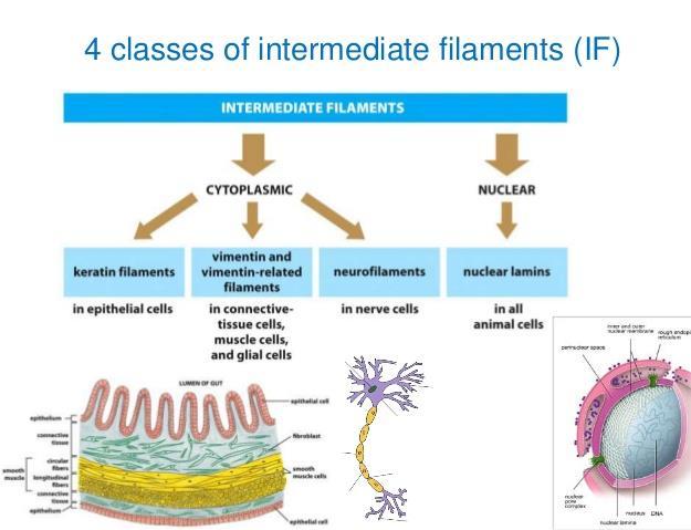
16 3) Intermediate filaments A family of structures with dimensions (8-12 nm) intermediate between actin filaments and microtubules. Several different types of monomeric protein subunits form intermediate filaments. Intermediate filaments are like ropes with many long strands to provide tensile strength. The subunits of the intermediate filaments are fibrous proteins. Intermediate filaments can be divided into several different categories.
17

 To position its")
18 Function 1) To provide internal mechanical support for the cell. 2) To position its organelles.

19
20 Diseases Examples of the diseases: Primary ciliary dyskinesia Dilated cardiomyopathy