ACCEPTED VERSION. The Author Published by Oxford University Press. All rights reserved.
|
|
|
- Cameron Milo Cole
- 5 years ago
- Views:
Transcription
1 ACCEPTED VERSION Newman, M.; Wilson, L.; Verdile, G.; Lim, A.; Khan, I.; Nik, S.H.M.; Pursglove, S.; Chapman, G.; Martins, R.N.; Lardelli, M. Differential, dominant activation and inhibition of Notch signalling and APP cleavage by truncations of PSEN1 in human disease Human Molecular Genetics, 2014; 23(3): The Author Published by Oxford University Press. All rights reserved. This is a pre-copy-editing, author-produced PDF of an article accepted for publication in Human Molecular Gene following peer review. The definitive publisher-authenticated version: Newman, M.; Wilson, L.; Verdile, G.; Lim, A.; Khan, I.; Nik, S.H.M.; Pursglove, S.; Chapman, G.; Martins, R.N.; Lardelli, M., Differential, dominant activation and inhibition of Notch signalling and APP cleavage by truncations of PSEN1 in human disease, Human Molecular Genetics, 2014; 23(3): , is available online at: PERMISSIONS Author Self-Archiving Policy Authors may upload their accepted manuscript PDF ("a post-print*") to institutional and/or centrally organized repositories, but must stipulate that public availability be delayed until 12 months after first online publication in the journal. Please note that a special policy applies for articles published under the Oxford Open initiative (see below). 7th October,
2 HMG Advance Access published September 18, Differential, dominant activation and inhibition of Notch signalling and APP cleavage by truncations of PSEN1 in human disease Morgan Newman *,1, Lachlan Wilson *,1, Giuseppe Verdile 2,3,4, Anne Lim 1, Imran Khan 2,3, Seyyed Hani Moussavi Nik 1, Sharon Pursglove 5, Gavin Chapman 5, Ralph Martins 2,3, Michael Lardelli,1 1 Discipline of Genetics, School of Molecular and Biomedical Science, University of Adelaide, SA 5005, Australia 2 Centre of Excellence for Alzheimer's Disease Research and Care, School of Medical Sciences, Edith Cowan University, Joondalup, WA 6027, Australia 3 School of Psychiatry and Clinical Neurosciences, University of Western Australia, Crawley, WA, 6009, Australia 4 School of Public Health, Curtin University, Bentley, WA 6102, Australia 5 Developmental Biology Division, Victor Chang Cardiac Research Institute, NSW 2010, Australia Author for correspondence Corresponding author: Michael Lardelli, Discipline of Genetics, School of Molecular and Biomedical Sciences, The University of Adelaide, SA 5005, Australia. Tel. (+61 8) , Fax (+61 8) , michael.lardelli@adelaide.edu.au * Contributed equally to this work The Author Published by Oxford University Press. All rights reserved. For Permissions, please journals.permissions@oup.com
3 2 Abstract PRESENILIN1 (PSEN1) is the major locus for mutations causing familial Alzheimer s disease (FAD) and is also mutated in Pick s disease of brain, familial acne inversa and dilated cardiomyopathy. It is a critical facilitator of Notch signalling and many other signalling pathways and protein cleavage events including production of the Amyloidβ (Aβ) peptide from the AMYLOID BETA A4 PRECURSOR PROTEIN (APP). We previously reported that interference with splicing of the zebrafish orthologue of PSEN1 creates dominant negative effects on Notch signalling. Here we extend this work to show that various truncations of human PSEN1 (or zebrafish Psen1) protein have starkly differential effects on Notch signalling and cleavage of zebrafish Appa (a paralogue of human APP). Different truncations can suppress or stimulate Notch signalling but not Appa cleavage and vice versa. The G183V mutation possibly causing Pick disease causes production of aberrant transcripts truncating the open reading frame after exon 5 sequence. We show that the truncated protein potentially translated from these transcripts avidly incorporates into very stable Psen1- dependent higher molecular weight complexes and suppresses cleavage of Appa but not Notch signalling. In contrast, the truncated protein potentially produced by the P242LfsX11 acne inversa mutation has no effect on Appa cleavage but, unexpectedly, enhances Notch signalling. Our results suggest novel hypotheses for the pathological mechanisms underlying these diseases and illustrate the importance of investigating the function of dominant mutations at physiologically-relevant expression levels and in the normally heterozygous state in which they cause human disease rather than in isolation from healthy alleles.
4 3 Introduction The human PRESENILIN proteins, PSEN1 and PSEN2, form the central components of - secretase complexes that are required for the intra-membrane cleavage of over 70 different substrates including the APP protein from which the Aβ peptide is derived (1). Aggregates of Aβ peptide are the major component of the neuritic plaques that are diagnostic of Alzheimer s disease (AD). Activation of -secretase activity appears to require the autoproteolytic cleavage of PRESENILIN proteins followed by association of the resulting N- and C-terminal fragments within -secretase complexes (2). These complexes consist additionally of the proteins NICASTRIN, PSENEN and APH1A or APH1B (reviewed by (3)). Alternatively, a variety of evidence supports that PSEN1 plays an important role in macroautophagy (4). Lee and colleagues have shown data supporting that an effect of PSEN1 on autophagy is via a -secretase-independent role of the PSEN1 holoprotein in acidification of lysosomes (4). However, other published data conflict with this (5, 6). The possibility exists that dysfunctional autophagy due to mutations in PSEN1 may contribute to inherited (familial) AD (FAD) although this is currently debated (see (7) and resulting correspondence). Over a decade of research has focussed on the role of mutations in the PRESENILINS in altering the -secretase cleavage of APP to support varieties of Aβ peptide. Much of this work has been conducted in cultured cells such as HEK293 cells and using cell-free assays where -secretase is solublised using the non-denaturing detergent CHAPS (3-[(3- cholamidopropyl)dimethylammonio]-1-propanesulfonate, (8)). Currently, the dominant idea is that FAD mutations promote the formation of more aggregation-prone forms of Aβ such as the 42 amino acid residue (aa) form relative to the more common (and possibly protective (9)) 40 aa form. This supposedly occurs in endosomes and at the plasma membrane (10, 11). However, recent discoveries raise caveats over these ideas. Winkler et al. (12) have shown that the thickness of the lipid bilayer (affected by its lipid makeup) can influence the site of - secretase cleavage of APP and so alter Aβ size. Since cell-free assays involve solubilisation of the -secretase complex, this must change the lipid environment of -secretase and so probably affects Aβ product size. Also, Area-Gomez et al (13) showed in an elegant paper in 2009 that, in mouse brain, the majority of Psen1 protein, all detectable Psen2 protein, and most -secretase activity resides in a detergent-impermeable lipid-raft like region of the endoplasmic reticulum named the mitochondrial associated membranes (MAM). The implications of this revision of our understanding of the subcellular distribution of PRESENILIN protein and -secretase activity have not yet been examined. The gene PSEN1 is the major locus for mutations causing FAD. Interestingly, all of the more than 180 different mutations in PSEN1 known to cause FAD allow retention of the C- terminal sequence of the protein. This and the absence of FAD mutations in other components of -secretase complexes is consistent with involvement of an autophagy function of the PSEN1 holoprotein in AD pathology. The only disease-causing mutation of PSEN1 involving simple truncation of the open reading frame (rather than partial interference with splicing resulting in multiple transcript isoforms) is the P242LfsX11 mutation causing
5 4 the inherited skin disease familial acne inversa (FAI) (14). P242LfsX11 causes a frameshift in exon 7 resulting in a premature termination codon. This mutation causes decreased PSEN1 transcript levels in lymphocytes (14) probably due to nonsense mediated decay (NMD) of transcripts of the mutant allele. Nevertheless, the mutant transcript can still be detected implying that low levels of truncated PSEN1 protein may be produced. Similarly, the G183V mutation that may cause Pick disease of brain affects the splice donor site of exon 6 producing a transcript encoding a full-length protein with a missense mutation (G183V, (15)) and also transcripts lacking either sequence from exon 6 or exons 6 and 7 (16). These transcripts would have frameshifted open reading frames putatively encoding proteins prematurely truncated after exon 5 sequence. Interestingly, in a mouse knock-in model of this mutation, Watanabe et al. (17) observed aberrantly splicing of PSEN1 only in brain. The aberrant transcripts are subject to NMD but can, nevertheless, be detected. When transfected into cultured HEK293 cells or into MEF cells lacking endogenous PRESENILINS, the G183V full length transcript itself gave only mild effects on Aβ production (15) or no effects on Notch cleavage (17) respectively. This is consistent with the lack of AD pathology seen for G183V. Watanabe et al. suggest that the Pick disease possibly resulting from G183V may be due to decreased overall -secretase activity but this contrasts with the observations of Wang et al. (14) where the P242LfsX11 FAI mutation also reduced PSEN1 levels but was not reported to produce Pick disease (and did not appear to cause FAD). Another change in PRESENILIN gene splicing associated with human disease is exclusion of exon 5 sequence from transcripts of PSEN2 in the brains of people with sporadic, late onset AD (18, 19). This produces a truncated isoform of the PSEN2 protein, PS2V. Interestingly, this isoform can stimulate APP cleavage and Aβ production when expressed in mouse Neuro 2a cells (20). We have also recently shown the PS2V transcript to be up-regulated in guinea pig brain under conditions of hypoxia and cholesterol loading (21). Up-regulation of this transcript was associated with cholesterol-enhanced amyloidogenic processing of APP. In mammals, PSEN1 protein is essential for Notch signalling since it is required for cleavage of Notch receptors within their single transmembrane domains after their extracellular domains are removed as a consequence of binding extracellular ligands. PSEN1-controlled - secretase activity releases the Notch intracellular domain (NICD) that subsequently enters the nucleus to regulate gene activity. Notch signalling is essential for maintaining stem cells in an undifferentiated state (22), for controlling cell fate decisions (reviewed by e.g. (23-26)) and is commonly dysregulated in cancer (27). Changes in Notch signalling have been suggested to underlie FAI pathology (14) and this is supported by the discovery of additional mutations causing FAI in other components of -secretase complexes (in contrast to FAD) (14). Rates of aberrant splicing have been seen to increase in ageing and cancerous cells (reviewed in (28, 29)). Aberrant splicing of PSEN1 in particular was observed in sporadic frontotemporal dementia ((30), of which Pick s disease of brain is one type). While NMD plays an essential role in the elimination of transcripts encoding aberrantly truncated proteins it can be incompletely effective under certain circumstances, e.g. when a premature termination codon occurs within 55 bp of a downstream exon-exon junction (31). The modular nature of many proteins where separate domains have separate activities means that
6 5 it is common for truncated proteins to possess a subset of their normal activities. This allows them to interfere dominantly with the function of normal full-length proteins. Thus it is important to consider the activities of the potential protein products of aberrant transcripts whenever detection of such transcripts suggests that truncated proteins may exist. Zebrafish embryos are an excellent system in which to analyse cellular responses to changes in gene expression. Unlike cells grown in culture, the cells of zebrafish embryos exist in an environment of normal substrates and cell-cell interactions. Simultaneous and subtle upregulation and down-regulation of gene expression is possible at levels consistent with normal cell physiology. In previous work we attempted to use the unique advantages of the zebrafish system to model particular rare FAD mutations in PSEN1 where protein coding exons are excluded but the reading frame is maintained. We did this by blocking splice acceptor sites in the zebrafish orthologue of PSEN1 (named psen1) using morpholino antisense oligonucleotides ( morpholinos ). Unexpectedly we generated low levels of intron inclusion rather than exon exclusion but some of the aberrant transcripts nevertheless had potently dominant negative effects on the activity of both psen1 and the zebrafish orthologue of PSEN2 (named psen2) (32). In the work described in this paper we confirm the predominant MAM-localisation of PSEN1 protein in mammalian (mouse) brain. We then use our zebrafish embryo-based in vivo assays of Notch signalling and APP cleavage to expand our previous study of the -secretase activity of PSEN1 truncations including PSEN1 truncations relevant to human diseases other than AD. We show that, at physiologically relevant levels, different truncations of PSEN1 can have starkly contrasting effects on zebrafish Notch signalling and Appa cleavage. Truncations resembling PS2V boost both Notch and Appa cleavage in a manner dependent on the presence of endogenous zebrafish Psen1 protein but not Psen2 protein. Truncations equivalent to some of the products of the G183V Pick disease mutation reduce Appa cleavage but do not affect Notch. They also bind full-length endogenous Psen1 and Psen2 and integrate into higher molecular weight complexes. In contrast, the truncation produced by the P242LfsX11 acne inversa mutation unexpectedly increases Notch signalling with no effect on Appa cleavage. Our discoveries imply that in vivo analysis of -secretase activity may give more realistic insights into the role of -secretase activity in human disease. Our observations also suggest transgenic approaches to allow manipulation of Notch signalling without affecting APP cleavage and vice versa. Results PSEN1 protein is concentrated in the MAM in brain The majority of FAD is due to mutations in the genes encoding the human proteins PSEN1 and PSEN2. In 2009 Area-Gomez et al. demonstrated that, in the mammalian (mouse) brain, the greatest concentration of PSEN1 protein, -secretase activity and -secretase cleavage of APP is localised to the MAM (13). They also showed that the MAM is difficult to permeabilise for immunohistochemistry using standard detergents since it has a lipid
7 6 constitution similar to that of lipid rafts (13, 33). Also, Winkler et al (12) recently showed that changes in the thickness of lipid bilayers can affect the cleavage of APP proteins by - secretase to produce different forms of A. However, much of the debate on the role of - secretase activity in AD is based on data obtained from cell-free assays where -secretase activity is solubilised using detergents. Such cell-free assays may not entirely reflect the - secretase activity found in vivo. To confirm independently the controversial MAMlocalisation of PSEN1 protein we replicated the sub-cellular fractionation of mouse brain by Area-Gomez et al. (13). As expected, this showed that PSEN1 is predominantly localised to the MAM with lower concentrations in the rest of the endoplasmic reticulum and in mitochondria (Supplemental Data Figure 1). Delimiting the region of Psen1 in which truncation produces dominant negative effects on Notch signalling In addition to their role in -secretase, PRESENILIN proteins are thought to have multiple cellular activities in e.g. phosphorylation of β-catenin (34, 35), Ca 2+ transport (36, 37) and macroautophagy (4). The involvement of the PRESENILINs in these other activities has not been studied in depth and it is unclear to what extent these activities interact with -secretase activity. Also, cell-free assays of -secretase activity have been shown to be very sensitive to preparation and assay conditions (discussed in (38)). Therefore, analysis of PRESENILIN activity in vivo may give a more realistic understanding of their roles in cell biology and disease than analysis in cell-free systems. -Secretase activity can be studied using cultured cells (such as HEK293 cells) transfected with assay or other constructs (e.g. (39)). However, due to their unusual environments, cultured cells commonly have unusual patterns of gene and protein expression (40, 41) while high-level expression of genes/proteins through transfection can produce physiologically irrelevant results such as the complete abolition of endogenous Psen protein by exogenous Psen (42). Fertilized zebrafish eggs represent an interesting alternative in vivo system in which to analyse gene activity since their large size allows easy, subtle and multifactorial manipulation of gene activity at the 1-cell stage. Also, their subsequent development through the various stages of embryogenesis allows changes in phenotype to be used as bioassays of changes in gene activity. -Secretase activity is required for the intra-membraneous cleavage of Notch receptors that facilitates transcriptional regulation of genes by Notch intracellular domains (reviewed by (43)). We (32, 44) and others ((45-48)) have previously observed changes in Notch signalling in zebrafish embryos by examining changes in the expression of the neurogenin1 gene that is a under transcriptional regulation by Notch (45). In previously published work examining PRESENILIN s -secretase activity we monitored spinal cord neurogenin1 transcript levels in zebrafish embryos at 24 hours post fertilisation C) to assess changes in Notch signalling. This led to discovery of dominant negative effects on Notch signalling caused by interference with splicing of zebrafish psen1 gene transcripts (32). These dominant negative effects occur upon interference with splicing to zebrafish psen1 exons cognate with human PSEN1 exons 7 and 8 but not 6 or 9. (Hereafter the zebrafish exons are referred to by their human cognate numbers, see Figure 1.) Injection into embryos of mrna encoding zebrafish
8 7 Psen1 protein truncated after exon 6 (equivalent to the putative product of the aberrant transcript produced by interference with splicing to exon 7 (32)) produced a similar dominant negative phenotype supporting that this effect is due to the formation of truncated Psen1 protein molecules. We have subsequently shown that our injection of synthetic mrna into zebrafish embryos gives total mrna levels less than threefold higher than normal (see Supplemental Data Figure 2). Thus, interference with the splicing of endogenous genes and injection of synthetic mrnas causes changes in gene activity in zebrafish embryos at physiologically relevant levels. Notch signalling is an important component of embryo development, stem cell maintenance and human diseases including some cancers. Truncations of the N-terminal fragment (NTF) of human PSEN1 feature in a number of human disease states (see the Introduction and below). Therefore, we sought to use our zebrafish model to analyse how truncations of the NTF in zebrafish Psen1 affect Notch signalling. Since the effects of interference with splicing are not entirely predictable we focussed instead on injection of synthetic mrnas. We synthesised mrnas encoding the entire NTF or forms of Psen1 truncated downstream of sequence encoded in exons 3, 4, 5, 6, 7, or 8 (Figure 1). These were injected into zebrafish embryos and their effects on neurogenin1 expression in the spinal cord (negatively regulated by Notch signalling) were analysed at 24 hpf. Interestingly, we observed both enhancement and suppression of Notch signalling. Injection of mrna encoding a truncation of Psen1 after exon 3 sequence (Psen1Δ>3) proved quite toxic to embryo survival unlike the other, longer truncations. Therefore, we did not analyse this truncation further. Injection of mrna coding for Psen1 truncated after exon 4 sequence (Psen1Δ>4) caused significant change in neurogenin1 transcription compared to embryos injected with a negative control mrna (Figure 2A). Surprisingly, Psen1Δ>4 acts in a dominant positive manner to decrease neurogenin1 transcription, presumably by increasing Notch signalling. In contrast, mrnas encoding Psen1 proteins truncated after exon 5, 6, 7, or 8 sequence behaved as expected from our previously published analysis using morpholinos (44). Truncation of Psen1 after exons 5 or 8 (Psen1Δ>5 and Psen1Δ>8) or the entire NTF caused no significant change in neurogenin1 transcription relative to embryos injected with a negative control mrna. However, mrnas encoding Psen1 truncated after exons 6 and 7 (Psen1Δ>6 and Psen1Δ>7) caused an increase in neurogenin1 expression indicative of decreased Notch signalling (Figure 2A). Our results, and those of our previous study using morpholinos ((32)) initially led us to believe that the region of psen1 flanked by exons 6 and 8 (i.e. exon 7) encoded a section of the Psen1 protein within which any truncation would produce dominant negative effects on Notch signalling. However, additional analysis showed this interpretation to be incorrect (see later).
9 8 Truncations of Psen1 produce distinct and overlapping effects on Notch signalling and cleavage of zebrafish Appa -Secretase cleaves over 70 protein substrates in addition to Notch receptors (reviewed by (1)). Among the most important of these for human disease is the AMYLOID PRECURSOR PROTEIN, APP. APP is cleaved by -secretase after initial cleavage by secretase or secretase in its N-terminal, luminal/extracellular domain close to the lipid bilayer (49-52). Zebrafish possess two paralogous genes, appa and appb, derived from an ancestral orthologue of human APP (53). We recently developed a zebrafish assay for -secretase cleavage of Appa that monitors cleavage of an Appa substrate truncated to resemble the putative 86 aa C-terminal product of β-secretase cleavage ( AppaC86::GFP, (38)). Mutations (54) and drugs (55) affecting Presenilin activity have been reported to have differential effects on Notch and APP cleavage. Therefore we exploited our Appa-based assay to investigate whether our truncations of Psen1 had similar or different effects on APP cleavage compared to Notch signalling. Injection of mrna encoding Psen1Δ>4 caused increased cleavage of Appa similar to the effect of this truncation on Notch signalling. However, whereas Psen1Δ>5 had no effect on Notch signalling, we found that it acted to inhibit -secretase cleavage of Appa (Figure 3A). Futhermore, in contrast to their dominant negative effect on Notch signalling, Psen1Δ>6 and Psen1Δ>7 did not affect Appa cleavage. Psen1Δ>8 and the entire NTF did not affect either Appa cleavage or Notch signalling in our zebrafish embryo system (Figure 3A). To support that our observations were the result of expression of truncated forms of Psen1 at physiologically relevant levels we attempted to reproduce the above effects on AppaC86::GFP substrate cleavage through directed interference with endogenous psen1 transcript splicing. Morpholinos that bind over the acceptor sites of exons 5, 7, 8, and 9 exist and can interfere with splicing of endogenous psen1 transcripts. These cause retention of the respective upstream introns (see Supplemental Data Figure 3) and, presumably, expression of transcripts with open reading frames prematurely terminated after exons 4, 6, 7 and 8 respectively. The assay results from morpholino injection are consistent with those seen for mrna injection (Figure 3C). The different effects of particular Psen1 truncations on Notch and Appa are consistent with the idea that differently composed -secretase complexes are responsible for Notch and APP cleavage (56, 57). Our observations also show that it should be possible to construct transgenes that specifically inhibit Notch cleavage but not APP cleavage and vice versa. Alternatively Psen1Δ>4 can be used to boost both Notch signalling and APP cleavage in cells, tissues and transgenic animals. Psen1Δ>4 requires endogenous, normal Psen1, but not Psen2 to increase -secretase activity Psen1Δ>4 is structurally equivalent to the PS2V isoform of human PSEN2 that is induced in neurons by hypoxia and is found at increased levels in the brains of people with sporadic,
10 9 late onset AD. Consistent with our results, PS2V has been shown to increase -secretase activity and Aβ levels when its expression is forced in the mouse neuro2a neuroblastoma cell line (20). However, Psen1Δ>4 and PS2V do not possess the NTF portion of the putative proteolytic site of -secretase that includes a conserved, essential Asparagine residue (Asp 257 in human PSEN1 or Asp 263 in human PSEN2). Truncations Psen1Δ>4, Δ>5, Δ>6, and Δ>7 also lack this residue and so would be incapable themselves of contributing directly to - secretase cleavage. An explanation for the ability of Psen1Δ>4 and PS2V to boost -secretase activity is that they are dependent on endogenous, normal Presenilin proteins to do so. Zebrafish embryos are a very versatile cellular system in which to dissect molecular interactions since it is possible to block and force expression of multiple gene activities simultaneously. To test the dependence of Psen1Δ>4 on endogenous Presenilin proteins we injected mrna encoding Psen1Δ>4 while simultaneously blocking translation of either endogenous Psen1 or Psen2 using morpholinos ( MoPsen1Tln and MoPsen2Tln respectively). Analysis of Notch signalling using the neurogenin1 expression assay was then performed by comparing trunk spinal cord neurogenin1 expression in these embryos to that in control embryos that had been injected either with negative control (inactive) morpholino MoCont or with the MoPsen1Tln or MoPsen2Tln morpholinos as appropriate to block endogenous Psen1 or Psen2 translation respectively (see Figure 4A). Psen1Δ>4 was unable to increase Notch signalling (neurogenin1 expression) significantly in embryos lacking endogenous full-length Psen1 (Figure 4A). In contrast, blockage of Psen2 translation could not block the ability of Psen1Δ>4 to boost Notch signalling (Figure 4A). This supports that Psen1Δ>4 acts through normal Psen1 in order to boost -secretase activity while acting to a lesser extent (if at all) through endogenous Psen2. Psen1Δ>4 expression also boosts cleavage of Appa (see above). To support that Psen1Δ>4 s dependence on endogenous Psen1 also applies for substrates other than Notch we tested its ability to cleave AppaC86::GFP in the absence of Psen1 or Psen2 translation. Consistent with the results for Notch above, Psen1Δ>4 could only increase AppaC86::GFP cleavage when Psen1 was present while blockage of Psen2 translation had no effect on the ability of Psen1Δ>4 to increase AppaC86::GFP cleavage (Figure 4B). Psen1 truncations incorporate into higher molecular weight complexes It is commonly observed that Presenilin proteins are incorporated into higher molecular weight (HMW) complexes that are sufficiently stable to withstand the highly denaturing conditions involved in protein resolution by SDS-polyacrylamide gel electrophoresis (PAGE, e.g. see (58-60)). Kim et al (59) previously showed that forced expression of the NTF of human PSEN1 in mouse embryonic fibroblasts (MEFs) resulted in the formation of such stable complexes. A simple model for the dominant effects of truncated Psen1 on Notch signalling and Appa cleavage is that this occurs by interference with formation of -secretase complexes. Our various truncated forms of zebrafish Psen1 have been tagged at their N- termini using the FLAG antibody tag. To observe whether the Psen1 truncations could incorporate into HMW complexes we expressed them in zebrafish embryos and then subjected embryo lysates to SDS-PAGE followed by western blotting against the FLAG tag
11 10 (Figure 5A). This revealed apparent complexes in the range of kda while few protein bands in the predicted monomer range of ~15-30 kda were observed. In particular, truncations Psen1Δ>5, Δ>7, Δ>8 and NTF avidly incorporated into apparent SDS-resistant complexes of approximately 200 kda or greater (Figure 5A). However, it is known that forced over-expression of proteins in cells can lead to the formation of non-specific SDSresistant aggregates (e.g. (61)). Since our analysis of Psen1Δ>4 has shown that its activity is dependent on the presence of endogenous Psen1 (see above) we sought to demonstrate that the Psen1 truncations were being incorporated into HMW complexes relevant to endogenous Psen1 activity. We analysed the incorporation of the truncations into SDS-resistant complexes with and without blockage of translation of endogenous Psen1 (Figure 5A). Distinct differences in complex formation were observed for truncations Psen1Δ>5, Δ>7, Δ>8 and NTF when these were expressed with or without endogenous Psen1. Apparently, the loss of endogenous Psen1 enhances the incorporation of the truncated Psen1 molecules into HMW complexes. This supports that the complex formation observed is specific and related, in some way, to Psen1 activity (although not necessarily γ-secretase activity). As an additional test that our truncated Psen1 proteins can interact with endogenous, normal Psen1 protein we performed immuno-precipitation analysis under non-denaturing conditions using ANTI-FLAG M2 Affinity Gel on embryos injected with mrna coding for either the most avid apparent interactor Psen1Δ>5 or (as a negative control) a form of GFP tagged at its N-terminal with FLAG. Immunoblotting of the lysates and precipitates clearly demonstrated a specific interaction of Psen1Δ>5 with Psen1 and, interestingly, also with endogenous Psen2 while GFP failed to interact with either endogenous protein (Figure 5B). This suggests that immuno-precipitation of our FLAG-tagged Psen1 truncations under non-denaturing conditions should co-precipitate interacting proteins and permit their identification by proteomics methods. Remarkably, loss of endogenous Psen1 apparently causes increased incorporation of Psen1Δ>5 into the very largest HMW complexes but a dramatic decrease in the incorporation of this truncation into an ~50 kda complex with a concomitant increase in levels of a smaller form at ~25 kda (while the monomer has a theoretical mass of ~17 kda). A similar behaviour is shown by Psen1Δ>8 and these are the two truncations that do not appear to influence Notch activity. The behaviour of Psen1Δ>5 is particularly interesting since this is the only truncation that inhibited Appa cleavage. This supports that Notch and APP are cleaved by - secretase activities in distinct complexes. Also, Psen1Δ>5 resembles the putative protein products of the abnormal transcripts caused by the G183V mutation of human PSEN1 that affects the splice donor site of exon 6. This is the only PSEN1 mutation suggested to cause FTD without associated AD pathology. Our analysis suggests that the transcript putatively encoding human PSEN1 truncated after exon 5 sequence may suppress APP cleavage and A peptide formation consistent with the lack of AD histopathology (A plaques). The ~50 kda complex seen for all the injected truncations (but not Psen1NTF or the negative control) does not appear to represent dimer formation since the Psen1Δ>5 truncation and larger truncated peptides have monomeric masses in the range of kda. Also, the mass of the ~50 kda complex does not appear to change with the size of the truncation injected.
12 11 Therefore, we suspect that the ~50 kda complex represents the protease-resistant core of a larger unstable entity that includes part or all of our FLAG-tagged Psen1 truncations. Truncations of human PSEN1 produce similar effects to their zebrafish equivalents when expressed in zebrafish embryos To confirm that our observations using zebrafish embryos are relevant to the human system we sought to express truncations of human PSEN1 in the human kidney-derived cell line HEK293. Truncations of human PSEN1 after exon 4, 5, 6, 7 and 8 sequence and with N- terminal HA antibody tags were synthesised in the pcglobin2 vector that can be used for mrna synthesis or to express genes downstream of a CMV promoter (62). The truncations of human PSEN1 were named hpsen1δ>4, 5, 6, 7 or 8 to correspond to the zebrafish Psen1 truncations. Unfortunately, repeated attempts to generate stably transformed cell lines expressing the human PSEN1 truncations failed. Apparently, the human PSEN1 truncations are too toxic to allow survival and growth of HEK293 cells, possibly because transfection gives excessive, non-physiologically relevant levels of expression. Therefore, we generated mrnas encoding each of the human PSEN1 truncations and expressed these in zebrafish embryos by mrna injection. Our assays for Notch signalling (Figure 2B) and Appa cleavage (Figure 3B) showed that the human PSEN1 truncations produce effects on these activities identical to their zebrafish Psen1 equivalents. This supports that our analysis of Presenilin activity in zebrafish is relevant to our understanding of this activity in the human system. The P242LfsX11 mutation of human PSEN1 causing acne inversa dominantly increases Notch signalling but not Appa cleavage. Recently Wang et al (2010) identified that mutations in three of the four protein components of the -secretase complex, PSEN1, NICASTRIN and PSENEN, can cause an inherited disease of hair follicles named acne inversa (AI). These researchers discovered only one AI mutation in PSEN1 - a single base deletion, c.725delc, causing a frameshift that results in the premature termination of the open reading frame. The mutant protein structure is denoted as P242LfsX11. This is the first known inherited mutation in PSEN1 exclusively causing truncation of the open reading frame. (The FAD mutations of PSEN1 all still allow synthesis of mrnas encoding entire open reading frames.) Unexpectedly, none of the families with inherited AI show familial Alzheimer s disease and Wang et al (14) suggested that AI may be caused by changes in Notch signalling affecting follicle biology (14). PSEN1 transcript levels are reduced by varying amounts in heterozygotes suggesting that nonsense mediated decay is destroying transcripts possessing the premature termination codon generated by this mutation. However, supplemental data published with the Wang et al. paper shows that low levels of transcript containing the P242LfsX11 mutation are still observable in lymphocytes from affected individuals. However, the P242LfsX11 mutation places a premature stop codon within 20 bp upstream of the next downstream exon junction (Figure 6A). Thus, the mutation potentially lies too close to the exon-exon junction for nonsense mediated decay to be completely effective (according to the bp rule (31)).
13 12 We were surprised to see that P242LfsX11 lies in exon 7 since we believed that truncating mutations in this region would have potently dominant negative effects on Notch signalling and that even minor levels of the mutant transcript might interfere fatally in embryo development i.e. they would be dominant lethal (32). Therefore, we tested the activity of the putative protein product of the P242LfsX11 mutation in our zebrafish system. Construct hpsen1-p242lfsx11 incorporating the P242LfsX11 mutation was generated in the pcglobin2 vector using the human PSEN1 gene and then mrna was synthesised for injection into zebrafish embryos. No effect was observed on Appa cleavage consistent with the fact that individuals with this AI mutation do not show early onset FAD (Figure 6B). Also, Notch signalling was observed to increase rather than decrease implying that Notch signalling in individuals heterozygous for P242LfsX11 may be greater than that expected if P242LfsX11 was a simple, loss-of-function mutation (Figure 6C). This discovery also means that it should be possible to construct transgenes that boost Notch signalling without increasing APP cleavage. Discussion The PRESENILIN proteins are essential to facilitate many signalling pathways and protein cleavage events. They modulate intracellular Ca 2+ ion levels (36, 37), regulate β-catenin turnover (34, 35) and influence autophagy (4). Therefore, changes in PRESENILIN activity can have widespread effects on cell function and are associated with numerous human diseases. In earlier work we showed that low levels of truncated zebrafish Presenilin molecules can exert potent dominant negative effects on Notch signalling (32). Truncated PRESENILIN proteins (or at least aberrantly spliced transcripts) occur either as natural isoforms (e.g. PS2V (18)), result from mutation (e.g. FAD (63), acne inversa (14)) or exist in particular disease states such as FTD (30). Thus it is important to understand their effects on cell biology. The discovery of the MAM as the primary subcellular location of the PRESENILIN proteins, -secretase activity and -secretase cleavage of APP in neural tissue (13) is revolutionising our understanding of the role of PRESENILIN mutations in FAD (33). We have now independently confirmed the predominant localisation to the MAM of mammalian Psen1 protein. The true sub-cellular distribution of the PRESENILINs remained unknown until recently due to the MAM s tight physical association with mitochondria and its peculiar lipid raft-like characteristics that prevented its permeabilisation by common immunohistochemistry detergents (13, 64). Also, Winkler et al. recently observed that the site of -secretase cleavage within APP is affected by the thickness of the lipid bilayer within which APP resides (12). This raises the question of how solubilisation of -secretase with detergents in cell-free assays affects observations of APP cleavage and the different forms of Aβ formed by different PRESENILIN mutants. Interestingly, Area-Gomez et al recently showed that changes in PRESENILIN activity affect movement of cholesterol into and out of MAM membranes (33). This raises the possibility that changes in the profile of A lengths observed in FAD PRESENILIN mutants may be the secondary effect of changes in the lipid
14 13 constitution of MAM membranes rather than a direct effect of the mutations on the cleavage interaction between a PRESENILIN molecule and an APP molecule within the -secretase complex itself. Of course, many of the experimental caveats regarding cell-free and cell culture approaches to analysis of -secretase would be obviated by the use of in vivo assays. Indeed, our in vivo assay for -secretase cleavage of APP (Appa) is unique in this regard. In disease states, mutant genes encoding truncated PRESENILIN open reading frames are always found concomitantly with wild-type PRESENILIN genes. However, previous analyses of truncated PRESENILINs have examined their biochemical activities in backgrounds lacking full-length proteins. Thus Laudon et al (65) transfected constructs expressing NTF truncations together with the C-terminal fragment (CTF) of PSEN1 into mouse BD8 cells lacking endogenous Psen1 and Psen2 and saw Notch and APP cleavage activity only for truncations with at least 288 amino acid residues (aa) which is nearly the complete NTF. Similarly, Kim et al (59) saw similar results for Notch cleavage when examining truncations of the PSEN1 NTF together with full-length CTF in mouse embryonic fibroblast cells (MEFs) lacking Psen1 and Psen2. The different results we obtained for the activity of Presenilin truncations in APP cleavage and Notch signalling compared to those above are likely because our analysis has examined the expression of truncated PRESENILINs in the presence of endogenous gene expression. This illustrates the importance of examining dominant mutations in their real, heterozygous state - in the presence of a normal allele rather than in isolation since the activity of the dominant mutation can be dependent on the function of the normal allele. Indeed, in our experimental system, when we blocked endogenous Psen1 expression in the presence of Psen1Δ>4 we saw that this truncation could no longer affect -secretase activity. This is expected since all the truncations except Psen1Δ>8 (which includes almost the entire NTF) lack the Asp 257 residue that is thought to be essential to the -secretase catalytic site (66). Thus, truncations Psen1Δ>4 and P242LfsX11 that both boost Notch signalling must do this by stimulating, (by an unknown mechanism/s), the activity of -secretase complexes comprised of normal Presenilin NTFs and CTFs. Interaction of truncated Presenilin NTFs with functional -secretase complexes is also supported by the data of Kim et al (59) who saw that incorporation of truncated NTFs into higher molecular weight complexes was greatest in cells possessing both PSEN1 and PSEN2 and was decreased in cells lacking one or both of these genes. For incorporation of our truncated forms of Psen1 into higher molecular weight complexes we also observed evidence for interactions with endogenous (non-mutant) Presenilin proteins. Future work will aim to exploit the FLAG-tagged N-termini of our truncated Psen1 proteins to identify interacting proteins by co-immunoprecipitation and proteomics analyses. The stimulation of Appa cleavage and Notch signalling by Psen1Δ>4 is consistent with the activity of the PS2V isoform that is an equivalent truncation of PSEN2. Forced expression of PS2V in mouse neuro 2a cells was seen to increase the production of Aβ peptide consistent with an increase in -secretase activity (20). Our observations suggest that PS2V will be found to require full-length Presenilin expression for its ability to increase -secretase activity. Since Psen1Δ>4 requires endogenous Psen1 but not Psen2 for its activity, it would
15 14 be interesting to know whether, by analogy, PS2V requires PSEN2 but not PSEN1 (or vice versa) to increase Aβ production. The G183V mutation of human PSEN1 is remarkable as the only known mutation of this gene suggested to produce an exclusively Pick disease FTD phenotype rather than FTD associated with AD. Dermaut et al (15) showed that the full-length PSEN1 protein incorporating the G183V missense mutation did not cause a greatly increased level of Aβ 42 to Aβ 40 production when transfected into HEK293 cells although total levels of Aβ production were not reported. This was consistent with brain histopathology showing Pick bodies and western analysis showing no detectable Aβ. In a comment on unpublished research regarding putative truncated PSEN1 from the aberrant transcript splicing caused by G193V, Dermaut et al (16) noted that, cellular gamma-secretase assays show that the truncated proteins behave as complete null alleles. However, no information was given on the cellular system used or whether endogenous full-length PSEN1 was present. Watanabe et al. (17) engineered the G183V mutation of human PSEN1 into the mouse Psen1 gene and observed aberrant splicing of Psen1 transcripts. However, the aberrant splicing was only observed in the brain and not other tissues. In the mouse brain, homozygosity for the G183V mutation reduced the levels of the normally spliced transcript by approximately half. (In humans, heterozygosity for G183V rather than homozygosity presumably means that the reduction in transcript levels would be less.) These researchers suggested that reduced -secretase activity in the brain may be responsible for the Pick s disease phenotype and noted that levels of Aβ are, unexpectedly, reduced by more than half in cell-free assays conducted using mouse brain. Their results supported that the reduction was due to the lower level of PSEN1 protein rather than the G183V mis-sense mutation of the coding sequence since G183V mutant PSEN1 protein had similar activity to wild type PSEN1 in transfected murine embryonic fibroblasts (MEFs) lacking endogenous Presenilin activity. When the same cells were transfected with a truncation after exon 5 sequence of the Psen1 open reading frame, Watanabe et al. saw no - secretase activity. However, if we assume that our observations of Psen1Δ>5 activity in the presence of endogenous, normal Presenilin are valid this explains some apparently anomalous observations (below). In our 2008 paper (32) we showed that a low levels of aberrantly splicing could, nevertheless, have potent dominant phenotypes. Thus, we should not assume that the low levels of Psen1Δ>5-like transcripts generated by aberrant splicing in the G183V mutant carriers have no effect. Indeed, the analyses described in this paper show that Psen1Δ>5 truncated protein is notable for its avid incorporation into very stable higher molecular weight complexes and its ability to bind full-length forms of Psen1 and Psen2. This may explain why Watanabe et al. saw ~50% reductions in both Psen1 transcript levels and Notch cleavage in their homozygous mutant mice but considerably more than a 50% reduction in Aβ 40 production (for Notch and Aβ synthesis measured by cell-free assays on mouse cortex). Also, the idea that FTD might be due to a reduction of human PSEN1 transcript levels is not consistent with the absence of any reports of FAD in the FAI mutant family bearing the P242LfsX11 mutation. These individuals show ~50% reduction in wild type PSEN1 transcript levels. This reduction presumably occurs in all cells of these individuals since it appears to be due to a
16 15 frameshift mutation affecting all the transcript products of the mutant allele. Also, aberrant PSEN1 transcript splicing has been observed in sporadic FTD brains (30) and, apparently, for the L113P FAD mutation of PSEN1 with features of FTD (67). Therefore, we suggest that truncated PSEN1 proteins, and in particular truncations similar to Psen1Δ>5, may play a decisive role in the development of FTD pathology where PSEN1 gene activity is involved. Whether Psen1Δ>5-like truncations perform this role via their effect on APP cleavage or by effects on other PRESENILIN activities (such as autophagy, Ca 2+ transport or -catenin phosphorylation) or via yet unknown activities not dependent on full-length PRESENILINs remains to be seen. We can begin to investigate this by analysing the higher molecular weight complexes into which Psen1Δ>5 becomes incorporated. In zebrafish embryos, suppression of Appa cleavage by Psen1Δ>5 was accompanied by an apparent dependence on endogenous Psen1 for complex formation but Psen1Δ>5 had no effect on Notch signalling. This would be consistent with Notch and APP cleavage being performed by different forms of -secretase complex and/or in different locations within the cell. There is experimental data to support both of these ideas since -secretase complexes comprised of different isoforms of Aph1 appear to show different proclivities to cleave Notch and APP (57) and Notch is thought to be cleaved by -secretase in a process involving endocytosis (reviewed by (68)) while cleavage of APP by -secretase has been observed to be predominantly in the mitochondrial associated membranes (MAM, (13)). Once again, it will be interesting to pursue the identity of the proteins observed to bind so strongly to Psen1Δ>5 since these may reveal more about the differential cleavage of -secretase substrates. The Psen1Δ>6 and Psen1Δ>7 mutations both inhibited Notch signalling but did not affect cleavage of Appa. However, the idea that these truncations might delimit a region of the PSEN1 NTF within which any truncation could dominantly inhibit Notch signalling was refuted by analysis of the truncation produced by the P242LfsX11 FAI mutation. We observed that this mutation actually boosted Notch signalling rather than inhibiting it. The discoverers of P242LfsX11 noted that levels of PSEN1 transcript are reduced in lymphocytes taken from people bearing this mutation (14)). However, the mutant transcript can still be detected at low levels (see the supplementary data published with (14)). This is not unexpected since the P242LfsX11 mutation generates a frameshift leading to a premature termination codon that lies within 20 nucleotides upstream of an exon-exon junction that is well within the range defined by the bp rule for inefficient nonsense mediated decay (31). Wang et al (14) suggest that the FAI phenotype is due to haploinsufficiency for genes encoding components of the γ-secretase complex i.e. a loss of function phenotype. Our result suggests that the effects of these mutations, or at least those of the P242LfsX11 mutation of PSEN1, may be more complex. The putative loss of Notch signalling caused by P242LfsX11 may be less than expected or (depending on the strength of the stimulation) the overall level of Notch signalling might possibly be increased. Any other cleavage events/pathways mediated by -secretase may be differentially affected compared to Notch. It will be informative to investigate the differential effects (if they exist) on -secretase activity of FAI mutations in the other components of the -secretase complex.
17 16 Despite the fact that -secretase activity is known to cleave over 70 different substrates (1) it is common to study the effects of Notch signalling using chemical inhibitors of -secretase (e.g. (69-71)). One justification for this simplistic approach is that the phenotypes from loss of Notch1 or Psen1 in mouse embryos are similar (72, 73) and so facilitation of Notch signalling may be the major role of -secretase during embryogenesis. Of course, it would be preferable to modulate -secretase activity in a more substrate-specific manner in order to dissect its role in development and disease. Our discovery that particular truncations of Psen1 differentially affects the functions of various -secretase substrates opens the door to a greater degree of substrate-specific manipulation of -secretase activity in transgenic animals. It will be valuable to investigate the differential effects of our truncations on -secretase substrates such as p75, E-cadherin and others. Materials and Methods Zebrafish husbandry and animal ethics Wild type zebrafish were maintained in a recirculated water system. All work with zebrafish was conducted under the auspices of the Animal Ethics Committee of the University of Adelaide. Construction of DNAs for expression of truncated zebrafish Psen1 and human PSEN1 cdnas encoding Presenilin truncations (including truncations equivalent to the NTF) were synthesised by PCR from zebrafish psen1 cdna or human PSEN1 cdna. All PCR oligonucleotide primer combinations used for constructing cdnas encoding Presenilin truncations are described in Table 1. The zebrafish Psen1 truncations are fused at their N- termini to a FLAG antibody tag, DYKDDDDK (74) that follows a start codon within a consensus Kozak sequence (75). The human PSEN1 truncations are fused at their N-termini to an HA tag (YPYDVPDYA (76)) that follows a start codon within a consensus Kozak sequence. The cdna encoding the putative protein product of the P242LfsX11 mutation is the human hpsen1 >7 construct into which the P242LfsX11 mutation has been introduced by QuikChange Site-Directed Mutagenesis (Stratagene Cloning Systems, La Jolla, CA, USA). As such, it includes the novel codons found downstream of this frameshift mutation before the premature termination codon. The cdna constructs for synthesis of the negative control zebrafish and human mrnas denoted as stop are full length cdnas including N- terminal FLAG or HA tag fusions respectively but with codon 4 of the Presenilin coding sequences mutated to the stop codon TAA. All sequences were cloned into the pcglobin2 vector (62) between the Bam HI and Eco RI restriction sites (for zebrafish cdnas) and Bam HI and Xho I restriction sites (for human cdnas) and their sequences confirmed by sequencing before their use for synthesis of mrna as previously described (32).
18 17 Injection of zebrafish embryos mrnas were injected as previously described (32). The morpholino antisense oligonucleotides (morpholinos) used in this study and their method of use have been described previously (32). In that paper, Morpholino MoPsen1Tln is named MoTln and MoPsen2Tln is named MoPS2Tln in (32). The inactive, morpholino, MoCont, was used as a negative control in embryo injections and to dilute the experimental morpholinos above as described in (32). Western immunoblot analyses of HMW complexes At 6 hpf embryos were dechorionated, deyolked and placed in sample buffer (2% sodium dodecyl sulfate [SDS], 5% β-mercaptoethanol, 25% v/v glycerol, M Tris HCl [ph 6.8], and bromphenol blue), heated immediately at 100 C for 5 min, and then stored at 20 C prior to separation on 10% SDS polyacrylamide gels. Proteins were transferred to nitrocellulose membranes using a semidry electrotransfer system. When probed with the anti- FLAG M2 monoclonal antibody (Sigma, Missouri, USA), the membranes were blocked with 5% w/v skim milk powder in TBST and then incubated with a 1 in 5,000 dilution of the antibody in TBST containing 1% w/v skim milk powder. The membranes were then washed four times in TBST and visualized with luminol reagents (Amresco, Ohio, USA) by exposure to X-ray films (GE Healthcare LTD, Amersham Hyperfilm TM ECL, UK) and the ChemiDoc MP imaging system (Bio-Rad, Hercules, CA, USA). For incubation with antiβ-tubulin antibodies (Antibody E7, Developmental Studies Hybridoma Bank, The University of Iowa, IA, USA), primary antibodies were diluted to 1 in 200 in TBST containing 1% w/v skim milk. The membranes were then washed in TBST and incubated with a donkey antimouse IgG secondary antibody (Jackson ImmunoResearch Laboratories, Inc.) diluted to 1 in 3,000 in TBST containing 2% w/v skim milk. After incubation with the secondary antibody the membranes were washed and visualised in the same fashion as for immunoblotting with the anti-flag antibody. Co-immunoprecipitation analyses Embryos at the one-cell stage were injected with mrna encoding Psen1Δ>5 or an N- terminally FLAG-tagged GFP molecule. At 6 hpf embryos were dechorionated, deyolked and lysed in cell lysis buffer (Sigma, Missouri, USA). For each FLAG-fusion protein sample a 40 l suspension of ANTI-FLAG M2 affinity gel (Sigma, Missouri, USA) was prepared and washed with TBS twice. The samples were incubated with the affinity gel overnight at 4 C with gentle mixing. After incubation the affinity gel protein sample mix was washed three times with TBS and was left in 10 l of sample buffer (2% sodium dodecyl sulfate [SDS], 25% v/v glycerol, M Tris HCl [ph 6.8], and bromphenol blue) prior to separation on 12% SDS polyacrylamide gels. Proteins were transferred to nitrocellulose membranes using a semi-dry electrotransfer system. The membrane was probed with the anti-flag M2 monoclonal antibody (Sigma, Missouri, USA) and the polyclonal antibodies raised against Psen1 CTF and Psen2 NTF (previously described in (77)).
19 18 qpcr Embryos at the one-cell stage were injected with mrnas encoding FLAG-tagged Psen1Δ >6 or zebrafish Psen1 stop negative control. Total RNA was extracted from whole embryos at 24 hpf using the QAIGEN RNeasy mini kit (QAIGEN, GmbH, Hilden, Germany). Total RNA was used to synthesise first-strand cdna by reverse transcription (Superscript ΙΙΙ kit; Invitrogen, Camarillo, USA). qpcr for psen1 and ef-1a expression was performed as described previously in Moussavi Nik et al Assays of Notch signalling and Appa cleavage Our assay for Notch signalling has previously been described (32) and involves comparison of the relative transcription of the Notch target gene neurogenin1 in the trunk region of negative control and treated embryos at 24 hpf embryos using in situ transcript hybridisation followed by chi-square contingency tests. Increases or decreases in Notch signalling are expressed values of p. Smaller values of p may indicate a stronger effect on Notch signalling but cannot be interpreted as directly correlated with the magnitude of the loss or gain of the signal. Raw data and statistical analysis is given in Supplemental Data Table 1. Our assay for γ-secretase cleavage of APP exploits a modified form of zebrafish Appa (see Figure 2A) and will be described in detail elsewhere (Wilson et al. manuscript in preparation). Briefly, mrnas encoding a set ratio of C86 fragment of zebrafish Appa fused to destabilised green fluorescent protein (dgfp, see Figure 2A), and free GFP are injected into embryos at the one-cell stage. At 12 hpf yolks are removed from embryos. These are then lysed and subjected to SDS polyacrylamide gel electrophoresis and western immunoblotting with anti-gfp antibody (Rockland Immunochemicals Inc. Gilbertsville, PA, USA) for detection of GFP. Variations in relative APP cleavage due to various treatments are observed by comparing the C86:dGFP bands on blots after normalising against the free GFP bands. The C86:dGFP band intensity was quantified on a Typhoon trio (Amersham Bioscience Corp., Piscataway, NJ, USA). Note that the γ-secretase cleavage products of C86:dGFP are too unstable to be detected. Assays were completely replicated three times and the statistical significance of changes in relative intensity were assessed by one-way ANOVA with the Bonferroni Post Hoc test. Raw data and statistical analysis is given in Supplemental Data Table 2.
20 19 Acknowledgements The β-tubulin (E7) monoclonal antibody was obtained from the Developmental Studies Hybridoma Bank developed under the auspices of the NICHD and maintained by The University of Iowa, Department of Biology, Iowa City, IA, USA. Funding This work was supported by the Australian National Health and Medical Research Council (NHMRC, Project Grant to ML and RM and Project Grant to GC), the Cancer Council of South Australia (research grant to ML), The Australian Research Council (ARC) Centre for the Molecular Genetics of Development (research funds to ML), the School of Molecular and Biomedical Sciences of the University of Adelaide (support of ML and MN), the late Douglas Anders (funds to ML), grants from the McCusker Alzheimer s Disease Research Foundation, the Department of Veterans Affairs, and Hollywood Private Hospital (to RM), funds from McCusker Alzheimer s Disease Research Foundation, particularly the generous support from Helen Sewell (to GV), Edith Cowan University s Strategic Research Grant (to GV) and the Australian Research Council (Discovery Project Grant DP to GC). We are indebted to Lindsay Carthew for his generous support of MN. Conflict of Interest Statement The authors declare no conflict of interest.
21 20 References 1 Lleo, A. and Saura, C.A. (2011) gamma secretase substrates and their implications for drug development in Alzheimer's disease. Curr. Top Med. Chem., 11, Fukumori, A., Fluhrer, R., Steiner, H. and Haass, C. (2010) Three amino acid spacing of presenilin endoproteolysis suggests a general stepwise cleavage of gamma secretasemediated intramembrane proteolysis. J. Neurosci., 30, St George Hyslop, P. and Fraser, P.E. (2012) Assembly of the presenilin gamma /epsilonsecretase complex. J. Neurochem., 120 Suppl 1, Lee, J.H., Yu, W.H., Kumar, A., Lee, S., Mohan, P.S., Peterhoff, C.M., Wolfe, D.M., Martinez Vicente, M., Massey, A.C., Sovak, G. et al. (2010) Lysosomal proteolysis and autophagy require presenilin 1 and are disrupted by Alzheimer related PS1 mutations. Cell, 141, Zhang, X., Garbett, K., Veeraraghavalu, K., Wilburn, B., Gilmore, R., Mirnics, K. and Sisodia, S.S. (2012) A Role for Presenilins in Autophagy Revisited: Normal Acidification of Lysosomes in Cells Lacking PSEN1 and PSEN2. J. Neurosci., 32, Coen, K., Flannagan, R.S., Baron, S., Carraro Lacroix, L.R., Wang, D., Vermeire, W., Michiels, C., Munck, S., Baert, V., Sugita, S. et al. (2012) Lysosomal calcium homeostasis defects, not proton pump defects, cause endo lysosomal dysfunction in PSEN deficient cells. J. Cell Biol., 198, Kelleher, R.J., 3rd and Shen, J. (2010) Genetics. Gamma secretase and human disease. Science, 330, Li, Y.M., Lai, M.T., Xu, M., Huang, Q., DiMuzio Mower, J., Sardana, M.K., Shi, X.P., Yin, K.C., Shafer, J.A. and Gardell, S.J. (2000) Presenilin 1 is linked with gamma secretase activity in the detergent solubilized state. Proc. Natl. Acad. Sci. U S A, 97, Giuffrida, M.L., Caraci, F., De Bona, P., Pappalardo, G., Nicoletti, F., Rizzarelli, E. and Copani, A. (2010) The monomer state of beta amyloid: where the Alzheimer's disease protein meets physiology. Reviews in the neurosciences, 21, Koo, E.H., Squazzo, S.L., Selkoe, D.J. and Koo, C.H. (1996) Trafficking of cell surface amyloid beta protein precursor. I. Secretion, endocytosis and recycling as detected by labeled monoclonal antibody. J. Cell Sci., 109 ( Pt 5), Yamazaki, T., Koo, E.H. and Selkoe, D.J. (1996) Trafficking of cell surface amyloid betaprotein precursor. II. Endocytosis, recycling and lysosomal targeting detected by immunolocalization. J. Cell Sci., 109 ( Pt 5), Winkler, E., Kamp, F., Scheuring, J., Ebke, A., Fukumori, A. and Steiner, H. (2012) Generation of Alzheimer disease associated amyloid beta42/43 peptide by gamma secretase can be inhibited directly by modulation of membrane thickness. J. Biol. Chem., 287, Area Gomez, E., de Groof, A.J., Boldogh, I., Bird, T.D., Gibson, G.E., Koehler, C.M., Yu, W.H., Duff, K.E., Yaffe, M.P., Pon, L.A. et al. (2009) Presenilins are enriched in endoplasmic reticulum membranes associated with mitochondria. Am. J. Pathol., 175, Wang, B., Yang, W., Wen, W., Sun, J., Su, B., Liu, B., Ma, D., Lv, D., Wen, Y., Qu, T. et al. (2010) Gamma secretase gene mutations in familial acne inversa. Science, 330, Dermaut, B., Kumar Singh, S., Engelborghs, S., Theuns, J., Rademakers, R., Saerens, J., Pickut, B.A., Peeters, K., van den Broeck, M., Vennekens, K. et al. (2004) A novel presenilin 1 mutation associated with Pick's disease but not beta amyloid plaques. Ann. Neurol., 55, Dermaut, B., Kumar Singh, S., Rademakers, R., Theuns, J., Cruts, M. and Van Broeckhoven, C. (2005) Tau is central in the genetic Alzheimer frontotemporal dementia spectrum. Trends Genet., 21, Watanabe, H., Xia, D., Kanekiyo, T., Kelleher, R.J., 3rd and Shen, J. (2012) Familial frontotemporal dementia associated presenilin 1 c.548g>t mutation causes decreased
22 mrna expression and reduced presenilin function in knock in mice. J. Neurosci., 32, Sato, N., Hori, O., Yamaguchi, A., Lambert, J.C., Chartier Harlin, M.C., Robinson, P.A., Delacourte, A., Schmidt, A.M., Furuyama, T., Imaizumi, K. et al. (1999) A novel presenilin 2 splice variant in human Alzheimer's disease brain tissue. J. Neurochem., 72, Smith, M.J., Sharples, R.A., Evin, G., McLean, C.A., Dean, B., Pavey, G., Fantino, E., Cotton, R.G., Imaizumi, K., Masters, C.L. et al. (2004) Expression of truncated presenilin 2 splice variant in Alzheimer's disease, bipolar disorder, and schizophrenia brain cortex. Brain Res Mol. Brain Res., 127, Sato, N., Imaizumi, K., Manabe, T., Taniguchi, M., Hitomi, J., Katayama, T., Yoneda, T., Morihara, T., Yasuda, Y., Takagi, T. et al. (2001) Increased production of beta amyloid and vulnerability to endoplasmic reticulum stress by an aberrant spliced form of presenilin 2. J. Biol. Chem., 276, Sharman, M., Moussavi Nik, S.H., Chen, M., Ong, D., Wijaya, L., Laws, S.M., Taddei, K., Newman, M., Lardelli, M., Martins, R.N., Verdile, G. (2013) The Guinea Pig as a Model for Sporadic Alzheimer s Disease (AD): The Impact of Cholesterol Intake on Expression of AD Related Genes. PLoS One, (in press). 22 Gustafsson, M.V., Zheng, X., Pereira, T., Gradin, K., Jin, S., Lundkvist, J., Ruas, J.L., Poellinger, L., Lendahl, U. and Bondesson, M. (2005) Hypoxia requires notch signaling to maintain the undifferentiated cell state. Dev. Cell, 9, Tung, J.J., Tattersall, I.W. and Kitajewski, J. (2012) Tips, Stalks, Tubes: Notch Mediated Cell Fate Determination and Mechanisms of Tubulogenesis during Angiogenesis. Cold Spring Harb Perspect Med., 2, a Thompson, P.K. and Zuniga Pflucker, J.C. (2011) On becoming a T cell, a convergence of factors kick it up a Notch along the way. Semin Immunol., 23, Sato, C., Zhao, G. and Ilagan, M.X. (2011) An Overview of Notch Signaling in Adult Tissue Renewal and Maintenance. Curr. Alzheimer Res. 26 Hester, S.D., Belmonte, J.M., Gens, J.S., Clendenon, S.G. and Glazier, J.A. (2011) A multi cell, multi scale model of vertebrate segmentation and somite formation. PLoS Comput Biol, 7, e South, A.P., Cho, R.J. and Aster, J.C. (2012) The double edged sword of Notch signaling in cancer. Semin Cell Dev. Biol. 28 Meshorer, E. and Soreq, H. (2002) Pre mrna splicing modulations in senescence. Aging Cell, 1, Fackenthal, J.D. and Godley, L.A. (2008) Aberrant RNA splicing and its functional consequences in cancer cells. Dis. Model Mech., 1, Evin, G., Smith, M.J., Tziotis, A., McLean, C., Canterford, L., Sharples, R.A., Cappai, R., Weidemann, A., Beyreuther, K., Cotton, R.G. et al. (2002) Alternative transcripts of presenilin 1 associated with frontotemporal dementia. Neuroreport, 13, Nagy, E. and Maquat, L.E. (1998) A rule for termination codon position within introncontaining genes: when nonsense affects RNA abundance. Trends Biochem. Sci., 23, Nornes, S., Newman, M., Verdile, G., Wells, S., Stoick Cooper, C.L., Tucker, B., Frederich Sleptsova, I., Martins, R. and Lardelli, M. (2008) Interference with splicing of Presenilin transcripts has potent dominant negative effects on Presenilin activity. Hum. Mol. Genet., 17, Area Gomez, E., Del Carmen Lara Castillo, M., Tambini, M.D., Guardia Laguarta, C., de Groof, A.J., Madra, M., Ikenouchi, J., Umeda, M., Bird, T.D., Sturley, S.L. et al. (2012) Upregulated function of mitochondria associated ER membranes in Alzheimer disease. EMBO J, 31,
23 34 Kang, D.E., Soriano, S., Xia, X., Eberhart, C.G., De Strooper, B., Zheng, H. and Koo, E.H. (2002) Presenilin couples the paired phosphorylation of beta catenin independent of axin: implications for beta catenin activation in tumorigenesis. Cell, 110, Soriano, S., Kang, D.E., Fu, M., Pestell, R., Chevallier, N., Zheng, H. and Koo, E.H. (2001) Presenilin 1 negatively regulates beta catenin/t cell factor/lymphoid enhancer factor 1 signaling independently of beta amyloid precursor protein and notch processing. J. Cell Biol., 152, Nelson, O., Tu, H., Lei, T., Bentahir, M., de Strooper, B. and Bezprozvanny, I. (2007) Familial Alzheimer disease linked mutations specifically disrupt Ca2+ leak function of presenilin 1. J Clin. Invest., 117, Tu, H., Nelson, O., Bezprozvanny, A., Wang, Z., Lee, S.F., Hao, Y.H., Serneels, L., De Strooper, B., Yu, G. and Bezprozvanny, I. (2006) Presenilins form ER Ca2+ leak channels, a function disrupted by familial Alzheimer's disease linked mutations. Cell, 126, Wilson, L. and Lardelli, M. (2013) The Development of an in vivo gamma Secretase Assay using Zebrafish Embryos. J. Alzheimers Dis. 39 King, G.D., Cherian, K. and Turner, R.S. (2004) X11alpha impairs gamma but not betacleavage of amyloid precursor protein. J. Neurochem, 88, Montzka, K., Lassonczyk, N., Tschoke, B., Neuss, S., Fuhrmann, T., Franzen, R., Smeets, R., Brook, G.A. and Woltje, M. (2009) Neural differentiation potential of human bone marrowderived mesenchymal stromal cells: misleading marker gene expression. BMC neuroscience, 10, Neumann, E., Riepl, B., Knedla, A., Lefevre, S., Tarner, I.H., Grifka, J., Steinmeyer, J., Scholmerich, J., Gay, S. and Muller Ladner, U. (2010) Cell culture and passaging alters gene expression pattern and proliferation rate in rheumatoid arthritis synovial fibroblasts. Arthritis research & therapy, 12, R Thinakaran, G., Harris, C.L., Ratovitski, T., Davenport, F., Slunt, H.H., Price, D.L., Borchelt, D.R. and Sisodia, S.S. (1997) Evidence that levels of presenilins (PS1 and PS2) are coordinately regulated by competition for limiting cellular factors. J. Biol. Chem., 272, Fortini, M.E. (2009) Notch signaling: the core pathway and its posttranslational regulation. Dev. Cell, 16, Nornes, S., Newman, M., Wells, S., Verdile, G., Martins, R.N. and Lardelli, M. (2009) Independent and cooperative action of Psen2 with Psen1 in zebrafish embryos. Exp. Cell Res., 315, Geling, A., Steiner, H., Willem, M., Bally Cuif, L. and Haass, C. (2002) A gamma secretase inhibitor blocks Notch signaling in vivo and causes a severe neurogenic phenotype in zebrafish. EMBO Rep., 3, Cornell, R.A. and Eisen, J.S. (2002) Delta/Notch signaling promotes formation of zebrafish neural crest by repressing Neurogenin 1 function. Development, 129, Campbell, W.A., Yang, H., Zetterberg, H., Baulac, S., Sears, J.A., Liu, T., Wong, S.T., Zhong, T.P. and Xia, W. (2006) Zebrafish lacking Alzheimer presenilin enhancer 2 (Pen 2) demonstrate excessive p53 dependent apoptosis and neuronal loss. J. Neurochem., 96, Itoh, M., Kim, C.H., Palardy, G., Oda, T., Jiang, Y.J., Maust, D., Yeo, S.Y., Lorick, K., Wright, G.J., Ariza McNaughton, L. et al. (2003) Mind bomb is a ubiquitin ligase that is essential for efficient activation of Notch signaling by Delta. Dev. Cell, 4, Lemere, C.A., Lopera, F., Kosik, K.S., Lendon, C.L., Ossa, J., Saido, T.C., Yamaguchi, H., Ruiz, A., Martinez, A., Madrigal, L. et al. (1996) The E280A presenilin 1 Alzheimer mutation produces increased A beta 42 deposition and severe cerebellar pathology. Nat. Med., 2, Duff, K., Eckman, C., Zehr, C., Yu, X., Prada, C.M., Perez tur, J., Hutton, M., Buee, L., Harigaya, Y., Yager, D. et al. (1996) Increased amyloid beta42(43) in brains of mice expressing mutant presenilin 1. Nature, 383,
24 51 Borchelt, D.R., Thinakaran, G., Eckman, C.B., Lee, M.K., Davenport, F., Ratovitsky, T., Prada, C.M., Kim, G., Seekins, S., Yager, D. et al. (1996) Familial Alzheimer's disease linked presenilin 1 variants elevate Abeta1 42/1 40 ratio in vitro and in vivo. Neuron, 17, De Strooper, B., Saftig, P., Craessaerts, K., Vanderstichele, H., Guhde, G., Annaert, W., Von Figura, K. and Van Leuven, F. (1998) Deficiency of presenilin 1 inhibits the normal cleavage of amyloid precursor protein. Nature, 391, Musa, A., Lehrach, H. and Russo, V.A. (2001) Distinct expression patterns of two zebrafish homologues of the human APP gene during embryonic development. Dev. Genes Evol., 211, Gong, P., Vetrivel, K.S., Nguyen, P.D., Meckler, X., Cheng, H., Kounnas, M.Z., Wagner, S.L., Parent, A.T. and Thinakaran, G. (2010) Mutation analysis of the presenilin 1 N terminal domain reveals a broad spectrum of gamma secretase activity toward amyloid precursor protein and other substrates. J. Biol. Chem., 285, Fraering, P.C., Ye, W., LaVoie, M.J., Ostaszewski, B.L., Selkoe, D.J. and Wolfe, M.S. (2005) gamma Secretase substrate selectivity can be modulated directly via interaction with a nucleotide binding site. J. Biol. Chem., 280, Mastrangelo, P., Mathews, P.M., Chishti, M.A., Schmidt, S.D., Gu, Y., Yang, J., Mazzella, M.J., Coomaraswamy, J., Horne, P., Strome, B. et al. (2005) Dissociated phenotypes in presenilin transgenic mice define functionally distinct gamma secretases. Proc. Natl. Acad. Sci. U S A, 102, Serneels, L., Van Biervliet, J., Craessaerts, K., Dejaegere, T., Horre, K., Van Houtvin, T., Esselmann, H., Paul, S., Schafer, M.K., Berezovska, O. et al. (2009) gamma Secretase heterogeneity in the Aph1 subunit: relevance for Alzheimer's disease. Science, 324, Podlisny, M.B., Citron, M., Amarante, P., Sherrington, R., Xia, W., Zhang, J., Diehl, T., Levesque, G., Fraser, P., Haass, C. et al. (1997) Presenilin proteins undergo heterogeneous endoproteolysis between Thr291 and Ala299 and occur as stable N and C terminal fragments in normal and Alzheimer brain tissue. Neurobiol. Dis., 3, Kim, H., Ki, H., Park, H.S. and Kim, K. (2005) Presenilin 1 D257A and D385A mutants fail to cleave Notch in their endoproteolyzed forms, but only presenilin 1 D385A mutant can restore its gamma secretase activity with the compensatory overexpression of normal C terminal fragment. J. Biol. Chem., 280, Capell, A., Grunberg, J., Pesold, B., Diehlmann, A., Citron, M., Nixon, R., Beyreuther, K., Selkoe, D.J. and Haass, C. (1998) The proteolytic fragments of the Alzheimer's diseaseassociated presenilin 1 form heterodimers and occur as a kda molecular mass complex. J. Biol. Chem., 273, Zhang, Y.B., Howitt, J., McCorkle, S., Lawrence, P., Springer, K. and Freimuth, P. (2004) Protein aggregation during overexpression limited by peptide extensions with large net negative charge. Protein Expr. Purif., 36, Ro, H., Soun, K., Kim, E.J. and Rhee, M. (2004) Novel vector systems optimized for injecting in vitro synthesized mrna into zebrafish embryos. Mol. Cells, 17, De Jonghe, C., Cruts, M., Rogaeva, E.A., Tysoe, C., Singleton, A., Vanderstichele, H., Meschino, W., Dermaut, B., Vanderhoeven, I., Backhovens, H. et al. (1999) Aberrant splicing in the presenilin 1 intron 4 mutation causes presenile Alzheimer's disease by increased Abeta42 secretion. Hum. Mol. Genet., 8, Schon, E.A. and Area Gomez, E. (2010) Is Alzheimer's disease a disorder of mitochondriaassociated membranes? J. Alzheimers Dis., 20 Suppl 2, S Laudon, H., Karlstrom, H., Mathews, P.M., Farmery, M.R., Gandy, S.E., Lundkvist, J., Lendahl, U. and Naslund, J. (2004) Functional domains in presenilin 1: the Tyr 288 residue controls gamma secretase activity and endoproteolysis. J. Biol. Chem., 279,
25 66 Wolfe, M.S., Xia, W., Ostaszewski, B.L., Diehl, T.S., Kimberly, W.T. and Selkoe, D.J. (1999) Two transmembrane aspartates in presenilin 1 required for presenilin endoproteolysis and gamma secretase activity. Nature, 398, Raux, G., Gantier, R., Thomas Anterion, C., Boulliat, J., Verpillat, P., Hannequin, D., Brice, A., Frebourg, T. and Campion, D. (2000) Dementia with prominent frontotemporal features associated with L113P presenilin 1 mutation. Neurology, 55, Pratt, E.B., Wentzell, J.S., Maxson, J.E., Courter, L., Hazelett, D. and Christian, J.L. (2011) The cell giveth and the cell taketh away: an overview of Notch pathway activation by endocytic trafficking of ligands and receptors. Acta Histochem, 113, Cheng, H.T. and Kopan, R. (2005) The role of Notch signaling in specification of podocyte and proximal tubules within the developing mouse kidney. Kidney Int., 68, Li, S., Zhang, X., Wang, Y., Ji, H., Du, Y. and Liu, H. (2012) DAPT protects brain against cerebral ischemia by down regulating the expression of Notch 1 and Nuclear factor kappa B in rats. Neurol. Sci. 71 Kitzmann, M., Bonnieu, A., Duret, C., Vernus, B., Barro, M., Laoudj Chenivesse, D., Verdi, J.M. and Carnac, G. (2006) Inhibition of Notch signaling induces myotube hypertrophy by recruiting a subpopulation of reserve cells. J. Cell Physiol., 208, Shen, J., Bronson, R.T., Chen, D.F., Xia, W., Selkoe, D.J. and Tonegawa, S. (1997) Skeletal and CNS defects in Presenilin 1 deficient mice. Cell, 89, Wong, P.C., Zheng, H., Chen, H., Becher, M.W., Sirinathsinghji, D.J., Trumbauer, M.E., Chen, H.Y., Price, D.L., Van der Ploeg, L.H. and Sisodia, S.S. (1997) Presenilin 1 is required for Notch1 and DII1 expression in the paraxial mesoderm. Nature, 387, Einhauer, A. and Jungbauer, A. (2001) The FLAG peptide, a versatile fusion tag for the purification of recombinant proteins. J. Biochem. Biophys. Methods, 49, Kozak, M. (1987) An analysis of 5' noncoding sequences from 699 vertebrate messenger RNAs. Nucleic Acids Res., 15, Field, J., Nikawa, J., Broek, D., MacDonald, B., Rodgers, L., Wilson, I.A., Lerner, R.A. and Wigler, M. (1988) Purification of a RAS responsive adenylyl cyclase complex from Saccharomyces cerevisiae by use of an epitope addition method. Mol. Cell Biol., 8, Nornes, S., Groth, C., Camp, E., Ey, P., Lardelli, M., McCarty, R. and Tamme, R. (2003) Developmental control of Presenilin1 expression, endoproteolysis, and interaction in zebrafish embryos. Exp. Cell Res., 289,
26 25 Legends to Figures Figure 1 Structure of Presenilin1 protein and truncations. The upper part of the figure shows the relationship of psen1 exons to protein structure. Alternating thick and thin areas on the protein strand indicate the sequence coded by successive exons (numbered after their cognate human exons). Horizontal grey lines indicate the region of the lipid bilayer. D indicates the position of aspartate residues critical for the catalytic site (shown by a star). The arrowhead indicates the site of endoproteolysis that creates the N-terminal fragment (NTF) and C- terminal fragment (CTF) and the N- and C-termini are indicated. The cytoplasmic loop domain and the approximate locations of the G183V mutation putatively causing Pick disease and of the P242LfsX11 acne inversa mutation are indicated. Zebrafish Psen1 and human PSEN1 truncations were fused at their N-termini to FLAG or HA antibody tags respectively. The lower part of the figure shows the putative structure of each truncation of Psen1 and summarises their observed effects on Notch signalling and Appa cleavage. indicates increase, indicates decrease, 0 indicates no significant change. Figure 2 Assays of relative Notch signalling activity in the presence of truncations of the NTF of Psen1. For the Notch assay, comparisons of neurogenin1 expression in the trunk region of the developing spinal cord at 24 hpf were made between embryos injected with non-translatable Psen1 mrna (i.e. as the negative control) and embryos injected with mrnas encoding truncations of the NTF of (A) zebrafish Psen1 and (B) human PSEN1 (see Materials and Methods and (32)). In A, Psen1 >4 stimulates Notch signalling while Psen1 >6 and >7 decrease it. Psen1 >5, >8 and non-truncated NTF show no significant effect (ns). In B, the same effects are observed for the human PSEN1 truncations. Figure 3 Assays of zebrafish Appa cleavage at 12 hpf after injection of mrnas encoding truncations of the NTF of (A) zebrafish Psen1, (B) human PSEN1 or with morpholinos putatively generating similar truncations by interference with splicing of endogenous zebrafish psen1 transcripts (C). The assay measures levels of the Appa C86 fragment that is a substrate for γ- secretase (see Materials and Methods). In A, Psen1 >4 increases Appa cleavage while Psen1 >5 and the γ-secretase inhibitor DAPT decrease it. Other truncations, the entire NTF (NTF) and a negative control mrna in which codon 4 of the full-length psen1 coding sequence is mutated to a stop codon ( stop ) have no significant effect. Similar effects are observed using human PSEN1 truncations in B. In C, morpholino antisense oligonucleotides blocking splice acceptor sites are used to interfere with zebrafish psen1 transcript splicing as
27 26 described in (32) putatively resulting in truncated peptides similar to those indicated below within parentheses. Not all Psen1 truncations can be created in this way. 5Ac hybridises to the acceptor site of exon 5 etc.. Error bars show standard errors of the means. -ve* represents embryos injected with the inactive, control morpholino MoCont. **, p < 0.01; ***, p < 0.001; ****, p<0.0001; ns, not significant (i.e. p 0.01). Figure 4 Zebrafish Psen1Δ>4 acts through endogenous Psen1 to increase Notch signalling and Appa cleavage. A, Assays of Notch signalling activity in zebrafish embryos at 24 hpf after injection of mrna encoding either zebrafish Psen1Δ>4 alone (Δ>4), Psen1Δ>4 mrna with the MoPsen1Tln morpholino blocking endogenous Psen1 translation (Δ>4 Psen1), or Psen1Δ>4 mrna with the MoPsen2Tln morpholino blocking endogenous Psen2 translation (Δ>4 Psen2). For the assay, comparisons of neurogenin1 expression in the trunk region of the developing spinal cord at 24 hpf were made against control embryos injected with either the MoPsen1Tln or MoPsen2Tln morpholino to block translation of mrna from the relevant endogenous presenilin gene (as indicated) or with an inactive, negative control morpholino (all other results). All control embryos were also injected with zebrafish stop negative control mrna (see Materials and Methods). B, Assays of Appa cleavage in embryos injected with the mrna encoding the assay substrate and with either zebrafish Psen1Δ>4 mrna alone, Psen1Δ>4 mrna with the MoPsen1Tln morpholino blocking endogenous Psen1 translation, Psen1Δ>4 mrna with the MoPsen2Tln morpholino blocking endogenous Psen2 translation (Δ>4 Psen2), with inactive morpholino MoCont only (-ve cont.), or incubated in γ-secretase inhibitor DAPT. All assays are conducted relative to embryos injected only with the substrate mrna. Figure 5 Truncations of the Psen1 NTF are incorporated into higher molecular weight complexes. A, Embryos were injected with mrnas encoding the NTF truncations and with either negative control morpholino MoCont or with a morpholino suppressing Psen1 translation MoPsen1Tln (see lanes marked with *). Embryos were then lysed at 6 hpf and their proteins separated by SDS PAGE before immunoblotting with anti-flag antibody. Psen1 >5 ( >5 ) and Psen1 >8 ( >8 ) particularly show decreased formation of two bands/complexes at ~50kDa (arrowheads) and increased apparent formation of smaller putative complexes (arrows) and higher molecular weight complexes when endogenous Psen1 is reduced. The ~50 kda complexes may include breakdown products of the truncations since their size does not vary with the differences in truncation size. However, the specificity of the anti-flag antibody detection of these complexes is illustrated by their absence when endogenous Psen1 expression is blocked in embryos with Psen1 >5 or Psen1 >8 and by the lack of detectable staining for injection of the zebrafish negative control mrna stop in which codon 4 of the full-length psen1 coding sequence is mutated to a stop codon ( stop, see Materials and
28 27 Methods). Ponceau S staining of the blots before antibody exposure confirmed similar amounts of total embryo proteins present in each lane (not shown) and this is also illustrated by staining for β-tubulin (β-tub). B, The constructs coding for zebrafish Psen1 truncations include N-terminal FLAG antibody tags allowing co-immunoprecipitation of associated proteins. mrna coding for Psen1 >5 (FLAG- Psen1 >5) or for N-terminally FLAG-tagged GFP (FLAG-GFP) was injected into zebrafish embryos before lysis under non-denaturing conditions at 6 hpf and immunoprecipitation using ANTI-FLAG M2 Affinity Gel. Western blotting of the immunoprecipitate revealed binding of endogenous Psen1 and Psen2 proteins only to FLAG- Psen1 >5 and not to FLAG-GFP. As expected, this procedure conducted on uninjected embryos as a negative control showed no co-immunoprecipitated proteins. Figure 6 A, Diagram showing the single nucleotide change in exon 7 of human PSEN1 (light grey shading) that causes the P242LfsX11 mutation and generates a premature stop codon (dark grey shading) within 15 nucleotides of the downstream exon 7 / exon 8 boundary. The novel theoretical C terminal of the truncated protein caused by P242LfsX11 is shown in bold text. Nucleotide sequence in alternating exons is shown in alternating bold and regular text. Codons are indicated using the single amino acid code. Nucleotide and codon numbers are derived from ENSEMBL transcript PSEN B, Assay of zebrafish Appa cleavage at 12 hpf after injection of mrna encoding the truncated protein putatively formed by the P242LfsX11 mutation. This showed no significant change (ns) relative to a negative control in which embryos were injected with mrna in which codon 4 of the full-length PSEN1 coding sequence is mutated to a stop codon ( stop, see Materials and Methods). In contrast, the positive control, DAPT treatment, decreased γ-secretase cleavage of Appa (see legend to Figure 3 for an explanation). The stop control showed no significant change (ns) relative to untreated embryos. Bars show standard errors of the means. C, Assay of relative Notch signalling activity (neurogenin1 expression) in the trunk region of the developing spinal cord of zebrafish embryos at 24 hpf after injection of mrna encoding the truncated protein putatively formed by the P242LfsX11 mutation. The mutant boosts Notch signalling relative to embryos injected with the stop control (p = ).
29 28 Tables Table 1 PCR primer and morpholino sequences PCR primers Sequence (5 3 ) Amplifies with To form zfex4 GGAATTCTTACTGCTGTCCGTCCTTCTG zfforwardtag zebrafish Psen1Δ>4 zfex5 GGAATTCTTACTTGTAGCATCTGTACTT zfforwardtag zebrafish Psen1Δ>5 zfex6 GGAATTCTTACAAATAAATTAAGGAG zfforwardtag zebrafish Psen1Δ>6 zfex7 GGAATTCTTAGTAGACTGAAATAGCAGC GAGGA zfforwardtag zebrafish Psen1Δ>7 zfex8 GGAATTCTTAGGAGTAGATGAGCGCTGG GA zfforwardtag zebrafish Psen1Δ>8 zfntf GGAATTCTTACATATTGAAGAGCCACAC CAT zfforwardtag zebrafish Psen1NTF zftaa CGGGATCCACCATGGACTACAAAGACGA CGACGACAAAATGGCTGATTAAGTGCAG zffulllengthrev zebrafish stop negative control zfforwardtag CGGGATCCACCATGGACTACAAAGACGA CGACGACAAAATGGCTGATTTAGTGCAG zfex4, zfex5, zfex6, zfex7, zfex8, zfntf zebrafish Psen1Δ>4, Δ>5, Δ>6, Δ>7, Δ>8, NTF zffulllengthrev GGAATTCTTATATGTAGAACTGATGGAC G zftaa zebrafish stop negative control huex4 CCGCTCGAGCTATAGCTGCCCATCCTTC huforwardtag human hpsen1δ>4 huex5 CCGCTCGAGCTACTTATAGCACCTGTATT TATACA huforwardtag human hpsen1δ>5 huex6 CCGCTCGAGCTACACTTCCCCCAAGTAA ATGAATG huforwardtag human hpsen1δ>6 huex7 CCGCTCGAGCTAATCATATACTGAAATC ACAGCCAA huforwardtag human hpsen1δ>7 huex8 CCGCTCGAGCTATGAGGAGTAAATGAGA GCTGG huforwardtag human hpsen1δ>8
30 29 huntf GGGAGCTCTTACATATTCACCAACCACA CCAT huforwardtag human hpsen1ntf hutaa huforwardtag CGGGATCCACCATGTACCCATACGATGT TCCAGATTACGCTATGACAGAGTAACCT GCACCG CGGGATCCACCATGTACCCATACGATGT TCCAGATTACGCTATGACAGAGTTACCT GCACCG hufulllengthrev huex4, huex5, huex6, huex7, huex8, huntf. human stop negative control human hpsen1δ>4, Δ>5, Δ>6, Δ>7, Δ>8, NTF hufulllengthrev CCGCTCGAGCTAGATATAAAATTGATGG AATGC hutaa human stop negative control QuikChange Site-Directed Mutagenesis primers c725delcforward Sequence (5 3 ) CAAGTACCTCCTGAATGGACTGCGTGGC TC Used with primer c725delcreverse Produces P242LfsX11 acne inversa mutation c725delcreverse GAGCCACGCAGTCCATTCAGGAGGTACT TG c725delcforward P242LfsX11 acne inversa mutation Morpholinos Sequence (5 3 ) Function MoCont CCTCTTACCTCAGTTACAATTTATA Negative control morpholino MoPsen1Tln ACTAAATCAGCCATCGGAACTGTGA Inhibits endogenous Psen1 translation MoPsen2Tln GTGACTGAATTTACATGAAGGATGA Inhibits endogenous Psen2 translation MoPsen15Ac GGTGTAGATCCTGCAGAAAAGAAGA Inhibits transcript splicing to the acceptor site of exon5 MoPsen16Ac GCTTGAATCACCTGCAAAAAACAAA Inhibits transcript splicing to the acceptor site of exon6
31 30 Abbreviations AD APH1A APP ARC CMV CTF ER FAD FAI FTD GFP HA HMW hpf kda MAM morpholino NHMRC NTF PAGE PSEN1,2 / PSEN1,2 psen1,2 / Psen1,2 PSENEN TBST Alzheimer s disease ANTERIOR PHARYNX DEFECTIVE 1, C. ELEGANS, HOMOLOG OF, A AMYLOID BETA A4 PRECURSOR PROTEIN Australian Research Council cytomegalovirus C-terminal fragment (of a Presenilin protein) endoplasmic reticulum familial Alzheimer s disease familial acne inversa frontotemporal dementia Green Fluorescent Protein Haemagglutinin high molecular weight hours post fertilisation kilodaltons mitochondrial associated membranes morpholino antisense oligonucleotide National Health and Medical Research Council (of Australia) N-terminal fragment (of a Presenilin protein) polyacrylamide gel electrophoresis human PRESENILIN1,2 gene / PRESENILIN1, 2 protein zebrafish presenilin1,2 gene / Presenilin1,2 protein PRESENILIN ENHANCER 2, C. ELEGANS, HOMOLOG OF Tris Buffered Saline with Tween buffer


32 31

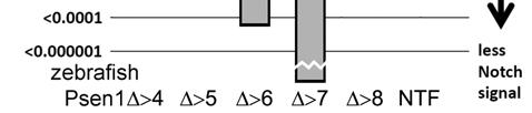

33 32


34 33


35 34


36 35


37 36