Nature Immunology: doi: /ni.3015
|
|
|
- Josephine Wilson
- 5 years ago
- Views:
Transcription

1
2 Supplementary Figure 1 Role of RIP1-RIP3 and PGAM5 in RNA virus induced inflammasome activation. (a) LDH release from LPS-primed BMDMs from wild-type mice (WT), Rip3 -/- or Nlrp3 -/- mice infected with VSV for 6 hours or stimulated with nigericin for 30 min. (b) qpcr analysis of Ifn-β expression in non-primed BMDMs from WT, Rip3 -/- or Nlrp3 -/- mice infected with VSV as indicated doses for 6 hours. (c) Immunoblot analysis of RIP1 expression in BMDMs transfected with sirna against Rip1 for 48 hours. (d) Immunoblot analysis of pro-il-1β or NLRP3 expression in BMDMs transfected with sirna against Rip1 and stimulated with Pam3CSK4 for 6 hours. (e) Immunoblot analysis of PGAM5 in THP-1 cells stably expressing shrna against PGAM5. (f, g) IL-1β secretion from THP-1 cells stably expressing shrna against PGAM5 infected with VSV for 6 hours (f), or stimulated with MSU for 4 hours or nigericin for 30 min (g). (h) Confocal microscopy analysis in LPS-primed BMDMs from Rip3 +/+ or Rip3 -/- mice infected with VSV for 2 hours or stimulated with nigericin for 30 min, and then stained with MitoSOX and DAPI. Scale bar, 50 µm. (i) Confocal microscopy analysis in LPS-primed BMDMs pretreated with Nec-1s (30 µm) for 1 hour and then infected with VSV for 2 hours or stimulated with nigericin for 30 min. After treatment, the cells were stained with MitoSOX and DAPI. Scale bar, 50 µm. NS, P > 0.05 (two-way ANOVA (a,b,f,g). Data are representative of at three independent experiments (c-e, h, i) or are from three independent experiments (with biological duplicates in each) (a, b, f, g; mean and s.e.m.).
3
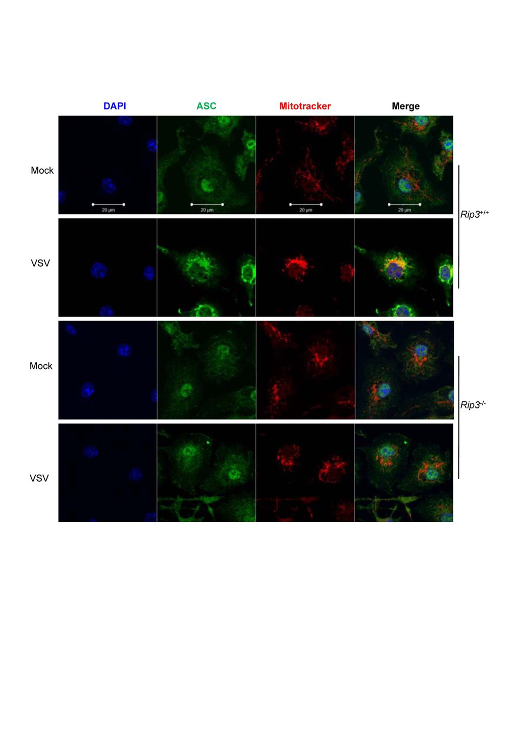
4 Supplementary Figure 2 RIP3 is required for VSV-induced translocation of ASC to mitochondria. Confocal microscopy analysis in LPS-primed BMDMs from Rip3 +/+ or Rip3 -/- mice infected with VSV for 2 hours and then stained with anti-asc antibody, Mitotracker red and DAPI. Data are representative of at three independent experiments.

5
6 Supplementary Figure 3 RIP3 is required for VSV-induced translocation of DRP1 to mitochondria. Confocal microscopy analysis in LPS-primed BMDMs from Rip3 +/+ or Rip3 -/- mice infected with VSV for 2 hours or stimulated with nigericin for 30 min, and then stained with anti-drp1 antibody, Mitotracker and DAPI. Scale bar, 20 µm. Data are representative of at three independent experiments.
7
8 Supplementary Figure 4 RIP1 is required for VSV-induced translocation of DRP1 to mitochondria. Confocal microscopy analysis in LPS-primed BMDMs pretreated with Nec-1s (30 µm) for 1 hour and then were infected with VSV for 2 hours or stimulated with nigericin for 30 min. After treatment, the cells were stained with anti-drp1 antibody, Mitotracker and DAPI. Scale bar, 20 µm. Data are representative of at three independent experiments
9
10 Supplementary Figure 5 MLKL is not required for VSV-induced translocation of DRP1 to mitochondria. Confocal microscopy analysis in LPS-primed BMDMs from Mlkl +/+ or Mlkl -/- mice were infected with VSV for 2 hours or stimulated with nigericin for 30 min, and then stained with anti-drp1 antibody and Mitotracker. Scale bar, 20 µm. Data are representative of at three independent experiments.

11
12 Supplementary Figure 6 Role of DRP1 or FIS1 in mitochondrial damage and inflammasome activation. (a) Immunoblot analysis of BMDMs transfected with sirna against Drp1 for 48 hours. (b) Confocal microscopy analysis of LPS-primed BMDMs transfected with sirna against Drp1 and then infected with VSV for 2 hours or stimulated with nigericin for 30 min and followed by staining with MitoSOX and DAPI. Scale bar, 50 µm. (c) IL-1β secretion from LPS-primed BMDMs transfected with sirna against Drp1 and then stimulated with MSU, poly (A:T) for 4 hours or ATP, nigericin for 30 min. (d) Immunoblot analysis of BMDMs transfected with sirna against Fis1 for 48 hours. (e, f) Confocal microscopy analysis of LPS-primed BMDMs transfected with sirna against Fis1 and infected with VSV for 2 hours or stimulated with nigericin for 30 min and followed by staining with Mitotracker red. Representative pictures (e) or qualification (f) was shown. Scale bar, 50 µm. * P < 0.001, NS P > Data are representative of at three independent experiments (a, b, d, e) or are from three independent experiments (with biological duplicates in each) (c, f; mean and s.e.m.).

13
14 Supplementary Figure 7 Role of RIP3 and DRP1 in CCCP-induced mitochondrial fission and IL-1β production. (a) Immunoblot analysis of DRP1 phosphorylation in LPS-primed BMDMs stimulated with CCCP (10 µm) or infected with VSV for 1 hours. (b) IL-1β secretion from LPS-primed BMDMs from Rip3 +/+ or Rip3 -/- mice were stimulated with CCCP for 5 hours. (c) Confocal microscopy analysis of LPS-primed BMDMs from Rip3 +/+ or Rip3 -/- mice stimulated with CCCP (10 µm) for 2 hours, and then stained with anti-drp1 antibody and Mitotracker. Scale bar, 25 µm. (d) IL-1β secretion from LPS-primed BMDMs transfected with sirna against Drp1 and then stimulated with CCCP (10 µm) for 5 hours. (e, f) Confocal microscopy analysis of LPS-primed BMDMs transfected with sirna against Drp1 and stimulated with CCCP (10 µm) for 2 hours, and followed by staining with Mitotracker red and DAPI. Representative pictures (e) or qualification (f) was shown. Scale bar, 25 µm. NS P > 0.05, * P<0.01 (two-way ANOVA), * P<0.001 (two-way ANOVA). Data are representative of at three independent experiments (a, c, e) or are from three independent experiments (with biological duplicates in each) (b, d, f; mean and s.e.m.).
15
16 Supplementary Figure 8 Role of RNA sensors in VSV- or dsrna-induced activation of the NLRP3 inflammasome. (a, b) IL-1β secretion from LPS-primed BMDMs from wild-type mice, Rig-I -/- or Mda5 -/- mice infected with VSV (a) or transfected with poly (I:C) (b) for 6 hours. (c) qpcr analysis of Ifn-β expression in non-primed BMDMs from wild-type mice, Rig-I -/- or Mda5 -/- mice infected with VSV for 6 hours. (d) Immunoblot analysis of THP-1 cells stably expressing shrna against Rip1 or DHX33. (e) IL-1β secretion from THP-1 cells stably expressing shrna against Rip1 or DHX33 infected with VSV for 6 hours or stimulated with nigericin for 30 min. (f, g) IL-1β secretion from Pam3CSK4-primed BMDMs from Tlr3 +/+ or Tlr3 -/- mice infected with VSV (f) or stimulated with poly (I:C) transfection (g) for 6 hours. NS P > 0.05 (unpaired t-test (a, b, c, g), NS P > 0.05 (two-way ANOVA (f)), * P < 0.01 (two-way ANOVA (e)), ** P < (two-way ANOVA (c)). Data are representative of at three independent experiments (d) or are from three independent experiments (with biological duplicates in each) (a-c, e-g; mean and s.e.m.).
17 Supplementary Table 1. sirna sequences. Target mrna Drp1 (1) Drp1 (2) Rip1 (1) Rip1 (2) Fis1 (1) Fis1 (2) sirna sequences (5' - 3') GGCAAUUGAGCUAGCGUAUAUTT CUAUAAUGCAUGCACUAUUUATT GCCAAAUCUAAGCCAAAUGUATT CGUGACUUUCACAUUAAGAUATT CCUGGUUCGAAGCAAAUACAATT CCUGGUUCGAAGCAAAUACAATT