Lecture 3 MOLECULAR REGULATION OF DEVELOPMENT
|
|
|
- Elijah Hancock
- 5 years ago
- Views:
Transcription
1 Lecture 3 E. M. De Robertis, M.D., Ph.D. August 16, 2016 MOLECULAR REGULATION OF DEVELOPMENT GROWTH FACTOR SIGNALING, HOX GENES, AND THE BODY PLAN
2 Two questions: 1) How is dorsal-ventral (D-V) cell differentiation regulated by morphogen gradients. BMP signal transduction. 2) How is antero-posterior (A-P) pattern regulated by Hox genes? - Colinearity - Activation by retinoic acid. Retinoid receptors. Conserved gene networks control Evolution and Development. Evo-Devo. Fig. 1

3 1) During development groups of inducing cells called organizing centers secrete graded growth factor signals. The concentration gradient of a diffusible morphogen can induce multiple cell differentiation fates at different concentrations. Fig. 2
 embryo. Fig.")
4 Dorsal-Ventral (D-V) patterning can be best studied in the frog (Xenopus) embryo. Fig. 3

5 The best example of a morphogen is the gradient of BMP signaling that controls D-V tissue differentiation. Fig. 4
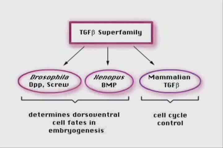
6 A morphogen gradient of BMP activity induces differentiation of mesodermal cell types. BMP signaling is maximal in the ventral side. BMP signaling ventral Lateral plate Somite Notochord dorsal Bone Morphogenetic Proteins are growth factors discovered here at UCLA by Dr. Marshall Urist; BMPs are members of the TGFβ superfamily Fig. 5
7 Genes specifically expressed in the dorsal blastopore lip (Spemann organizer) of the gastrula were cloned. Organizer-specific Genes. Chordin, Noggin Fig. 6

8 Chordin mrna is expressed in Spemann s organizer. Chordin protein is secreted and diffuses in the embryo. Fig. 7
9 Chordin is a BMP antagonist that binds BMP growth factors in the extracellular space, preventing their binding to cell surface receptors. Chordin generates a BMP4 activity gradient at gastrula. Another protein, Noggin, has similar activity. Secreted antagonists diffuse and are used in development to generate morphogen gradients. Chordin inhibits Ventral Dorsal Fig. 8
 activate cell surface receptors called Serine-Threonine kinases. Smads and DNA-binding partners. BMP4 TGFβ I will try to show a movie of this.")
10 Signal transduction: membrane receptors transduce the signal by phosphorylating and activating transcription factors. TGFβ family members (30 different ligands in humans) activate cell surface receptors called Serine-Threonine kinases. Smads and DNA-binding partners. BMP4 TGFβ I will try to show a movie of this. No need to remember any details; it is just to illustrate that activated cell membrane receptors can cause changes in gene expression. Fig. 9
11 Fig. 10


12 The BMP gradient of activity can be visualized in the Xenopus gastrula as a gradient of phosphorylated Smad1 (maximal in the ventral side). Dorsal Blastopore Side view Ventral Transverse section at the level of white arrows Fig. 11


13 Epidermis CNS Ventral Dorsal Mesoderm Endoderm Spemann s Organizer At gastrula, graded BMP4 activity is established by a dorsal source of Chordin and Noggin (two BMP antagonists secreted by the dorsal organizing center) and a ventral source of BMP4. All germ layers are affected coordinately; is there one or multiple gradients? Fig. 12

, which is present in all vertebrate embryos. Plouhinec et al.")
14 Chordin forms a gradient Chordin antibody staining shows long-range diffusion of a gradient of endogenous Chordin protein in the narrow space between ectoderm and mesoderm (called Brachet s cleft in Xenopus), which is present in all vertebrate embryos. Plouhinec et al., PNAS 2013 Fig. 13
15 The morphogen gradient induces different tissues in mesoderm and ectoderm (because the DNA-binding partners are different in each layer). BMP signaling Mesoderm differentiation BMP signaling Ectoderm differentiation Lateral plate Somite Notochord Epidermis Neural crest CNS Fig. 14
. Both mechanisms are used in development. This is how organizing centers work in embryonic induction.")
16 Conclusion; a morphogen gradient can be generated by a source of growth factor (such as BMP) or by a localized source of inhibitor (such as Chordin). Both mechanisms are used in development. This is how organizing centers work in embryonic induction. Fig. 15
17 Cell-cell communication is controlled by surprisingly few signal transduction pathways: 1) TGFβ/BMP Serine/Threonine kinase receptors 2) Receptor Tyrosine kinases such as FGF, EGF, IGF, Insulin 3) G protein-coupled receptors (7-transmembrane receptors) 4) Wnts 5) Sonic Hedgehog 6) Notch 7) Nuclear hormone receptors Only a few signaling pathways pattern the embryo, but there are hundreds of differentiated cell types in the human body. The same signals can trigger different types of cell differentiation responses in cells of different developmental history (because of different combinations of DNA-binding partners). Each of these signal transduction pathways has been liked to human cancer. We now turn to Hox genes. Fig. 16
18 A-P patterning outline: 2a) Hox genes: colinearity between the gene order in genomic DNA and the body plan 2b) Hox genes and Retinoic acid 2c) Hox genes in Evolution and Development (Evo-Devo) Fig. 17
19 2a) Hox genes. Homeotic transformations change one body region into the likeness of another Homeotic transformations in humans. A cervical vertebra transformed into a thoracic one with ribs (0.5%). 1-3% of humans have a lumbar 13 th rib. Fig. 18
20 Homeotic Mutations the Homeobox story Fig. 19


21 Edward B. Lewis Walter J. Gehring Homeotic genes specify body segment identity in Drosophila. Edward Lewis predicted Hox genes would be duplicated. Walter Gehring found that Hox genes had a highly-conserved segment. Fig. 20
22 In 1984 in a collaboration with Walter Gehring we cloned the first Hox gene from a vertebrate. Shown here is Southern blot from a Xenopus phage λ clone DNA band that cross-hybridized at low stringency with three different Drosophila homeobox probes. Carrasco, McGinnis, Gehring and De Robertis, Cell 1984 Fig. 21
23 Homeobox refers to nucleic acid (180 nucleotides). Homeodomain refers to protein (60 aa). The homeodomain is a 60 aa helixturn-helix DNA-binding domain Define Hox, homeobox that is very conserved during evolution. It fits into the major groove of the DNA. The term homeobox is reserved for the nucleic acid sequences that encode homeodomains. Since they are highly conserved we were able to clone them by low-stringency hybridization across species. Fig. 22

24 Conserved Hox gene complexes are similarly arranged in the genome between Drosophila and mammals (from De Robertis et al., Scientific American, 1990) Fig. 23
25 Humans have four Hox complexes, containing 39 Hox genes. Hox complexes arose from two whole-genome duplications of an ancestral complex consisting of 13 genes (13 x 4=52; therefore some Hox paralogues were lost in evolution). They display colinearity: a) Spatial colinearity: the more anteriorly expressed genes are in one end, the more posterior ones at the other end of the gene complex. b) Temporal colinearity: genes on one end of the complex are expressed first, those on the other (posterior) end are turned on last. c) Anterior Hox genes are activated sequentially by retinoic acid. Fig. 24
26 Extensive conservations between Drosophila and the four human Hox complexes. Colinearity. High RA response Low RA response De Robertis, Cell 132, (2008) Fig. 25
27 Spatial and temporal colinearity: order of Hox genes in DNA follows the A-P axis, anterior genes expressed first. Why have Hox genes stayed together in gene complexes? Perhaps due to common regulation of gene expression. Fig. 26

28 Hox knockouts in mice cause homeotic transformations, in this case an extra rib in the lumbar region (HoxC-8 mutant). Treatment with retinoic acid can also cause lumbar ribs. Your patient this week has 13 ribs. We next turn to retinoic acid teratogenesis. Fig. 27
29 2b) Retinoic acid activates HOX genes sequentially in cultured human teratocarcinoma cells mrna amount Fig. 28
30 Retinoic acid receptor (RAR) is a nuclear protein that binds to DNA and to retinoic acid. A very different mode of action from the cell surface receptors discussed above. How do Retinoic acid receptors work? RAR binds to DNA sequences called RA response elements (RAREs) and its transcriptional activity is regulated by a ligand-binding domain. DBD LBD Dimerization ligand coactivator Fig. 29
31 Retinoic acid receptor is a DNA-binding protein that works as a ligandactivated transcription factor. Many hydrophobic hormone receptors important in medicine work in this way. Fig. 30
32 Hox complexes have a retinoic acid receptor response element (RARE) in the DNA before paralogue 1. This DNA enhancer element controls expression of many genes in the complex. In retinoic acid teratogenesis, Hox gene expression borders move into more anterior regions. RARE Fig. 31
33 RA activates Hox gene expression ectopically in more anterior regions, causing RA embryopathy. Figure Patterns of Hox gene expression in the hindbrain and the pattern of neural crest cell migration into the pharyngeal arches. Hox genes are expressed in overlapping patterns starting at specific rhombomere boundaries. These genes confer positional value along the anterior posterior axis of the hindbrain and determine the identity of the rhombomeres. Paralogous genes have the same expression borders. Retinoic acid causes the expression of Hox genes in pharyngeal arch P1 and midbrain where they are never expressed, with devastating consequences. P1 gives rise to a maxillary and a mandibular branch, explaining why retinoic acid causes cleft palate and micrognathia. Ectopic Hox gene expression in the midbrain could cause the oculomotor changes seen in this week s patient. Fig. 32

34 2c) The common ancestor Urbilateria used Hox genes and Chordin/BMP to pattern the embryo. 30 of the 35 animal phyla are bilaterans. BMP Hox Chd Ur = primeval Fig. 33
35 Evo-Devo: Urbilateria had a Hox gene complex of at least 7 genes. The Chordin/BMP D-V gene system was also conserved. Evolution used conserved gene systems to develop new morphologies. Developmental control genes placed evolutionary constraints on the types of animal shapes that evolved by Natural Selection. Variation had to be compatible with the ancestral developmental gene networks that determine body shape. BMP Hox Chd High RA response Low RA response Fig. 34

36 Bird s eye overview Langman s Medical Embryology Fig. 35