FLUORESCENCE. Matyas Molnar and Dirk Pacholsky
|
|
|
- Emily McLaughlin
- 5 years ago
- Views:
Transcription

1 FLUORESCENCE Matyas Molnar and Dirk Pacholsky 1
2 Information This lecture contains images and information from the following internet homepages

3 3
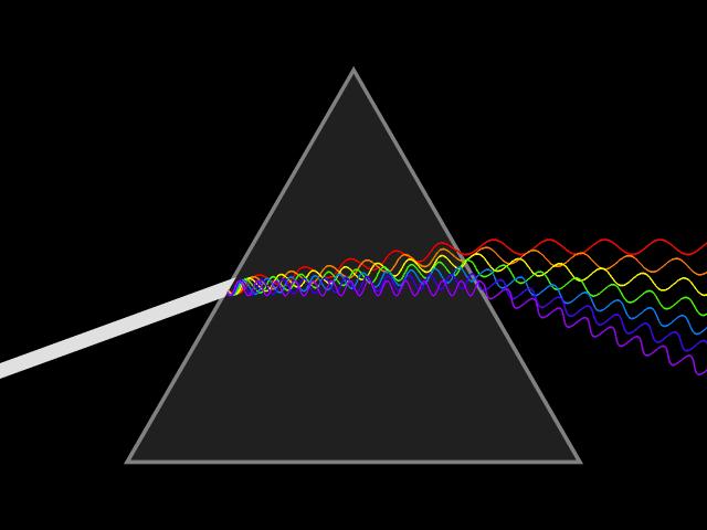
4 Light phenomenon 4


5 Wave or particle?... Wave! 5

6 Wave or particle?... Wave! 6

 light is 1.")

7 Wave or particle?... Wave! λ E Light travels in vacuum C = km/sec Seems to slows down in denser matter. Refractive index n = C/v (v= speed in matter) light is 1.5 x slower at n=1.5 Light changes direction between different dense materials : shorter λ refract more than longer λ Optic lenses, effects in sample... shorter wavelength (blue) has higher energy 7

8 Wave or particle?... Particle! Principle of fluorescence Light behaves sometimes as quantized energy pockets, light has particle behavior. 8
9 Fluorescence basics 9


10 Fluorescence basics Definition: Fluorescence is the emission of light by a substance that has absorbed light or other electromagnetic radiation. Usually the emitted light has a longer wavelength, and therefore lower energy than the absorbed radiation. Emission of light happens in time scale of nano second so to speak immediately Compared to Phosphorescence: - specific type of photoluminescence related to fluorescence. Unlike fluorescence, a phosphorescent material does not immediately emit light. Absorbed radiation may be re-emitted for up to several hours after original excitation. (wikipedia ;) ) 10

11 Fluorescence basics s : 1 femto sec s : 10 fs s : 100 fs s : 1 pico sec s : 10 ps s : 100 ps 10-9 s : 1 nano sec 10-8 s : 10 ns 10-7 s : 100 ns 10-6 s : 1 micro sec 10-5 s : 10 µs 10-4 s :100 µs 10-3 s : 1 milli sec s : 10 ms 10-1 s :100 ms 10 0 s : 1 second 11


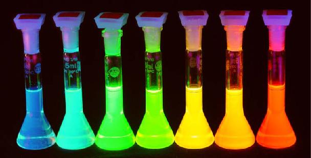
12 Fluorescence basics Examples of fluorescent probes Principle of fluorescence Principle of fluorescent microscope Excitation-Emission filter cube 12
13 Fluorescence: The spectra Normalized Intensity Stokes Shift Alexa 488 QY 0.92 Alexa 555 QY Alexa 488 X axis: λ in nm Y-axis: Intensity or probability of event that A) fluorophore absorbs the light for excitation (dashed line) and B) Fluorophore emits ligth (full line) Ex peak at 100% em peak at 100 %, ex 20% em 20 %, same range of emission Stokes shift: gap between ex-peak and em peak = (loss of energy, dissipation ) important for separation of excitation and emission light in microscope etc Other important features of fluorophores: Extinction coefficient: absorbtion efficiency of a photon at particular wavelength Quantum yield: proportion of photons emitted at λ em to those absorbed at λ em nm 13
14 Stokes shift Stokes shift is the energy difference between the lowest energy peak of absorbance and the highest energy of emission. The Stokes shift is an extra for observing fluorescence; without it there would be (almost) no way to distinguish between excitation and emitted light Probes with varying Stokes shifts are very useful for multicolor applications Fluorescnece Intensity Fluorescein molecule Stokes Shift is 25 nm 495 nm 520 nm Excitation Emission Wavelength 14
15 Parameters for fluorescence efficiency Extinction Coefficient ε refers to a single wavelength (usually the absorption maximum) ε measure of how efficiently a substance absorbs light of a certain wavelength Quantum Yield Q f is a measure of the integrated photon emission over the fluorophore spectral band The ratio of photons emitted to photons absorbed 15
 Reduce excitation intensity Use antifade reagents (not compatible with viable cells)")
16 Photobleaching, quenching Defined as the irreversible destruction of an excited fluorophore Methods for countering photobleaching Illuminate for shorter times Use high magnification, high NA objective Use wide emission filters more signal to capture (may create problems with multiple probes) Reduce excitation intensity Use antifade reagents (not compatible with viable cells) 16

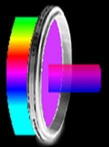
17 Filter and dichroic mirror 17


18 Filter and dichroic mirror 18
19 Dealing with fluorescence Cell sample Cell images merged RGB image 19
20 Dealing with fluorescence ex Cell sample Cell image* Excitation 350 nm excitates Blue and Green, using BP filter collects them both. *Remember: the camera is color blind. You decide with your choice of filter what it will see. 20
21 Dealing with fluorescence ex Cell sample Cell image* Excitation 350 nm excitates Blue and Green, using BP filter collects only the blue. *Remember: the camera is color blind. You decide with your choice of filter what it will see. 21
22 Dealing with fluorescence ex Cell sample Cell image* Excitation 480 nm excitates Green and Red, using BP filter collects only the green. *Remember: the camera is color blind. You decide with your choice of filter what it will see. 22
 were transduced with CellLight Golgi-GFP, CellLight Mitochondria-RFP Hoechst 33342 Imaging was performed on live cells using a DeltaVision Core microscope + standard DAPI/FITC/TRITC")
23 Specific Organelle Probes Gibco human aortic smooth muscle cells (HASMC, Cat. No. C0075C) were transduced with CellLight Golgi-GFP, CellLight Mitochondria-RFP Hoechst Imaging was performed on live cells using a DeltaVision Core microscope + standard DAPI/FITC/TRITC filter sets. Gallery/Image-Detail html 23
 Peak emission at 509 nm contains a p-hydroxybenzylidene-imidazolone chromophore generated by oxidation of the Ser- Tyr-Gly at positions 65-67 of the primary sequence Very stable Major application")
24 Fluorescent protein GFP - Green Fluorescent Protein GFP is from the chemiluminescent jellyfish Aequorea victoria excitation maxima at 395 and 470 nm (quantum efficiency is 0.8) Peak emission at 509 nm contains a p-hydroxybenzylidene-imidazolone chromophore generated by oxidation of the Ser- Tyr-Gly at positions of the primary sequence Very stable Major application is as a reporter gene for assay of promoter activity requires no added substrates Now in the enhanced form of egfp, eyfp, ecfp
25 Important points Fluorescence is the primary information source for confocal microscopes and flow cytometry equipment Fluorescence emission is longer than the exciting wavelength Dye molecules must be close to, but below saturation levels for optimum emission Fluorescence probes must be appropriate for the excitation source and the sample of interest Correct optical filters must be used for multiple color fluorescence emission 25

26 THANKS FOR YOUR ATTENTION! 26