PREPARED FOR: U.S. Army Medical Research and Materiel Command Fort Detrick, Maryland
|
|
|
- Isabel Jefferson
- 5 years ago
- Views:
Transcription
1 AD Award Number: W81XWH TITLE: Spectroscopic Photoacoustic Tomography of Prostate Cancer PRINCIPAL INVESTIGATOR: Xueding Wang CONTRACTING ORGANIZATION: University Of Michigan Ann Arbor, MI REPORT DATE: March 2008 TYPE OF REPORT: Annual PREPARED FOR: U.S. Army Medical Research and Materiel Command Fort Detrick, Maryland DISTRIBUTION STATEMENT: Approved for Public Release; Distribution Unlimited The views, opinions and/or findings contained in this report are those of the author(s) and should not be construed as an official Department of the Army position, policy or decision unless so designated by other documentation.
2 REPORT DOCUMENTATION PAGE Form Approved OMB No Public reporting burden for this collection of information is estimated to average 1 hour per response, including the time for reviewing instructions, searching existing data sources, gathering and maintaining the data needed, and completing and reviewing this collection of information. Send comments regarding this burden estimate or any other aspect of this collection of information, including suggestions for reducing this burden to Department of Defense, Washington Headquarters Services, Directorate for Information Operations and Reports ( ), 1215 Jefferson Davis Highway, Suite 1204, Arlington, VA Respondents should be aware that notwithstanding any other provision of law, no person shall be subject to any penalty for failing to comply with a collection of information if it does not display a currently valid OMB control number. PLEASE DO NOT RETURN YOUR FORM TO THE ABOVE ADDRESS. 1. REPORT DATE 2. REPORT TYPE Annual 3. DATES COVERED 15 Feb Feb TITLE AND SUBTITLE 5a. CONTRACT NUMBER Spectroscopic Photoacoustic Tomography of Prostate Cancer 5b. GRANT NUMBER W81XWH c. PROGRAM ELEMENT NUMBER 6. AUTHOR(S) 5d. PROJECT NUMBER Xueding Wang 5e. TASK NUMBER 5f. WORK UNIT NUMBER xdwang@umich.edu 7. PERFORMING ORGANIZATION NAME(S) AND ADDRESS(ES) 8. PERFORMING ORGANIZATION REPORT NUMBER University Of Michigan Ann Arbor, MI SPONSORING / MONITORING AGENCY NAME(S) AND ADDRESS(ES) 10. SPONSOR/MONITOR S ACRONYM(S) U.S. Army Medical Research and Materiel Command Fort Detrick, Maryland SPONSOR/MONITOR S REPORT NUMBER(S) 12. DISTRIBUTION / AVAILABILITY STATEMENT Approved for Public Release; Distribution Unlimited 13. SUPPLEMENTARY NOTES 14. ABSTRACT Major research accomplishment during the first year of this research project includes 1) fabrication of a fast speed noninvasive PAT prostate imaging system by using a stand-alone commercial ultrasound system; 2) testing the performance of this imaging system through the experiments on phantoms and ex vivo canine prostates; and 3) fabrication and optimization of a specially designed PVDF array transducer for high sensitive broad bandwidth detection of photoacoustic signals. 15. SUBJECT TERMS Prostate cancer diagnosis, imaging system, photoacoustic tomography, ultrasonography 16. SECURITY CLASSIFICATION OF: 17. LIMITATION OF ABSTRACT a. REPORT U b. ABSTRACT U 18. NUMBER OF PAGES c. THIS PAGE U UU 13 19a. NAME OF RESPONSIBLE PERSON USAMRMC 19b. TELEPHONE NUMBER (include area code) Standard Form 298 (Rev. 8-98) Prescribed by ANSI Std. Z39.18
3 TABLE OF CONTENTS Page INTRODUCTION...1 BODY...1 KEY RESEARCH ACCOMPLISHMENTS...10 REPORTABLE OUTCOMES...10 CONCLUSION...10
4 INTRODUCTION The ultimate goal of this research is to adapt spectroscopic photoacoustic tomography (SPAT) to prostate cancer imaging and evaluation, and fill the existing void in the early diagnosis, localization and accurate grading of prostate cancer, as well as in future treatment planning and therapeutic monitoring. As an essential step toward the ultimate goal, the objective of this funded project is to develop a laboratory prototype photoacoustic tomography (PAT) system that enables simultaneous photoacoustic and ultrasound imaging of prostates. BODY High speed PAT prostate imaging system The specific aims of this two-year project include: 1) design and develop a noninvasive PAT system with near-infrared (NIR) laser light and an ultrasonic transducer array for imaging prostates. This system is capable of both functional photoacoustic imaging and ultrasound imaging; 2) examine and optimize this imaging system through experiments on phantom samples; 3) validate the performance of this system for photoacoustic and ultrasound imaging of normal canine prostates; and 4) explore the sensitivity of this system in detection regional hemodynamic changes in canine prostates, including blood oxygen saturation and blood volume. During the first year, we were focused on the accomplishment of aim 1 and aim 2, i.e. the fabrication and testing of a fast speed noninvasive PAT prostate imaging system. Unlike any former pioneering studies, the PAT system fabricated by our group is integrated with a stand-alone commercial ultrasound (US) without affecting the original US imaging functions. This innovative design has very unique advantages. First, this arrangement makes it convenient to take advantage of state-of-the-art US technologies. For the first time, high quality US and PAT images of a prostate can be acquired simultaneously, with one shown in real-time by the US unit and the other shown rapidly by a connected PC. Second, with this arrangement, US and PAT of a prostate are scanned with the same acquisition system at the same time, which may not only benefit the image co-registration but also help improve the reconstruction and interpretation of PAT images by using a priori morphological and tissue acoustic information. Third, this arrangement enables highly accelerated imaging speed by acquiring signals from 64 channels of the US system simultaneously. The high temporal resolution, which is essential for accurate quantified imaging of metabolic and physiological parameters in prostate tissues, makes the designed PAT system worthy of preclinical and clinical studies. We expect that, by presenting both optical and mechanical contrasts and comprehensive diagnostic information, this dual-modality system may lead to substantially improved sensitivity and specificity in imaging prostate cancer in comparison with conventional transrectal US. Zonare ultrasound In the past year, we have purchased a z.one ultrasound system from Zonare Medical Systems, Inc. Unlike most commercial US units, the z.one provides a good research platform for PAT studies by its ability to synchronize with the laser, access real-time RF signals 1

5 channel-by-channel, and store a frame of RF data for signal averaging. We are one of the research collaborators of Zonare. The strong support from them has significantly contribute to this research. Working together with Larry Mo, Ph.D. and Glenn McLaughlin, Ph.D. at Zonare Medical Systems, Inc, we have achieved high speed photoacoustic image acquisition by using the z.one and a customized L10-5 probe. L10-5 is a 128-element linear array working at 5 MHz central frequency with 0.3-mm pitch and 6-mm height. By acquiring signals simultaneously with 64 independent channels of z.one, which are multiplexed to form 128 element array apertures, the PAT system powered by the z.one has an imaging speed 64 times faster than conventional PAT systems employing single transducer. The z.one also enables on-line signal processing with adjustable parameters such as TGC and demodulation frequency that can be optimized for PAT imaging mode. The cine buffer of the z.one is sufficient to store up to 1000 frames when each frame has an imaging depth of 6 cm, a beam number of 64 and a sampling frequency of 5 MHz. The channel IQ data can be downloaded from the cine buffer to a pc rapidly for off-line signal processing and image reconstruction. For photoacoustic imaging using the z.one, the trigger-out signal from the z.one is sent to the laser (Nd:YAG pumped OPO, Opotek) for accurate synchronization. Currently, the start and the stop of signal acquisition are controlled manually. We are still working with Zonare to further optimize the software control of the z.one and make the data acquisition on PAT model faster and more convenient. Z.One L10-5 probe Vessel Laser beam Fig. 1 Schematic of the setup for PAT of a vessel by using the z.one ultrasound system. To examine the performance of PAT by using the z.one and the L10-5 probe, experiments on phantoms and ex vivo canine prostates have been conducted. The first experiment was to examine the spatial resolution which can be achieved by the z.one working with the L10-5 probe. The schematic of the setup is in Fig. 1, where a laboratory coordinate system helps to indicate the orientations. Because the distribution of the array was along the x-axis and the orientation of the vessel was along the y-axis, the vessel formed a point target in the 2D image taken along the x-z plane. The phantom vessel was optically transparent microtubing (50 micron inner diameter) filled with fresh canine blood. The vessel was illuminated by the laser beam with an input energy density of 20 mj/cm 2 at 532-nm wavelength which was lower than the ANSI safety limit. The acquired photoacoustic signals by the first and the second 64 channels are shown in Fig. 2(A). The signals were generated by single laser pulse without averaging. Therefore, considering the 10 Hz repetition rate of the laser, the maximal frame rate of the current system is 10/2=5 Hz when the signal-to-noise ration (SNR) is Y X Z 2
 shows an example of the signal acquired by single array element, where a SNR larger than 20 db can be observed.")
 and the vertical direction (i.e. x-axis) which will enable us to find the spatial resolution achieved in this experiment.")
, we can achieve axial resolution of 0.")
6 sufficient. Fig. 2(B) shows an example of the signal acquired by single array element, where a SNR larger than 20 db can be observed. By using a back-projection algorithm, 2D PAT image of the vessel is reconstructed, as shown in Fig. 2(C). With this image, we can then study the amplitude along the horizontal direction (i.e. z-axis) and the vertical direction (i.e. x-axis) which will enable us to find the spatial resolution achieved in this experiment. As shown in Figs. 2(D) and (E), for PAT using the z.one system with the L10-5 probe and when the target is 34 mm from the surface of the probe (i.e. f number is about 1), we can achieve axial resolution of 0.63 mm and lateral resolution of 1.8 mm. A 800 IQ data Amplitude (a.u.) Data point B C 3
7 Normalized amplitude Axial Resolution 0.63 mm Relative amplitude Lateral resolution 1.8 mm D Position (mm) E Position (mm) Fig. 2 (A) Photoacoustic signals from the vessel acquired by the first and the second 64 channels of the z.one ultrasound. (B) An example a-line photoacoustic signal acquired by single array element (the first element in the second 64 channels). (C) Reconstructed 2D photoacoustic image of the vessel. (D) Normalized amplitude in the image through the center of the vessel along the horizontal direction (i.e. z-axis). The measured FWHM of 0.63 mm indicates the axial resolution achieved in this experiment. (E) Normalized amplitude in the image through the center of the vessel along the vertical direction (i.e. x-axis). The measured FWHM of 1.8 mm indicates the lateral resolution achieved in this experiment. In the second experiment to examine the image quality can be achieved by this system, a porcine gel phantom containing two vessels placed in the 2D imaging plane were fabricated. The photograph of this phantom is in Fig. 3(D). Fig. 4(A) shows the imaging geometry, where the light illumination is along the z-axis and the imaged cross section is in the x-y plane. Without moving the sample or the probe, 2D image of the sample can be taken through a single scan. In this case, the signals are acquired with a limited view angle. For better receiving of photoacoustic signals through all the directions, the probe or the sample can be rotated around the center of the imaging plane. As indicated in the literatures, a full-view detection of photoacoustic signals will lead to much improve image quality. In this experiment, to realize full-view detection, the sample is rotated a π/4 angle around its center after each scan. Therefore, to cover 2π view angle, 8 scans evenly distributed around the sample are performed. As an example, Fig. 3(A) shows the 2D image of this sample taken by single scan. Although imaged with limited view angle, major part of the vessels is still presented clearly, especially when the orientations of the vessels are nearly perpendicular to the ultrasound beam path. Fig. 3(B) is the image taken with 8 scans, where the vessels are presented with much improved consistency. For both Figs. 3(A) and (B), the back-projection algorithm is used for image reconstruction. This experiment indicates that, when performed with a limited view angle, the image quality can be achieved by this PAT system is spatially dependent. For better consistency in describing the anatomical information, large solid angle for signal detection is preferred. Fig. 3(C) presents the intensity profile along the yellow dash line in the image in Fig. 3(B). The FWHM indicates that the spatial resolution achieved in this experiment is 0.65 mm which matches the axial resolution observed in the first experiment
 2D PAT image of two vessels acquired with the probe at one position (i.e. single scan with limited view angle without rotating the sample).")
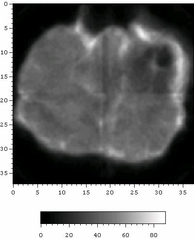
8 A Relative amplitude (a.u.) mm Position (mm) C D Figure 3. (A) 2D PAT image of two vessels acquired with the probe at one position (i.e. single scan with limited view angle without rotating the sample). (B) 2D PAT image of two vessels acquired with the probe at 8 positions around the sample (i.e. full view detection by rotating the sample). (C) The intensity profile along the yellow dash line in the image in (B). (D) Photograph of the sample. The third experiment was performed on ex vivo canine prostates. Fresh prostates harvested from sacrificed dogs were embedded in gel phantoms and imaged using our PAT system working at 720 nm. After the first set of images of the prostate were taken, we induced a lesion in the prostate by injecting 0.1 ml fresh canine blood in the target position using a 20G1 1/2 needle. The distance from the lesion to the surface of the prostate illuminated by the laser beam is 5-10 mm. After the lesion was induced, the second set of images of the same cross section in the prostate was taken. Fig. 4(C) and (D) show gray scale US images of B 5
 and (H) where US compounding images present the anatomical structure of the prostate more clearly than those acquired through signal scan, we still cannot see any changes as a result of the")
 and (F) are PAT images of the target cross section in the prostate before and after the generation of the lesion, where the color bars in (E) and (F) are the same.")
9 the target cross section in the prostate before and after the generation of the lesion, where we cannot see any obvious changes in the images caused by lesion generation. Even in Figs. 4(G) and (H) where US compounding images present the anatomical structure of the prostate more clearly than those acquired through signal scan, we still cannot see any changes as a result of the lesion generation due to the limited sensitivity of US to the changes in blood volume. Fig. 4(E) and (F) are PAT images of the target cross section in the prostate before and after the generation of the lesion, where the color bars in (E) and (F) are the same. Due to the limited sensitivity of the L10-5 probe, the anatomical structure of the prostate cannot be seen in the PAT images. However, due to the strong optical absorption of blood, the lesion can be recognized clearly in Fig. 4(F). The lesion has been presented more successfully in Fig. 4(J) which was acquired with full view scanning angle (i.e. scanned at 8 positions around the sample for better image reconstruction). This experiment has proven the high sensitivity of PAT to tissue hemodynamic changes which are believed to play significant roles in prostate cancer generation and progression. X Laser beam Y Z Sample Linear probe Inclusion (vessels or prostate) A B C D 6
10 E F G H 7
11 I J Figure 4. (A) Photoacoustic imaging geometry. (B) Photography of the imaged cross section in the canine prostate, where the induced lesion is close to the center of the prostate. (C) and (D) Gray scale US images of the target cross section in the prostate before and after the generation of the lesion. These two images were acquired with the probe at one position. (E) and (F) PAT images of the target cross section in the prostate before and after the generation of the lesion. These two images were acquired with the probe at one position (i.e. single scan with limited view angle). (G) and (H) Compounding US images of the target cross section before and after the generation of the lesion. For spatial compounding, 8 images taken along 8 positions around the target cross section were acquired and added incoherently. (I) and (J) PAT images of the target cross section in the prostate before and after the generation of the lesion. These two images were acquired with the probe at 8 positions around the sample (i.e. full view detection by rotating the sample). Specially designed array transducers In the past year, the PAT system has been developed and tested by using the Zonare L10-5 probe which is an ordinary transducer for ultrasonography. Unlike ultrasound imaging, PAT needs to detect laser generated ultrasound signals which are much weaker than the signals for conventional ultrasonography. Therefore, the sensitivity of the probe is one of the key parameters determining the image quality in PAT experiments. Besides sensitivity, the receiving bandwidth of the probe is another parameter which also significantly affects the performance of PAT. The ordinary transducer for ultrasonography cannot satisfy the requirements of PAT. As a major part of the system development, we are fabricating and testing a specially designed PVDF array transducer working with the researchers at the NIH Resource on Medical Ultrasonic Transducer Technology at University of Southern California. 8

12 Unlike ordinary PZT transducers for ultrasonography, this PVDF array will enable both high receiving sensitivity and broad receiving bandwidth. USC investigators have successfully built and tested one 128-channel receive-only array. This array (see Fig. 5) was built using a 100 µm thick layer of co-polymer P(VDF-TrFE) as the active material and a custom designed flexible circuit and printed circuit board connector for interconnect. The array has a pitch of 0.3 mm and an elevation height of 10 mm. With a concave surface, the array is elevationally focused at 3 cm which is the target imaging depth for in vivo Figure 5. Specially designed PVDF array for PAT. imaging of prostate cancer. As shown in Fig. 6, a representative array element s two-way response was centered at 8 MHz with 80% -6dB bandwidth. According to our measurements, this array enables a very broad receiving bandwidth of 125% which will significantly benefit photoacoustic imaging by enabling satisfactory spatial resolution. An additional two arrays are currently near completion. One of these arrays incorporates a 50 µm thick P(VDF-TrFE) layer and will be used to test our theory that an improved electrical impedance match to the receive preamplifiers will result in a larger SNR. A B Figure 5. (A) Pulse-echo waveform for single element of the PVDF array. (B) Frequency spectrum of the pulse-echo waveform. USC investigators are also currently fabricating a 16-channel preamplifier test-board. This board will be used to evaluate several commercial preamplifiers which are candidates for the 128-channel design. The goal is to provide the highest possible SNR and bandwidth. After the test-board is evaluated, the appropriate single channel preamplifier circuit design will be used in the layout of the 128-channel version to be delivered by June This 9
13 compatible 128-channel receive preamplifier board will be used to connect the PVDF array to the z.one ultrasound system for image generation. With a fixed gain of 20 db and electrical impedance matching, this preamplifier board will further enhance the sensitivity for PAT of prostate cancer. KEY RESEARCH ACCOMPLISHMENTS 1) Fabricate a fast speed noninvasive PAT prostate imaging system by using a stand-alone commercial ultrasound system. 2) Test the performance of this imaging system through the experiments on phantoms and ex vivo canine prostates. 3) Fabricate and optimize a specially designed PVDF array transducer for high sensitive broad bandwidth detection of photoacoustic signals. REPORTABLE OUTCOMES 1) The presentation entitled Photoacoustic tomography of human peripheral joints, given at the SPIE Photonics West Conference, BiOS 2008: International Biomedical Optics Symposium was supported in part by this DoD grant. 2) We are also preparing a paper describing the performance of this fast-speed PAT system for prostate cancer imaging which will be submitted to 2008 IEEE International Ultrasonics Symposium. CONCLUSION During the first year, we were focused on the fabrication and testing of a fast speed noninvasive PAT prostate imaging system. The performance of this system has been examined through a series of experiments on phantom and ex vivo tissues. We are still working for the improvement of this imaging system by optimizing the SNR and receiving sensitivity of a specially designed PVDF array and a compatible preamplifier board. After that, the system will be ready for proposed experiments on canine prostates in vivo. 10