Spectral-karyotyping- SKY
|
|
|
- Louisa Conley
- 5 years ago
- Views:
Transcription

1 Spectral-karyotyping- SKY
2 Content: 1) Introduction 2) Technical tips: 3) Frequently Asked questions 4) Spectral-karytoyping protocol
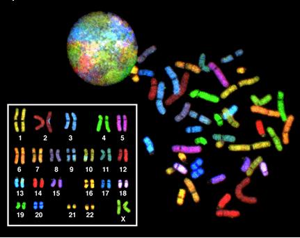
3 1) Introduction SKY is a powerful technique that improves karyotype analysis by identifying chromosomal aberration not previously detected by G-banding alone. Traditional karyotyping, although generally informative, is limited in its ability to detect cryptic translocations, marker chromosomes and to resolve complex karyotypes. While FISH provide accurate means to locate specific aberrations this technique is hard to use as a screening tool. Spectral karyotyping on the other hand, which allows the simultaneous visualization of all human chromosomes in different colors, is a powerful tool to identify subtle chromosomal rearrangements, marker chromosomes, complex karyotypes and HSR and DMs. The detection of these chromosomal aberrations is of great importance for precise diagnosis, risk assessment and genetic counseling either in clinical cytogenetics or in cancer analysis.
4 2) Technical tips: 2.1 Samples preparation One of the main reasons for SKY to fail is the quality of the samples. For optimal SKY results, metaphases should be well spread and free from extra nuclear cytoplasm. Cytoplasm is seen as a gray area surrounding the nucleus or the metaphase. Test specimens should be evaluated using phase-contrast microscopy prior to hybridization to determine if they are optimum for FISH and SKY. Suggested guidelines for slides for FISH/SKY are indicated below: Cells should be evenly distributed across the slide. Areas of cell clumping indicate poor slide preparation or sample preparation. Fewer cells indicate the cell concentration is too low for optimum enumeration. Nuclei and metaphase chromosomes should not be phase bright. Phase bright cells indicate slide preparation conditions were not optimum. Always optimize slide preparation conditions using a good quality lymphocyte preparation prior to preparing the slides. There should be no visible cytoplasm surrounding nuclei and metaphases. Cytoplasm can preclude hybridization and lead to inefficient hybridization and inaccurate results Cell Spreading If the nuclei and metaphases in the samples are surrounded by cytoplasm, there are a number of techniques that may help you clean it up. The main reason for cytoplasm contamination is failure of the cells to spread properly. Spreading of the sample is dependent on the temperature and humidity in the laboratory and the condition of the fixative of the sample at the exact day of performing the spreading. For optimal spreading the temperature should be 24 C, the humidity between 48-52% and the fixative freshly made. If the temperature and the humidity are not optimal the correct condition can be simulated by filling a sink with hot water (60 C) and placing a rack over the sink. Place the slides on the rack and drop the sample on the slides from a distance of 30cm. Allow the slides to completely dry before removing them.
5 The control of the spreading is mainly due to the action of the evaporating fixative. When conditions are correct, the drop of fixative will spread across the slide and reach a maximum spread. The solvent will then move back towards the sample slightly, and then evaporate. To improve cell spreading, drop the sample in the normal way watch the fixative and once it has reach the maximum spread drop another drop of fresh fixative into the sample and allow it to dry. The fixative should be made from high grade reagents. We recommend to use Methanol and Glacial acetic acid from Merck (cat# and , respectively) The control of spreading is a function of the evaporating fixative. Check that you are using the correct fixative of 3 parts methanol, 1 part glacial acetic acid (Carnoy s fixative). If you have doubt about the condition of the fixative, centrifuge the sample and replace the fixative before preparing the slides. To improve the results the last fixative can be made with 3 part of methanol and 2 parts of glacial acetic acid. The condition of the microscope slides is also critical for adequate spreading. If it is not clean then the fixative will not move across its surface easily, and the cells will not spread well. It is recommended that the slides will be purchase preplanned for example Super frost slides from Menzel Glaser or from BDH. If purchasing of precleaned slides is not possible, then cleaning in 80% methanol/20% concentrated HCL mixture for 24 hours and washing in tap water for 2 minutes. Slides can be stored in 70% methanol or in fixative at -20 C until use. Before slides are required remove them from the cold methanol or the fixative, wiped with a tissue and spot the cells on the cold slides. If slides are already spotted then a quick incubation in 70% glacial acid for 2 minutes and dehydration in ethanol series can help.
6 2.1.2 Tips for optimizing the slide preparation conditions: The most effective method for obtaining good clean sample is the adjustment of the hypotonic treatment. It is possible to increase the incubation time in the hypotonic solution, trying a different concentration salt solution or using a different solution 1M Sodium Citrate instead of 0.075M KCL. Another critical step in the sample process is the initial fixation. After the hypotonic step centrifuge the cell and removes the upper solution, mix the sample and add gently the first drop of fixative, allow it to run down the side of the tube and mix vigorously with your hands or with a vortex. Add few more drops in the same way and then add the large volume 5-8ml. This procedure will eliminate the clumps of cells which appear granular to the eye. 2.2 Pretreatment If under phase illumination there is still cytoplasm residual then enzymatic treatment is recommended especially for SKY slides. Mouse preparation usually does no need any pretreatment. In the SKY protocol two procedures using Pepsin or Trypsin are written. We recommend using the Trypsin procedure which is shorter and gives good results. 2.3 Denaturation In order for the DNA probe to hybridize to the target DNA on the slide, they must first become single stranded. Proper denaturation of the sample and the probe is critical for optimum hybridization. If denaturation of the sample and the probe is insufficient then the hybridization is damaged. Two procedures are used for denaturation: A. The sample is denatured separately from the probe by heating the sample in a hot formamide solution either in a hot bath or on a hot plate. The probe is then denatured by heat too and the two are brought together for hybridization. B. Codenaturation: the sample and the probe are denatured simultaneously. The probe is put on the sample, seal with a robber cement and they are denature together on a hot plate at 75 C for 5 minutes. The denaturation step is the most critical step in the whole FISH/SKY process. Controlling the temperature and the time is important in order to achieve good results. If the chromosomes look fuzzy, the edges of the chromosomes are no longer sharp and the morphology of the chromosomes is destroyed then the temperature was too high. In this case lower the temperature and the time. There are sensitive samples that need only 65 C for 1 minute for denaturation. On the other hand old samples need longer denaturation time and elevated temperature, these parameters used will vary according to the age and type of the sample used.
7 2.4 Washing If the stringent washes are not at the correct composition there can neither be too much background, or there can be no signal at all. There are two procedures for the washing step: 1) Formamide wash and 2) rapid wash without Formamide, which is recommended. 1) Formamide wash: The wash is done at 45 C with deionised formamide from a higher grade with a ph=7.0 2) Rapid wash: When using the formamide free washes the temperature should be high C and the ph should be adjusted. Tip: Sometimes there are yellow fluorescent particles causing a kind of dust over the slide, this is usually caused by the powder in the Latex gloves. It is recommended to use powder free gloves.
8 3) Frequently Asked Questions about- SKY *Can I perform SKY on slides that have been G-banded? *Can I perform SKY more than once on the same slide? *Can I perform FISH after SKY on the same slide? *Do I have to perform a pretreatment step for every type of slides? *The chromosome morphology was destroyed, why? *The slide has a green haze over it and all the chromosomes are green and not colorful, why? *How should I store my samples before hybridization? *For how long can I keep my slides after SKY analysis? *What is the content of vials no.2-4? * What can be done to improve my results if the Cy dyes are weak? *For how long can I keep the kit? *Can I use other types of pretreatment? *Can I use a rapid wash without Formamide? *Can I hybridize my slides for only hours? * Can I denature the slide and the probe together (codenaturation)? *What is the pink region on the top of chromosome X and erthe centromere? *Why are there different colors on the tips of all the acocentric chromosomes? (chromosomes 13-15, 21 and 22) *Can I perform SKY on slides that have been G-banded? Yes, but the results are not always satisfactory. It is recommended to use fresh slides (less than one week). Protocol for G-band to SKY 1. If there is oil on yours slides dip them briefly (2-5 minutes) in Xelene or Hemo-De (Fisher, ) 2. Dip slides in fixative (Methanol:Acetic acid) for 2 min and then in degrading series of ethanol : 100%, 80% and 70%. Air dry. 3. Denature the slides for only 30 seconds. 4. Continue with the regular protocol (no pretreatment is needed).
9 *Can I perform SKY more than once on the same slide? Yes, if the signals are faint and you have a green haze on the slides (probably as a result of inadequate pretreatment process) you can rehybridize your slides once more with a SKY probe. Wash the slides twice in 4XSSC/0.1% Tween 20 for 5 min each wash. Rinse in running water and dehydrate in ethanol series 70%, 80%, and 100%. Air-dry. Continue with the regular protocol without any pretreatment step. Denature the slides for only 30 seconds. *Can I perform FISH after SKY on the same slide? Yes, if you want to confirm your results or to map a unique probe you can rehybridize your slide with a single probe. Wash the slides twice in 4XSSC/0.1% Tween 20 for 5 min. Rinse in running water and dehydrate in ethanol series 70%, 80%, and 100%. Air-dry. Continue with the regular protocol without any pretreatment step. Denature the slides for only 30 seconds. *Do I have to perform a pretreatment step for every type of slides? Residual cytoplasm will result in faint signals and a green haze over your slide after SKY hybridization, therefore if there is cytoplasm around the metaphases under phase contrast a pretreatment step is recommended. If the metaphases look clean, then do not perform any pretreatment step just start with denaturation. *The chromosome morphology was destroyed, why? Usually this happens when the denaturation step is too strong. Denaturation time and temperature can vary between different cell types. Sensitive cell lines need only 65C for 1-2 minutes to denature. * The slide has a green haze over it and all the chromosomes are green and not colorful, why? Usually this happens when the pretreatment step was not strong enough and residual cytoplasm was left on the slide. If the slide is important you can hybridize it again with a SKY probe. In this case no pretreatment is needed and the slides denaturation should be for only 30 seconds. *How should I store my samples before hybridization? The best way is to store the cells in fixative at 20C and to drop fresh slides 1-2 days before hybridization. It is not recommended to keep slides in 4C. *For how long can I keep my slides after SKY analysis? Slides can be kept at 20C for several months.
10 *What is the content of vials no.2-4? Vial 2 contains a blocking solution to reduce the background; this step is an optional step and can be omitted from the process. Vial 3 and 4 contain antibodies conjugated to Cy5 and Cy5.5. * What can be done to improve my results if the CY dyes are weak? You can repeat the detection step. Remove carefully the cover slip and wash the slides for 5 minutes in 4XSSC/).1%Tween 20 at room temperature. Continue with step 5 in the detection step. *For how long can I keep the kit? Under proper condition (at 4C, vial no. 1 can be stored at 20C) the kit is stable for 6 months, after this period the antibodies (vials 2-4) should be replaced. The Sky Paint- DNA kit (without vials 3 and 4) and the CAD (concentrated antibodies) kit is stable for 9 months under proper conditions (stored at -20C) *Can I use other types of pretreatment? Yes, Proteinaze K can be used. In our hand a brief incubation in Trypsin (1/2-1/5 of the concentration that is used for G-banding) is also very useful to remove residual cytoplasm. Protocol for Trypsin pretreatment 1. Wash the slides briefly in Earl s medium. 2. Put l of Trypsin/EDTA (5g/l Trypsin&2gr/l EDTA) to 50 ml Earl s medium at RT. 3. Incubate the slides for seconds in the Trypsin solution. 4. Wash in water and dehydrate in ethanol series: 70%, 80% and 100% for 2 min each wash. 5. Air-dry the slides and continue with denaturation. *Can I use a rapid wash without Formamide? Yes, you can wash your SKY slides in 0.5XSSC for 5 min at 72C and then for 2 min at 4XSSC/0.1%Tween20. *Can I hybridize my slides for only hours? Yes, you can hybridize your SKY slide for only hours and usually get very good results.
11 *Can I denature the slide and the probe together? Yes, you can use codenaturation for the slide and the probe. In this case no pretreatment: Protocol for Codenaturation 1. Put 10l from the probe on the slides, cover with a cover glass and close with rubber cement. 2. Put on a hotplate at 75C for 1.5 minute 3. Transfer immediately to the incubator at 37C for hours 4. Continue with the washing step *What is the pink region on the top of chromosome X and under the centromere? The pink color that you see on chromosome X is not a contamination, these are the pseudoautosomal regions at chromosome Xp22.3 (called PAR1 2.6M) and at chromosome X q21.3 (XY-HR 4M). These two regions have similar sequences on the X and Y chromosomes. Since the SKY kit has also chromosome Y labeled in pink, these sequences hybridized also to the two pseduautosomal regions on the X chromosome. The fact that you see these sequences indicates that your hybridization is clean and good. This landmark can be used for the assessment of the hybridization quality and resolution limits. *Why are there different colors on the tips of all the acrocentric chromosomes? (Chromosomes 13-15, 21 and 22) The short arm of all the acrocentric chromosomes has secondary constrictions on the short arms that connect very small pieces of DNA, called stalks and satellites, to the centromere. The stalks contain genes which code for the ribosomal RNA. These genes have similar sequences on all 5 chromosomes and therefore have a mixture of dyes in these regions that are different from the long arm of these chromosomes.
12 Intended use: SPECTRAL KARYOTYPING PROTOCOL For Research Use Only - Not for use in Clinical Diagnosis The Spectral Karyotyping Reagents are designed to enable simultaneous visualization of all chromosomes in different colors. The distinction between the dyes can be performed only with the SKY spectral imaging system from Applied Spectral Imaging. The following procedure is intended for hybridization of the Spectral Karyotyping Reagents on a normal metaphase slide preparation. Slide quality is one of the most important factors affecting the degree of hybridization. It is highly recommended that sample slides are viewed under phase contrast before, during and after pretreatment steps to ensure a successful hybridization. Sample slides that are sparse, have visible cytoplasm surrounding the metaphase spreads, or were aged at room temperature for more than 2 weeks are not recommended for use. For long term storage, dehydrate and store slides with a desiccant at -20C, or store the cells in fixative at -20C and drop slides 1-3 days before hybridization. Analyte Specific Reagents-supplied by ASI: Vial 1 Vial 2 Vial 3 Vial 4 Vial 5 SpectralKaryotyping(Human/Mouse/Rat) Reagent Blocking Reagent Cy5 Staining Reagent Cy5.5 staining Reagent Anti-fade-DAPI Reagent STORE ALL REAGENTS AT +4 0 C 10µl/slide 100µl/slide 100µl/slide 100µl/slide 20µl/slide Reagents Required/ Not Supplied: 20XSSC (prepare 1XSSC, 2XSSC, 4XSSC) Distilled water Formamide (molecular biology grade) 70%, 80%, 100% Ethanol Tween 20 Trypsin/ETDA Earl s Medium (or any other medium that is used with Trypsin for G-banding)
13 DAY 1 Reagent Preparation: 1. Ethanol series Prepare 70%, 80% and 100% ethanol and place in Coplin jars (room temp). Prepare same series and place at -20C. 2. Denaturation solution Add 35 ml formamide, 10 ml distilled H2O, 5ml 20X SSC (final concentration is 70% formamide/2x SSC). Adjust ph to 7.0 using HCL, heat to 72C. Day 2 1. Rapid washing (0.4XSSC) Add 2 ml 20XSSC 98 ml distilled water Total: 100ml Mix well and heat to 72C. 2. Washing solution III (4 X SSC/0.1%Tween 20) Add 100 ml 20X SSC 400 ml distilled water 0.5ml Tween 20 Total: 500 ml Mix well and heat to 45C. Caution: Always wear gloves and safety glasses when working with any reagents and chemicals. Follow all laboratory safety guidelines when using this procedure.
14 Protocol *Please note that the hybridization time for Spectral karyotyping reagent is 16-36hrs. Day 1 Prepare and select samples for hybridization. Look at slides under phase microscope. Select the best area for hybridization and mark it (about 1/3 of the slide). If no cytoplasm is observed and the metaphases look clean and nice, continue with the denaturation step. If there is cytoplasm then a short pretreatment using Trypsin is recommended A) Trypsin pretreatment 1. Wash slides briefly in Earl s medium. 2. Put ml of Trypsin/EDTA (5g/l Trypsin&2gr/l EDTA) in 50 ml Earl s medium at RT. 3. Incubate the slides for seconds in the Trypsin solution. 4. Wash in water and dehydrate in ethanol series: 70%, 80% and 100% for 2 min each wash. 5. Air-dry the slides and continue with denaturation. B) Chromosome denaturation 6. Heat 40ml of denaturation solution to 72C (2C) in a glass Coplin jar. Place slides in the solution for 1.5 minutes. DO NOT OVERDENATURE, some samples denature in 60 seconds. Slide warmer can also be used for denaturation: put 100l of the denaturation solution on the slide, cover with a cover glass and put on a slide warmer at 74C for 1.5 minutes. 7. Immediately place slides in Cold 70%, 80% and 100% ethanol, 2 min. each. Air-dry. C) Probe denaturation and hybridization 8. Centrifuge briefly the content of the Spectral karyotyping Reagent (vial # 1 supplied by ASI). Note: Some red precipitation or clumps may normally be visible in this vial 9. Mix well the content of the vial, including the red precipitation, by pipeting up and down for several times. Take 10l for each slide, put in an Ependorf tube and denature the probe by incubation at 80C in a water bath for 7 min. 10. Put in a water bath at 37C for mins. 11. Add 10l from the denature Spectral karyotyping Reagent to the denaturized chromosome preparation. 12. Place an 18 x 18mm2 glass cover slip over the probe mix, being careful not to trap air bubbles under the cover slip. Seal the edges with rubber cement. Transfer the slide to a humidified chamber or container and place in incubator or baking oven set at 37C for hours.
15 Day 2 D) Detection Note: During the whole procedure the slides should remain wet and protected from direct light. 1. Remove slides from the humidified chamber and carefully remove the rubber cement. 2. Put slides in a Coplin jar containing rapid washing solution (0.4XSSC) at 72C (2C) for 2 5 min. 3. Dip slides in washing solution III (4XSSC/ 0.1%. Tween 20) for 1 min. Optional step: Apply 80l of blocking reagent (vial # 2 - supplied by ASI), place a plastic cover slip (24X60m m2) and incubate at 37C for 30 min. 4. Tilt slides and allow fluid to drain. Apply 80l of Cy5 Staining Reagent (vial # 3 - supplied by ASI). Place a plastic cover slip (24X60mm2) and incubate at 37C for 40 minutes. 5. Wash slides 3 times in washing solution III (4XSSC/0.1% Tween 20) at 45C for 2 minutes each wash in a water bath. 6. Apply 80l of Cy5.5 Staining Reagent (vial # 4 -supplied by ASI), place a plastic cover slip (24X60mm2) and incubate at 37C for 40 minutes. 7. Repeat step Tilt slides and allow fluid to drain. Put 20l from the Anti-fade-DAPI Reagent (vial # 5 -supplied by ASI); place a cover glass (24X60mm2) over the surface. Try to remove any air bubbles that may have formed. 9. The slides are now ready for imaging with the HiSKY GenASIs HyperSpectral imaging system from Applied Spectral Imaging.