Antibodies specific for channel catfish virus cross-react with Pacific oyster, Crassostrea gigas, herpes-like virus
|
|
|
- Caroline Berry
- 5 years ago
- Views:
Transcription


1 Antibodies specific for channel catfish virus cross-react with Pacific oyster, Crassostrea gigas, herpes-like virus Rm Le Deuff, T Renault, N Cochennec To cite this version: Rm Le Deuff, T Renault, N Cochennec. Antibodies specific for channel catfish virus cross-react with Pacific oyster, Crassostrea gigas, herpes-like virus. Veterinary Research, BioMed Central, 1995, 26 (5-6), pp <hal > HAL Id: hal Submitted on 1 Jan 1995 HAL is a multi-disciplinary open access archive for the deposit and dissemination of scientific research documents, whether they are published or not. The documents may come from teaching and research institutions in France or abroad, or from public or private research centers. L archive ouverte pluridisciplinaire HAL, est destinée au dépôt et à la diffusion de documents scientifiques de niveau recherche, publiés ou non, émanant des établissements d enseignement et de recherche français ou étrangers, des laboratoires publics ou privés.
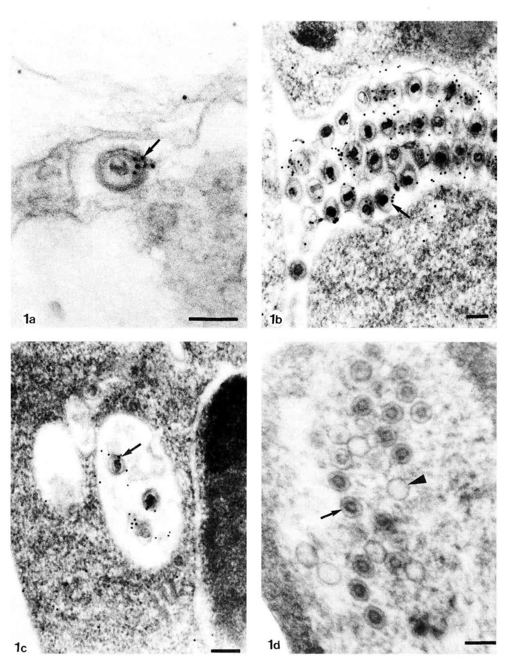
2 Antibodies specific for channel catfish virus cross-react with Pacific oyster, Crassostrea gigas, herpes-like virus RM Le Deuff, T Renault N Cochennec IFREMER, laboratoire de génétique, aquaculture et pathologie, unité de recherche en pathologie et immunologie générales, BP 133, La Tremblade, France Herpes-like viruses were first described in the larval Pacific oyster, Crassostrea gigas, during the summer of 1991 in France (Nicolas et al, 1992) and New Zealand (Hine et al, 1992). Since these first reports, sporadic incidence with high mortality rates (90-100%) have occurred among C gigas larvae in several French hatcheries in 1992, 1993 and 1994 (Renault et al, 1994a). Moreover, a similar virus was found in association with sporadic high mortality rates (80-90%) of C gigas spat in France in 1993 (Renault et al, 1994b) and 1994 (Renault, personal communication). In addition, the pathogenicity of the herpes-like virus was demonstrated by inoculating virus suspensions into axenic cultures of C gigas larvae (Le Deuff et al, 1994). As a result of these reports and the economic importance of C gigas to shellfish culture, it seems important to characterize this virus and to develop sensitive diagnostic methods in order to study the epidemiology of the disease and prevent its potential spread. Due to the lack of marine mollusc cell lines, the study of the viral cytopathogen effects in homologous cell culture is impos- sible. Thus, in an effort to overcome the limitations of existing methodologies, the direct detection of the oyster herpesvirus was attempted using monoclonal and polyclonal antibodies specific for a fish herpes-like virus, the channel catfish virus (CCV). Such antibodies were expected to provide the basis for the development of sensitive diagnostic immunoassays for direct detection of herpesvirus in oyster tissues. Using the immunogold method, the monoclonal antibody specific for a CCV envelope glycoprotein was found to specifically label the enveloped virus particles (fig 1a) when diluted 1/10, but this labelling disappeared when the monoclonal antibody was diluted 1/100. The same results were obtained for oyster herpes-like virus infected spat and larvae from different origins and for CCV-infected channel catfish ovary (CCO) cells. One or more colloidal gold particles was found in association with the viral particle envelope both extracellularly (fig 1b) and in the cytoplasm (fig 1c). No labelling of unenveloped nucleocapsids or empty particles was observed (fig 1 d). * Correspondence and reprints
3
4

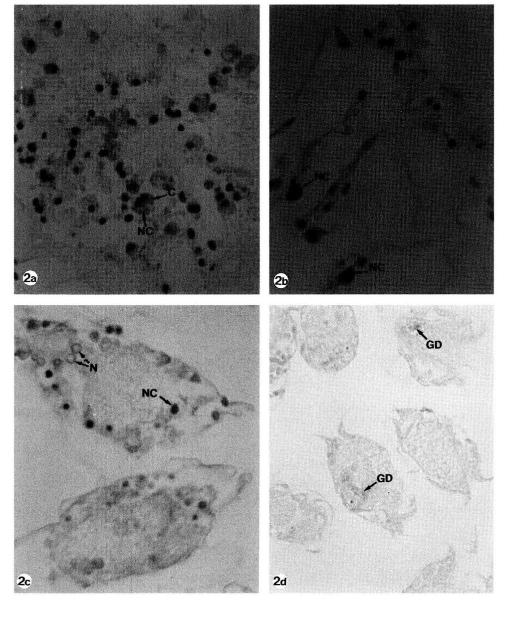
5 The binding of the monoclonal antibody could not be observed using immunohistochemical methods when the ascitic fluids were diluted 1/50 or 1/10. As no positive control was performed on the CCV-infected CCO cells, it was not possible to determine whether the antigens were too few in number to produce a visible labelling or if the fixatives used denatured the target antigens. Using the immunogold method, the polyclonal antibody diluted 1/10 produced a high level of background labelling on the CCVinfected CCO cells and the herpes-like virus infected oyster spat and larvae. When the polyclonal antibody was diluted 1/100, little residual labelling could be observed on the capsids. These results could be improved by using better blocking of unspecific binding sites and also by testing serial dilutions of the polyclonal antibody between 1/10 and 1/100. However, when immunohistochemical methods were used, the polyclonal antibody-diluted 1/200 specifically labelled cells in infected oyster spat, localized in the conjunctive tissues of the mantle, the digestive gland and the gills. Depending on cells, the labelling was localized on the nucleus, the cytoplasm or both (figs 2a,b). This was similar to that of the transmission electron microscope observations (Nicolas et al, 1992; Renault et al, 1994a) of virus particles localized in the cell nucleus and/or in the cytoplasm, depending on the stage of the viral replication cycle. Moreover, a few cells were labelled even in animals that had numerous lesions in histological examination. This is in agreement with the transmission electron microscope observations (Renault et al, 1994b) which revealed that few cells in the tissues of infected spat contained particles. lmmunohistochemistry on infected oyster larvae was difficult to interpret since it was not possible to completely eliminate the background effect due to unspecific tissue labelling. However, the labelling observed on healthy and herpesvirus-infected larvae was different. The background in healthy larvae was diffuse and mostly localized on the digestive gland (fig 2d). The labelling in the virus-infected oyster larvae was observed on specific cells in the mantle tissue (fig 2c). The results described here failed to provide a useful diagnostic tool for oyster herpesvirus infection of larvae and spat. However, this work could help to the taxonomic classification of this new agent and might further elucidate the relationships between viruses and their hosts (Hayashi et al, 1993). ACKNOWLEDGMENTS We wish to thank RP Hedrick for kindly providing the CCV antibodies. This work was supported by the Conseil General de la Charente-Maritime. REFERENCES Hayashi Y, Izawa H, Mikami T, Kodama H (1993) A monoclonal antibody cross-reactive with three salmonid herpesviruses. J Fish Dis 16, Hine PM, Wesney B, Hay B (1992) Herpesviruses associated with mortalities among hatchery-reared larval Pacific oysters Crassostrea gigas. Dis Aquat Org 12, Le Deuff RM, Nicolas JL, Renault T, Cochennec N (1994) Experimental transmission of herpes-like virus to axenic larvae of Pacific oyster, Crassostrea gigas. Bull Eur Ass Fish Pathot 14, Nicolas JL, Comps M, Cochennec N (1992) Herpes-like virus infecting Pacific oyster larvae, Crassostrea gigas. Bull Eur Fish Patholl2, Renault T, Le Deuff RM, Cochennec N, Maffart P (1994a) Herpesviruses associated with mortalities among Pacific oyster, Crassostrea gigas, in France-Comparative study. Rev Med Vet 145, Renault T, Cochennec N, Le Deuff RM, Chollet B (1994b) Herpes-like virus infecting Japanese oyster (Crassostrea gigas) spat. Bull Eur Fish Pathol 14, 64-66