|
|
|
- Amos Stephens
- 5 years ago
- Views:
Transcription
1 NeonatalHeartRateDetectionUsing AuxiliaryOpticalSystem MridulaGarimella,T.V.HimajaSree,AnushaBandari,IbrahimPatel Abstract: Fetal heart rate (FHR) monitoring is a proven means of assessing fetal health during the antenatalperiod.currently,theonlywidelyavailableinstrumentationforproducingthesedataisbasedon Doppler ultrasound, a technology that is unsuitable for longterm use. For nearly a century, it has been known that the fetal Photoplethysmography (FPPG) can be detected using electrodes placed on the maternalabdomen.thepaperdescribesthedesign,constructionanduseofacompact,longtermrecorder of three channels of 24 h antenatal trans abdominal data. Preliminary use of the recorder in around 400 short recording sessions demonstrates that FHR records of equivalent quality to those from Doppler ultrasound based instruments can be extracted from such data. The success of FHR derivation is, on average, around 65% of the recording period from around 20 weeks gestation (although this figure is reducedfromaround2832weeks,andthesuccessratesexhibitawiderangewhenindividualsubjectsare considered).theseresultsdemonstratethatthetechniqueoffers,notonlyameansofacquiringlongterm FHRdatathatareproblematictoobtainbyothermeans,butalsoamorepatientfriendlyalternativetothe Dopplerultrasoundtechnique. Keywords:Autonomicnervoussystem,ECG,Heartratevariability,FetalPhotoplethysmography,Doppler ultrasound,adaptivenoisecancelling(anc). 1. Introduction Nowadays there is a growing research interest in biomedical optics which utilizes the light to probe structure and function in biomedicine and leads to several noninvasive and nonionized diagnostic and therapeutic methods such as laser surgery, photodynamic therapy, laser Doppler flowery (LDF), and Photoplethysmography (PPG). To understand the fundamentals of such methods, it is necessary to investigate the phenomena of photontissue interaction. Photoplethysmography (PPG) is in this day and ageextensivelyuseandhasbeenacceptedbyphysiciansbecauseofitssimpledesignandrelativelylowcost perexamination.appgsystemwithniropticalsensorscanmeasurethe 1. She is pursuing her Bachelor s degree in BME Dr B. V. Raju Institution of Technology Narsapur, Medak,A.P. 2. She is pursuing her Bachelor s degree in BME Dr. B. V. Raju Institution of Technology Narsapur, Medak,A.P. 3. She is pursuing her Bachelor s degree in BME Dr.B. V. Raju Institution of Technology Narsapur, Medak,A.P. 4. Assoc.Prof.DeptofECE.Dr.B.V.RajuInstitutionofTechnologyNarsapur,Medak,A.P. bloodvolumechangesintheskinsurfacelayersbyregisteringtheattenuationchangeinthenearinfrared spectrum.itsbiophysicalprincipleisbasedonthefactthatthereisastrongcontrastinabsorptionofnir light between the bloodfilledvessels and the ambient bloodless tissue. Sothroughdetection of the light remitted from the skin, it is possible to measure the blood volume change of the skin. The PPG system provides a simple and noninvasive method to detect venous diseases at early stage through certain functionaltestslikevot(venousocclusiontest)andmpt(musclepumptest).onedisadvantageofppg isthatitcanonlymeasureonesmallareaatonetimeandthusverydifficulttogetaspatialdistributionof the blood volume change of the skin. Because of the spatial variation of the circulation system, it is necessarytogetamappingofthevenoushemodynamics.multichannelppgdevicehasbeendevelopedto monitor the skin perfusion at different sites, but it introduces more problems with sensor attachment, whichmakesthetestpersonuncomfortableandintroducingmoremovementartifacts.alsoahighspatial
2 resolution is impossible with multichannel PPG because the probesize of PPG is oftenmore than 1cm 2. Another disadvantage of PPG is that the test person has to be measured contactly, which restrict its applicationtosomeclinicalsituationsuchasmonitoringthewoundhealingprocess.inthispaper,anew noninvasive and noncontact method called Photoplethysmography Imaging (PPGI) is presented. A PPG ImagerwhicharrangesahighqualityCCDcamerawithauxiliaryopticalsystemcombinesthefeaturesof both classical PPG measurement and CCD imaging. It can visualize the structure of skin vessels and evaluate the venous hemodynamics as well as the arterial pulsation. PPGI avoids the time consuming scanning by using CCD as an array of photon detectors so that it can monitor the dynamic changes of dermalperfusionondifferentpartsofskinsurfacesimultaneouslyandflexiblyasshowninthefigure1. II.WorkingPrinciple Photoplethysmography(PPG)isasimpleandlowcostopticaltechniquethatcanbeusedtodetectblood volumechangesinthemicrovascularbedoftissue.itisoftenusednoninvasivelytomakemeasurements at the skin surface. The PPG waveform comprises a pulsatile ('AC') physiological waveform attributed to cardiac synchronous changes in the blood volume with each heartbeat, and is superimposed on a slowly varying ('DC') baseline with various lower frequency components attributed to respiration, sympathetic nervoussystemactivityandthermoregulation.althoughtheoriginsofthecomponentsoftheppgsignal arenotfullyunderstood,itisgenerally Fig.1:HardwaresetupforOFHRdetection. acceptedthattheycanprovidevaluableinformationaboutthecardiovascularsystem. An alternative to ultrasound is using the fetal electrocardiogram (FECG). Invasive FECG uses a scalp electrodeandremainsreservedtopredeliveryconditions.ontheotherhand,noninvasivefecggenerally needs 34 leads, which renders the procedure more complex from a practical perspective where many electrodesneedtoretainperfectohmiccontactwiththesubject'sbody.thefecgisgenerallyutilizedlater inpregnancyduetoitslowsignaltonoiseratio(snr),i.e.betweenthe28thand30thweekofgestation.it is worth mentioning that commercial devices operating on noninvasive FECG are not available at this moment. However, these techniques are expensive, require a high optical power and are difficult to implementduetosizeandpowerconsumptionlimitations.

3 III.HardwareDesignforPPGMeasurementofFetusHeartRate AconventionalreflectancePPGsensorconsistsofanLEDunitandphotodetectorinparallelconfiguration. Since LEDs emit light in a circular pattern, the detected light represents a fraction of the total reflected lightemittedtowardthephotodetector.toincreasetheincidentlight,weplacedthreeledssymmetrically aroundthephotodetector(seefig.2and3).lightemittedfromeachledhaditsowncircularpattern,and aportionofthelightfromeachledoverlappedatthephotodetector.theoutputsignalfromthesensor was converted into a voltage and amplified, then filtered with a band pass of Hz to separate the pulsatileaccomponent,andfilteredwithalowpasscutoffat0.1hztoseparatethedccomponent.theac anddcsignalsweredigitizedat400hzusingthemicroprocessor. Fig.2:BlockdiagramfortheproposedPPGmeasurementsystemthatincludes anadaptivelightintensitycontrolfunctionusingdccomponents. Fig.3:ConceptualdiagramsoftheproposedPPGsensorwiththreelightsources. (a)frontalview.(b)transverseview.
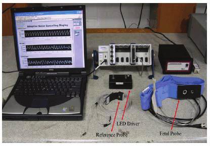
4 TheChallengeindesigningFHR III.Procedure Themainchallengeistodesignanddevelopalowpoweropticalfetalheartrate(FHR)monitor.Thesignal of interest is the photoplethysmogram (PPG), which is generated when a beam of light is modulated by blood pulsations. The light intensity is modulated by the mother as well as fetal blood circulations, producing a mixed signal which needs to be separated via advanced digital signal processing (DSP) techniques Solution is as the input power of the incident radiation leads to a lower SNR, the excitation signalisachoppedlightbeamandsynchronousdetectionisperformed.opticalfetalheartratedetection System. Fetalheartrate(FHR)detectionistheprimarymethodologyforantenataldeterminationoffetalwellbeing, assistingintheidentificationofpotentialhazardssuchashypoxiaanddistresstothefetus.theexpected outcome of this early detection is a reduced risk of fetal morbidity and mortality. Currently FHR can be detectedbyusingdopplerultrasound,wherethestandardpredeliverytestoffetalhealthisthefetalnon stress test (NST). These tests are routinely performed at the hospital, generally with continuouswave instruments.althoughcurrentultrasonicfhrdetectorsarebecominglessexpensiveandbulky,accurate sensoralignmentandsomedegreeofexpertisearestillrequiredtocorrectlyoperatethem.moreover,they aresensitivetomotionartifactandfinallycompletesafetyoflongtermexposureofthefetustoultrasound waveshasyettobeestablished,thereforeonlyshorttermtestingisactuallypracticed. An alternative to ultrasound is using the fetal electrocardiogram (FECG). Invasive FECG uses a scalp electrodeandremainsreservedtopredeliveryconditions.ontheotherhand,noninvasivefecggenerally needs 34 leads, which renders the procedure more complex from a practical perspective where many electrodesneedtoretainperfectohmiccontactwiththesubject'sbody.thefecgisgenerallyutilizedlater inpregnancyduetoitslowsignaltonoiseratio(snr),i.e.betweenthe28thand30thweekofgestation.it is worth mentioning that commercial devices operating on noninvasive FECG are not available this moment. Morerecently,opticalmethods,stillattheresearchstage,havebeenproposedwherehalogenandtungsten by a photomultiplier. In these works, the emphasize was on trans abdominal monitoring of fetal arterial blood oxygenation for pulseoximetry and by the same means FHR, with wavelengths in the nm and nm range to get optimum results. However, these techniques are expensive, require a high opticalpowerandaredifficulttoimplementduetosizeandpowerconsumptionlimitations.opticalfetal HeartRateDetectionSystem. Inthiswork,alowpoweropticaltechniqueisproposedbasedonthePPGtononinvasivelyestimatethe FHR.AbeamofLEDlight(<68mW)isshonetothematernalabdomenandthereforemodulatedbythe bloodcirculationofbothmotherandfetuswhereasmaximumpenetrationisachievedatawavelengthof 890 nm. This mixed signal is then processed by an adaptive filter with the maternal index finger PPG as referenceinput.thefigureshowstheopticalfetalheartratedetection(ofhr)systemblockdiagram Thefetalprobe(primarysignal)isattachedtothematernalabdomenusingaVelcrobelttoholdtheIR LEDandphotodetector,separatedby4cm.Thereferenceprobeisattachedtothemother sindexfingeras generallypracticedinpulseoxymetry.astheselectedirledcouldonlyemitamaximumopticalpowerof 68 mw, the OFHR system operates with an optical power less than the limit of 87 Mw specified by the International Commission on NonIonizing Radiation Protection (ICNIRP). In order to modulate the IRLED, the modulation signal is generated at a frequency of 725Hz usingsoftware subroutine through a counterport(niusb9474)totheleddriver. The diffused reflected light from the maternal abdomen, detected by the lownoise photo detector, is denotedasi(m1,f),wherem1andfdenotethecontributiontothesignalfromthemotherabdomenand
5 fetus,respectively.alownoise(6nv/hz1/2)transimpedanceamplifierisutilizedtoconvertthedetected currenttoavoltagelevel.thereferenceprobe(mother sindexfinger)consistsofanirledandasolid statephotodiodewithanintegratedpreamplifier.thesignalfromthisprobeisdenotedasi(m2),where M2referstothematernalcontribution.Synchronousdetectionisnotrequiredatthischannelasthefinger photoplethysmogramhasahighsignaltonoiseratio(snr). V.AnOpticalFHRDetectionSystem Our research team proposed a lowpower optical technique based on the photoplethysmogram (PPG) signal,whichisgeneratedwhenabeamoflightismodulatedbybloodpulsations,tononinvasivelyestimate thefhr.thedoctorortechnicianshinesabeamofledlight(lessthan68mw)atthematernalabdomen, modulatedbythebloodcirculationofthemotherandfetus.maximumlightwavepenetrationisachieved at a wavelength of 890 nm. This mixed signal can be processed by an adaptive filter using digital signal processingwiththematernalindexfingerppgasareferenceinput,figure1:ofhrsystemblockdiagram showingthehardwaremoduleshavebeenimplementedinembeddedsystems. TheteamdevelopedtheopticalFHR(OFHR)detectionsystemusingembeddedsystemsgraphicalsystem designsoftwareandnihardware.intheofhrsystem,reducingtheinputpoweroftheincidentradiation leadstoalowersignaltonoiseratio(snr),andtheexcitationsignalisachoppedlightbeam. At the receiver side, lownoise amplification and synchronous detection ensures conservation of the information with minimum noise power. A 24bit NI USB 9239 analogtodigital converter (ADC) minimizes the effects of quantization noise. Once digitized, the signal is processed via adaptive noise canceling(anc)techniquestoextractthefetalppgfromthemixedsignal. Weattachedthefetalprobe(primarysignal)tothematernalabdomenusingaVelcrobelttoholdtheIR LEDandphotodetectorseparatedby4cm.Weattachedthereferenceprobetothemother sindexfinger. Because the selected IRLED could only emit a maximum optical power of 68 mw, the OFHR system operateswithanopticalpowerlessthanthelimitof87mwspecifiedbytheinternationalcommissionon NonIonizingRadiationProtection(ICNIRP). VI.ResultsandDiscussion TomodulatetheIRLED,themodulationsignalisgeneratedatafrequencyof725Hzusingthesoftware subroutine through a NI 9474 counter port to the LED driver. As seen in Figure 4, the diffused reflected lightfromthematernalabdomen,detectedbythelownoisephotodetector,isdenotedasi(m1,f)sothat M1andFdenotethecontributiontothesignalfromthemother sabdomenandfetus,respectively.filtering and an adaptive noise cancelling (ANC) algorithm. The team used Lab View to implement the entire algorithm and part of the instrument. After preprocessing and applying the ANC algorithm, Lab View displays results for the fetal signal and the FHR. Figure 4 illustrates the laboratory prototype and the graphical user interface of the OFHR system and presents the maternal index finger PPG (top), the abdominalppg(middle),andtheestimatedfetalppg(bottom).figure5and6:thelaboratoryprototype andthegraphicaluserinterfaceoftheofhrsystem.
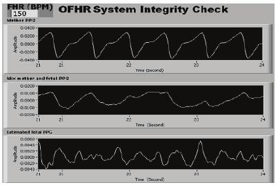
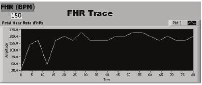
6 Fig.4:GraphicaluserinterfaceofOFHRsystem.Systemintegritycheckmenu. Fig.5:GraphicaluserinterfaceofOFHRsystem. Fig.6:FHRtracemenu.
7 Figure 7 illustrates the three selectable displays, including digital synchronous or lockin amplifier (LIA), adaptive noise cancelling (ANC), and heart rate trace. Figure 7: The three selectable displays, including digitalsynchronousorlockinamplifier(lia),adaptivenoisecancelling(anc),andheartratetrace.the purposeofthefirsttwodisplaysistoassistdevelopment,andthethirddisplayindicatesfhrvaluesversus time.theusercaneitherviewthedatalineorsaveitforfurtheranalysis.afterdevelopment,wetestedour system s functionality with a total of 24 data sets from six subjects at 37 ±2 gestational weeks from the UniversityKebangsaanMalaysiaMedicalCentre.TheUniversityEthicalCommitteereviewedandapproved thestudy,andallpatientswhoparticipatedprovidedwrittenconsent.allfetusesinthisstudywerefound to be healthy by an obstetrician and born without complication. In our study, we obtained a correlation coefficientof0.97(pvaluelessthan0.001)betweenopticalandultrasoundfhrwithamaximumerrorof4 percent.clinicalresultsindicatethatpositioningtheprobeoverthenearestfetaltissues,notrestrictedto theheadorbuttocks,improvessignalqualityanddetectionaccuracy.canbedetectedbyaphotodetector. Bloodabsorbsmostlymorelightthanthesurroundingtissuedoes,andthereforeareductionoftheamount ofbloodgivesanincreaseintheintensityofthedetectedlight.thewavelengthanddistancebetweenlight source and photo detector also determines the depth of penetration (69). Green light is suitable for measurementofsuperficialskinbloodflow,andinfrared(ir)ornearirisbetterformeasurementsofthe deeptissue(muscle)bloodflow(73). Fig.7:ANCBlockdiagram VII.Advantages LowPowerRequirement. LessExpensive. CommerciallyAvailable. MoreAccurateThanOtherFHRMeasurementTechniques. SmallComponents. VIII.Disadvantages HighlySophisticatedTechnology. ChanceofTroubleShootingisVeryHigh. IX.Conclusion OurresearchteamdevelopedanovelOFHRdetectionsystemusinglowcostandlowpowerIRlightanda commercially available silicon detector. With embedded systems, we rapidly and easily implemented the digitalsynchronousdetectionandadaptivefilteringtechniques.wemeasuredfhrresultswithacceptable
8 accuracy compared to the standard method of detection (Doppler ultrasound). Moreover, due to the noveltyofoursolution,weareintheprocessoffilingapatentforitscommercialuse. FutureDevelopment ResearchersaretakingplacetoachievemoreefficientanderrorlessFHRbyapplyingNanotechnology. X.References 1) GanKokBeng,EdmondZahedi,andMohd.AlauddinMohd.Ali,UniversityKebangsaanMalaysia, FacultyofEngineering 2) C.D.Kurth,J.M.Steven,D.Swedlow, Newfrontiersinoximetry AmJAnesthesiolvol23,pp , ) G.A.Dildy,J.A.Thorp,J.D.YeastandS.L.Clark,"Therelationshipbetweenoxygensaturationand ph in umbilical blood: implications for intrapartum fetal oxygen saturation monitoring," Am J ObstetGynecolvol.175,no.3Pt1,pp , ) J.L.ReussandD.Siker, Monitoringsiteandwavelengthselectionforfetalpulseoximetry, Assoc. Advanc.Med.Instr.AnnualMeeting,June ) A.K.Luttkus,J.H.Stupin,M.PorathandJ.W.Dudenhausen,"EvaluationOfsignalqualityofanew fetalpulsoximetrysystem(obs500).arecentdevelopmentofafetaloxisensorpositionedonthe fetalback,"jperinatmedvol.29,no.s1,pp.251, ) T.L.Rusch,R.SankarandJ.E.Scharf,"Signalprocessingmethodsforpulseoximetry,"ComputBiol Medvol.26,no.2,pp , ) D. E. Bahr and J. L. Reuss, Method and Apparatus for Processing a Physiological Signal, U. S. Patent6,339,715B1,January15, ) L.R.RabinerandB.Gold,TheoryandApplicationofDigitalSignalProcessing.EnglewoodCliffs, NJ:PrenticeHall, ) S.Palreddy, SignalProcessingAlgorithms, indesignofpulseoximeters,j.g.webster,ed.bristol andphiladelphia:instituteofphysicspublishing,1997,pp ) S.Nioka,M.Izzetoglu,T.Mawn,M.J.Nijland,D.A.Boas,andB.Chance, Fetaltransabdominalpulse oximeterstudiesusingahypoxicsheepmodel, J.MaternalFetalNeonatalMed.,vol.17,no.6,pp , ) B. Chance, Transabdominal examination monitoring and imaging of tissue, U.S. Patent 2005/ A1,Feb.17, ) A.M.Vintzileos,S.Nioka,andM.Lake, Transabdominalfetalpulseoximetryusingnearinfrared spectroscopy, Amer.J.Obstet.Gynaecol.,vol.192,pp , ) Zourabian,B.Chance,N.Ramanujam,R.Martha,andA.B.David, Transabdominalmonitoringof fetalarterialbloodoxygenationusingpulseoximetry, J.Biomed.Opt.,no.5,pp , ) E.ZahediandK.B.Gan, Applicabilityofadaptivenoisecancellationtofetalheartratedetection usingphotoplesthysmography, Comput.Biol.Med.,vol.38,no.1,pp.31 41,2008.