PRGF. An article by: Eduardo Anitua, MD, DDS; Vitoria, Spain (1) Isabel Andía, PhD; Vitoria, Spain (2) Mikel Sánchez, MD; Valencia, Spain (3)
|
|
|
- Magdalen Hardy
- 5 years ago
- Views:
Transcription
1
2 PRGF (Plasma Rich in Growth Factors) An article by: Eduardo Anitua, MD, DDS; Vitoria, Spain (1) Isabel Andía, PhD; Vitoria, Spain (2) Mikel Sánchez, MD; Valencia, Spain (3) (1) (2) (3) Private practice in implantology and oral rehabilitation. R+D Department, BTI I mas D. Arthroscopic Surgical Unit, U.C.A. Mikel Sánchez. PRGF (Plasma Rich in Growth Factors) is a system for obtaining platelet and plasma proteins; autologous proteins are obtained from the patient s own blood shortly before its therapeutic use. Its application accelerates the repair/regeneration mechanisms of various tissues. This article describes specific characteristics of PRGF, differentiating it from other systems and techniques available on the market. Included are descriptive preclinical studies on the content of the following growth factors: PDGF (Platelet-Derived Growth Factor), TGFß-1 (Transforming Growth Factor), EGF (Epermal Growth Factor), VEGF (Vascular Endothelial Growth Factor), IGF-I (Insulin-like Growth Factor Type I) and HGF (Hepatocyte Growth Factor). The relationship between the number of platelets and the concentration of these factors is analyzed. The complexity of interactions between proteins and their interactions with the different cell types prevents establishing a relationship between dose and clinical effect. The information derived from our research shows that an effective and sufficient concentration does not imply large doses. As examples, we show the effect that treatment with PRGF has on cellular proliferation when the donors have a different number of platelets and different concentrations of growth factors. Also, two clinical cases illustrate that the efficacy of these preparations does not strictly depend on the number of platelets. Key words: Growth Factors, Platelets, PRGF, Tissue Repair. Introduction Many times the objective of medical research is not so much to prolong life as to improve its quality. Similarly, in the field of implantology, research has led to the development of new geometric designs for implants, different surface treatments, new diagnostic imaging techniques, computerized systems to simulate the placement of implants and systems for obtaining and using autologous growth factors. All these factors have contributed to the achievement of very satisfactory clinical results. Osseointegration time has been reduced, and primary stability improved. Moreover, implant treatment has extended to more complex clinical situations, and currently different 2
3 surgical strategies and techniques can be applied to allow the restoration of situations where the placement of implants could not be consered until now. This has improved the quality of life for many patients. The situation is similar in the field of traumatology: articular prostheses have allowed patients affected by joint disease not to have to spend the rest of their lives in wheelchairs. But we should always keep in mind that the success of a treatment will be multifactorial and involves such things as surgical technique, implant or prosthesis design, imaging diagnostics, and the use of autologous biological techniques, like PRGF, designed to accelerate tissue repair and the regeneration process [1,2,3,4]. This involves using the patient s own proteins for regenerative purposes. The use of PRGF is not limited to oral surgery, but has quickly spread to other medical specialities [5,6]. New treatments are based on the use of proteins: these are the nucleus of human physiology and regulate the various mechanisms involved in the repair of distinct tissues. PRGF can be wely applied because its mechanism of action affects basic molecular and cellular processes, generally common to all tissues. Complexity of the System The human body has some 100 trillion cells that govern themselves through an exchange of chemical signals. The cells secrete these chemical signals that influence the behavior of other cells and, in turn, receive external signals through specific receptors in their cellular membrane. These are complex systems, difficult to understand completely due to the multiple interactions between the system components. These dynamic interactions are regulated by physical and chemical laws and serve to tell the cell what its immediate function is and if its situation is the correct one. All this information communicates to the cell where the cell is at any given moment, which cells surround it, and the next thing it should do. Currently, a great deal of research is directed toward improving understanding of the different processes and, in the case of regenerative medicine, to decipher the mechanisms of action involved in repair and regeneration. This is not a matter of invention, but of interpreting and using the information derived from research to achieve quicker and more efficient tissue regeneration. Until now, a large part of research has centered on in-depth study of the indivual signaling proteins. But the challenge is to know the system as a whole, i.e., the interactions of these proteins among themselves and with the distinct cell types. Two very clear facts must be kept in mind. First, repair is the result of the interaction of different cell types with multiple signalling proteins. Second, the biological condition of a tissue in repair/regeneration phase varies depending on the length of time since the injury and the distinct topography of the tissue. Platelets and Tissue Repair Platelets, most often studied for their role in hemostasis, have a very important physiological function which has recently been discovered and valated: they are protein carriers with an important role in tissue repair and regeneration. Platelets are small disco cellular elements, heterogeneous in size and density; they are cytoplasmic fragments of the megakaryocyte, a giant cell in medullary bone; they circulate through the blood stream for around 8-10 days. In addition to being involved in hemostasis for their pro-coagulant function, they contain various growth factors involved in the repair of different tissues. They act as a carrier for these growth factors and release them in the areas where there is tissue damage. GFs (Growth Factors) are stored inse, in special secreting granules, the α granules. Among others, some which are stored are: PDGF, TGFß-1, EGF and VEGF. These substances were synthesized by the megakaryocyte, since the platelet does not contain a nucleus or the necessary elements for protein synthesis. On the other hand, during its journey through the circulatory system, the platelet captures plasma proteins that it also stores inse its granules. It therefore contains a very complex mixture of proteins [7], some from its precursor cell, the megakaryocyte, 3

![The first publications on the use of autologous platelet proteins appeared in the late 90s in the area of oral and maxillofacial surgery [8,9,10,11,12].](/docs-images/95/125406666/images/4-1.jpg "In the years following, different systems for obtaining and preparing platelet concentrates for therapeutic purposes have become available on the market.")
 is a system for preparing platelets and plasma proteins; it has particular characteristics that differentiate it from other systems available on the market.")
4 and others captured by endocytosis from the blood stream. Platelet concentrates have become popular in the clinical environment as a tool that, along with surgery, allows quicker and more efficient tissue regeneration. The first publications on the use of autologous platelet proteins appeared in the late 90s in the area of oral and maxillofacial surgery [8,9,10,11,12]. In the years following, different systems for obtaining and preparing platelet concentrates for therapeutic purposes have become available on the market. Preparation protocols vary from system to system, along with the concentrations of the different integrating proteins. PRGF (Plasma Rich in Growth Factors) is a system for preparing platelets and plasma proteins; it has particular characteristics that differentiate it from other systems available on the market. The objective of this article is to describe PRGF and its principal characteristics. Characteristics of PRGF PRGF is a mixture of autologous proteins, prepared from a determined volume of platelet rich plasma (PRP). PRP is a term coined by hematologists to describe a plasma rich in platelets. According to hematologists criteria, PRP is plasma that contains more than ,000 platelets/ul. PRGF is the only technique described that prepares platelet-enriched plasma that does not contain leukocytes. PRGF always and exclusively uses the patient s own autologous proteins, prepared at the same time they are used. PRGF contains platelet and plasma growth factors involved in the repair process; it also contains sticky plasma proteins such as fibrin, fibronectin and vitronectin, among others. Preparation PRGF is prepared from a small volume of blood that is adapted to each specific clinical case. It can vary from 5-80 cm 3. To prepare the PRGF, blood is taken from a peripheral vein using sodium citrate as an anticoagulant. Other anticoagulants induce changes in the platelet morphology when the blood is collected into EDTA, the platelets swell and become spherical instead of disco. Another commonly used anticoagulant, ACD, has a lower ph (6.5) and interferes with platelet aggregation. The traditional preparation of PRP consists of a slow centrifugation, which allows the platelets to remain suspended in the plasma while the leukocytes and erythrocytes are displaced to the bottom of the tube. A rap centrifugation can cause mechanical forces and can raise the temperature which can induce changes in the ultrastructure and form of the platelet which, in turn, can initiate a partial activation, causing it to lose part of its content. The current systems for preparing platelet concentrations use various centrifuges, at different speeds. The final objective is to obtain a platelet pellet, but this precipita- Confocal microscopy of the PRGF clot Fig. 1a Fig. 1b Fig. 1a: Shows the network of the fibrin fibers marked with green fluorescence. Fig. 1b: Shows the network of the platelets marked in red. Fig. 2: Global image of PRGF clot in which the fibrin and platelets regroup. 4
5 Fig. 3: Diagram of the distribution of the different fractions of plasma, separated by centrifugation according to the PRGF protocol. tion is not selective and the precipitate itself includes all the leukocytes corresponding to the initial volume of blood. The PRGF is prepared with a single stage centrifugation. Using the parameters of time and speed established in the protocol, the platelets are concentrated in the cm 3 of plasma situated immediately above the red cells. Also, the leukocytes end up in a single layer (buffy coat) immediately on top of the erythrocytes; this allows collection of the PRGF without any contamination by white cells. In this way we obtain plasma without leukocytes and with a platelet count three times that in the perpheral blood. Intact platelets: By means of optical and electron microscopy and with monoclonal antibody techniques, we have proven that by using the PRGF protocol the platelets do not incur structural or morphological changes, because they are not activated during manipulation and so the content of their a granules remains intact. Platelet activation and coagulation: The secretion of growth factors begins with platelet activation. The PRGF protocol uses Ca 2+ to induce platelet activation and exocytosis of the α granules. Calcium acts as a necessary cofactor for platelet aggregation. Generation of thrombin: Ca 2+ intervenes at different stages of the coagulation cascade; in the last stage the activated Xa/Va complex transforms the prothrombin into thrombin; this is a specific process dependent on Ca 2+. Thrombin is the initiator of clot formation; it is responsible for the formation of the fibrin net containing the knots in which the platelets are found. (Figs. 1 and 2). Release of GFs: When the platelets are activated, a cascade of signals begins leading to the reorganization of the platelet cytoskeleton, the centralization of its secreting granules and exocytosis of small molecules and proteins coming from the three types of granules: dense granules, a granules and lysosomes. Dense granules contain small molecules such as ADP and serotonin; lysosomes contain degradative enzymes; a granules represent storage places for various proteins and their release is directed by precise molecular mechanisms. When Ca 2+ is used to induce platelet activation, the secretion of the GFs contained in the a granules is slow. To optimize the secretion process, the optimum concentration of Ca 2+ has been determined. When the Ca 2+, concentration increases excessively there is a decrease in exocytosis, probably due to the activation, of proteases present in the platelets that are dependent on divalent cations [13]. When a rap activation and coagulation is required, endogenous thrombin can be used (according to PRGF protocol). Differences in the kinetics of release of growth factors: When thrombin is used, the secretion of GFs is almost instantaneous (1-2 minutes); activation with Ca 2+ on the other hand, causes a slow release. One hour after the activation approximately 90-95% of GFs have been secreted. Consequently, the PRGF should not be activated until the time of its use and, once activated, it should be used immediately if activated with thrombin, or within 5-10 minutes after its slow activation with Ca 2+. Other systems for preparing platelet concentrations use very high concentrations of bovine thrombin (approximately 100 times the physiological concentration). The PRGF protocol DOES NOT use bovine thrombin in any case. Bovine thrombin may cause significant systemic adverse reactions that include anaphylaxis and coagulopathies from the production of anti-thrombin antibodies. 5
 contains a number of platelets, similar to the peripheral blood and is used for preparing the fibrin.")
 is the one containing a greater content of platelet GFs (Fig. 3).")

6 Quantitative description of PRGF: The PRGF protocol separates the plasma into three fractions with a distinct number of platelets and specific concentrations of GFs. (Figs. 4a - c). Fraction 1 (F1) contains a number of platelets, similar to the peripheral blood and is used for preparing the fibrin. The fibrin clot is more unstable the larger the number of platelets it contains; therefore, with fewer platelets this fraction proves a more stable fibrin. Fraction 3 (F3) is the one containing a greater content of platelet GFs (Fig. 3). Therapeutic dose and effect: There is a belief that in order to obtain a therapeutic PRP, the platelets must be concentrated to a maximum. Although some authors speculate that the minimum concentration of platelets to achieve a clinical effect should be one million platelets per microliter, there are no conclusive experimental results that support this hypothesis [14]. Although this criterion is assumed many times by clinicians with no questions as to its reality, we believe that it is at least a subject open to debate. When PRGF or another platelet concentrate is used, a multiple combination of proteins is applied [7] with varying interactions and effects on different cellular mechanisms. This seems to be an over - simplification for such a complex situation to believe that the more platelets the better and the greater the clinical result, or that these preparations are effective only above one million platelets per microliter. Based on our experiments, however, we believe that an effective and sufficient concentration does not involve large doses. In an injury site, there are different types of cells in different situations - some will be irreversibly damaged and will suffer a process of apoptosis; still other cells will be less damaged and will react to stimuli from the nearest microenvironment to maintain tissue homeostasis, i.e., the conditions prior to the injury. Fig. 4a Fig. 4 (a, b, c): Quantitative description (mean values ± standard error) of the different fractions of plasma. Fig. 4b Fig. 4c 6


7 Cells from the nearby tissues will begin a displacement in response to specific signals (chemotaxis) and inflammatory cells infiltrating from the blood stream and precursor cells will arrive. This is only a glimpse of the complexity in which the damaged tissue finds itself in the moments following injury. Preclinical studies: In preclinical studies carried out by our research department, concentrations of growth factors in PRGF have been studied along with their relationship with the number of platelets. Plasma from 55 healthy indivuals was analyzed: subjects included 22 females and 33 males between the ages of 18 and 60 years. Biological variability in the number of platelets and in the concentration of growth factors: Peripheral blood contains million platelets per ml, therefore, the range is very broad. PRGF in any given indivual, concentrates platelets around 3 times the number in the peripheral blood. The platelet population is not homogenous and there is a size distribution of a mean diameter of around 2 µm; the volume and area also present a distribution. Just as in all biological parameters, there is also a biological variability in the concentration of GFs that are inse the platelets of each indivual and there is also variability in the concentration of the plasma growth factors. Platelet growth factors: Platelet-derived growth factor (PDGF-AB), transforming growth factor (TGFß-1), epermal growth factor (EGF) and vascular endothelial growth factor (VEGF) are platelet secreted growth factors, are synthesized by the megakaryocyte and stored in the a granule of the platelets. Figures 5a, 5b, 5c and 5d show the number of platelets bese the concentration of these growth factors. In the case of VEGF (Fig. 5d) the relationship between the number of platelets and the concentration is weak, meaning that there are indivuals who have a high con- Fig. 5a Fig. 5b Fig. 5c Fig. 5d 7
 are very much interrelated and also related to the number of platelets.")
8 Fig. 5e Fig. 5f centration of VEGF and others who have a very low concentration. The concentration of VEGF is a characteristic of each indivual. However, the concentrations of PDGF-AB, TGFß-1 and EGF (Figs. 5a-c) are very much interrelated and also related to the number of platelets. This relationship is statistically significant to 99% certainty. A mathematical model exists that explains an 82% of the variability. In practical terms, this relationship means that for any indivual, a platelet concentrate will invariably contain a larger amount of TGFß-1 than PDGF-AB and, in turn, this will be more concentrated than the EGF. In short, the relationship between the concentrations of these factors is always similar for different indivuals: there will always be times more TGFß-1 than PDGF. This means that whatever number of platelets the preparation contains, there will always be times more TGFß-1 than PDGF-AB. This fact is very important and we will conserer it in the following discussion on platelet dose and therapeutic effect. Plasma growth factors: As seen in figures 5e and 5f, the concentrations of IGF-I and HGF have no relation to the number of platelets. The concentration of these factors inse the platelets is very small compared to the plasma concentration. This leads us to the conclusion that the relationship between the number of platelets and concentration for these GFs is very weak, since the contribution of platelets to the total concentration is very small. What are the repercussions of this variability in the clinical result? An effective and sufficient concentration does not imply large doses. Based on the belief that if a little is good then more is better, others seek to concentrate platelets up to 8-10 times (basal reference is always peripheral blood). Technically, this is very easy to achieve; all we need to do is to precipitate the platelets and resuspend the pellet in a small volume of platelet poor plasma. This can be done easily, so it is not a technical problem that leads us to favor a moderate platelet concentration more akin to human physiology. To measure the efficacy of something, an effect must be measured. As to the effect, which one do we measure? The GFs that concern us influence proliferation, inflammation, chemotaxis, differentiation, etc. They also deal with a complex mixture whose components show multiple interactions conditioned by relative concentrations and biological surroundings. An example will better illustrate the situation: PDGF is a factor that induces proliferation in mesenchymal type cells. TGFß-1, on the other hand, is an inhibitor of the proliferation of these same types of cells. According to findings from our research, regardless of the number of platelets we use, we always applied 2-3 times more TGFß-1 than PDGF-AB. This means that the quantitative relationship between two platelet factors that are recognized as pro- 8

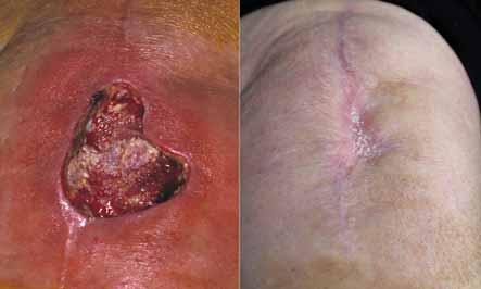
 Therapeutic dose and effect: Clinical Cases Will the clinical effect of these preparations depend on the number of platelets? Can the number of platelets predict clinical outcome?")

9 Fig. 6 Fig. 6: Relationship between the number of platelets of each donor and cellular proliferation. liferation modulators, one positive and the other negative, is going to be the same. This fact is independent of the number of platelets the preparation contains. We have proven this with experiments of in vitro cell cultures, using plasma with different numbers of platelets, but with the same PDGF-AB/TGFß-1 relationship. (Fig. 6) Therapeutic dose and effect: Clinical Cases Will the clinical effect of these preparations depend on the number of platelets? Can the number of platelets predict clinical outcome? Will those patients with a greater number of platelets have a better outcome after application of PRGF? To answer these questions, we chose the clinical case of an ulcer, since in ulcers the healing progress is seen from day-to-day and the therapeutic effect of these PRGF applications can be evaluated. Clinical case 1 Clinical case 1: A 73-year-old female patient with osteoarthritis in the left knee, fatty hypertrophy and resual edema secondary to thrombophlebitis. Clinical history reveals renal failure, hypothyroism, and chronic obstructive pulmonary disease (COPD). A total arthroplasty of the knee was performed that developed a deep skin necrosis in which the patellar tendon was left exposed. (Fig. 7a). The patient was treated with antibiotics and a bacterial culture showed that the necrosis was not caused by an infection. The surgeon recommended debrement and secondary closure. Over a period of 1 week, the wound was perfused with saline with no result and with a progressive increase in the size of the ulcer, which reached an approximate diameter of 6 cm (Fig. 7b). It was then deced to begin treatment with PRGF. Fig. 7a Fig. 7b Figs. 7a and b: The images show an ulcer approximately 6 cm in diameter and inflammatory reaction. 9


. Fig.")


10 Fig. 7c: Image of clotted PRGF covering the entire area of the ulcer. Fig. 7d: Progress of the ulcer in response to weekly treatment with PRGF (Image at 2 weeks). Fig. 7e Fig. 7f Figs. 7e and f: Images at the second and fourth week of treatment. Fig. 7g: Image of the ulcer at 5 weeks at the beginning of treatment with PRGF. Fig. 7h: The image shows complete healing of the ulcer after 7 weeks of treatment. 10
 in a population of 65 indivuals (mean values ± standard error) and concentrations of the aforementioned growth factors in the PRGF of the")
 in a population of 65 indivuals (mean values ± standard error) and concentrations of these growth factors in the PRGF of the treated patient.")
 and was totally cured by the seventh week (Fig. 7h).")
11 Fig. 8a: Number of platelets (millions/ml of PRGF) in a population of 65 indivuals (mean values ± standard error) and number of platelets in PRGF of treated patient. Fig. 8b: Concentration of PDGF-AB, TGFß-1, IGF-I (pg/ml of PRGF) in a population of 65 indivuals (mean values ± standard error) and concentrations of the aforementioned growth factors in the PRGF of the treated patient. Fig. 8c: Concentration of EGF, VEGF, HGF (pg/ml of PRGF) in a population of 65 indivuals (mean values ± standard error) and concentrations of these growth factors in the PRGF of the treated patient. After debrement, newly-activated PRGF was placed so that the clot would form in situ inse the necrotized area (Fig. 7c). This treatment was repeated weekly for 6 weeks. The ulcer healed from week to week (Figs. 7d, e, f and g) and was totally cured by the seventh week (Fig. 7h). The number of the patient s platelets in the PRGF was very low (Fig 8a) in relation to the population mean values. This anomaly in the number of platelets is related to her clinical history. The platelets in the PRGF, although scarce, were of a larger size. The result, 3 years post-intervention and PRGF treatment of the ulcer is stable and the area is completely regenerated. Clinical case 2 A 26-year-old male patient suffered traumatic amputation of the distal end of the index fingertip of the right hand. The patient was attended in a trauma center where two grafts were performed to close the wound (Figs. 9a, b, c and d). When treated at the hospital, amputation was elected due to repeated necrosis of the grafts and the patient was referred to us by a family member. PRGF treatment was initiated and applied weekly. Wound healing progressed positively from week to week (Figs. 9e - i) until it was totally cured at 5 weeks after commencement of treatment. 11


12 Fig. 9b Fig. 9b Figs. 9a - c: Situation following injury. Fig. 9c Fig. 9d: Initial 1 st application of PRGF. Fig. 9e: Exposed bone was remodelled with a gouge. The necrotic graft was painlessly eliminated without anesthesia. 12


13 Fig. 9f: Image of fingertip after 2 nd application. Fig. 9g: The image shows the cure after 3 weeks of treatment. Fig. 9h Fig. 9i Figs. 9h and 9i: Image of fingertip after 5th week of treatment. Observe remodelling of bone made with a gouge and repair of injury. Fig. 9j Fig. 9k Figs. 9j and 9k: Image one year after termination of treatment. The patient tells us that he uses the finger with total normalcy in daily life. 13
 in a population of 65 indivuals (mean values ± standard error) and concentrations of these growth factors in the PRGF of the patient with")
 in a population of 65 indivuals (mean values ± standard error) and concentrations of these growth factors in the PRGF of the patient with partial")

14 Fig. 10a: Number of platelets (millions/ml) of PRGF in a population of 65 indivuals (mean values ± standard error) and number of platelets in PRGF of patient with partial amputation of fingertip. Fig. 10b: Concentration of PDGF-AB, TGFß-1, IGF-I (pg/ml of PRGF) in a population of 65 indivuals (mean values ± standard error) and concentrations of these growth factors in the PRGF of the patient with partial amputation of fingertip. Fig. 10c: Concentration of EGF, VEGF, HGF (pg/ml of PRGF) in a population of 65 indivuals (mean values ± standard error) and concentrations of these growth factors in the PRGF of the patient with partial amputation of fingertip. Final conserations Two clinical cases treated with PRGF have been described. In both cases, otherconventional treatments were discontinued because they had been ineffective. Once the possibility of an infection that could impede the repair process had been discounted, the inability to repair could be due to the absence of cellular signals. The objective of PRGF is to prove those cellular signals in a matrix formed by sticky proteins such as fibrin, fibronectin and vitronectin. The combined proteins trigger angiogenesis and cellular proliferation. Furthermore, different types of cells will proliferate: cells reconstructing the localized injured tissue, as well as endothelial cells forming new vessels which transport precursor cells and the nutrients necessary for tissue regeneration. A very high number of platelets does not appear to be a requirement to start the repair mechanisms, as illustrated by the clinical cases. The use of PRGF offers conserable therapeutic benefits for treatment in a large number of clinical situations. Given the customary decrease of sequellae and the significant advantages for patients such as pain reduction, recovery of function and the fact that delayed healing or prolonged suffering caused by other more palliative treatments, even as irreversible as amputation in 14
, after consultation with the author and a detailed study of the PRGF technique, concluded that it cannot be consered a")
15 some cases, we clearly recognize a remarkably promising future for PRGF in regenerative medicine. Legal treatment The Spanish Health Ministry (Ministerio de Sanad Español), after consultation with the author and a detailed study of the PRGF technique, concluded that it cannot be consered a derived hemo therapy. Therefore, the requirements for its use are similar to those for other surgical procedures, regarding the quality of equipment, control of environmental conditions and TRAINING OF PERSONNEL that prepares and uses it and who should know its properties and limitations.it is used following the methodology of an autograft, i.e., using only the patient's own blood which prevents immunological responses of rejection and complications intrinsic to the use of heterologous or homologous materials and substances. Its use is immediate, so it does not require storage or transport. As with all new technological advances, these new tools take time to become fully integrated into society and there are always sectors among professionals who show a great resistance to their incorporation. It is event that, associated with these new therapeutic tools, there is the need to acquire new knowledge directed toward understanding the application and indications of these new treatments. Contact information: Dr. Eduardo Anitua Aldecoa San Antonio 15-3º; Vitoria, SPAIN; eduardoanitua@eduardoanitua.com Eduardo Anitua Biographical Data: - Graduate in Medicine and Surgery, University of Salamanca in Doctor of medicine and surgery. - Specialist in Stomatology Universad del País Vasco, continuing studies in many visits to the United States (Philadelphia, New York, Miami, San Francisco, Chicago) and in Europe (Italy, Germany, France, and Spain). - Delivered lectures at various Spanish and International universities as postgraduate assistant professor in Prosthetics and Occlusion at the Universad de Valencia, and postgraduate professor in Implantology at the universities in Seville, Murcia, Barcelona, and Madr. - Has given more than 500 seminars and lectures on implants, prosthetics, dental esthetics, and tissue regeneration in Spain and abroad (Europe, United States, Mexico, South America, Asia). - Director of the Continuing Education Program in Oral Rehabilitation given in Spain and other countries around the world for over 17 years. - Visiting professor at the dental schools at: Universad de Guatemala, Intercontinental de México, Javeriana de Colombia, Republic of Argentina, Uruguay, and Portugal (Oporto and Lisbon), Pennsylvania, Harvard, Boston, and Tufts. - Visiting professor at the Medical School of the Universad del País Vasco. - Scientific Director of BTI Biotechnology Institute. - Private practice in Implantology and Oral Rehabilitation in Vitoria, Spain - Director of Dental Dialogue. Bibiography [1] Anitua E, Andia Ortiz I. BTI implant system: The first implant system with a bioactive surface. Maxillaris 2001; 39: 2-7. [2] Anitua, Ardanza B, Paponneau A, et al. Clots from platelet-rich plasma promote bone regeneration in so doing reducing the time needed for dental implants and favouring their osteointegration. Blood 2001; 11: 242a. [3] Anitua E, Andía I. Un Nuevo enfoque en la regeneración ósea. Ed. Eduardo Anitua. Puesta al Día Publicaciones, Vitoria, [4] Anitua E. The use of plasma-rich in growth factors (PRGF) in oral surgery. Pract Proced Aesthet Dent 2001; 13: [5] Sanchez M, Azofra J, Aizpurua B, Andía I, Anitua E. Use of autologous plasma rich in growth factors in arthroscopic surgery. Cuader Artroscopia 2003; 10:12-19 [6] Sanchez M, Azofra J, Anitua E, Andía I, Padilla S, Santisteban J, Mujika I. Plasma rich in growth factors to treat an articular cartilage avulsion: a case report. Med Sci Sports Exerc 2003; 35: [7] Anitua E, Andia I, Ardanza B, Nurden P, Nurden AT. Autologous platelets as a source for healing and tissue regeneration. Thromb Haemost 2004; 91:4-15 [8] Whitman DH, Berry RL, Green DM. Platelet gel: an autologous alternative to fibrin glue with applications in oral and maxillofacial surgery. J Oral Maxillofac Surg. 1997; 55: [9] Anitua E. Factores de crecimiento derivados de las plaquetas: su utilización en la preparación de áreas futuras para implantes.31 Reunión Anual de la Sociedad Española de Periodoncia, SEPA; Noviembre 1997; Alicante, España, p.52 [10] Anitua E. Platelet Rich Plasma Growth Factor enhancement for developing future sites. XIII Annual Meeting Academy of Osseointegration; Marzo 1998; Atlanta, USA, p.77 [11] Anitua E. Plasma rich in growth factors: preliminary results of use in the preparation of future sites for implants. Int J Oral Maxillofac Implants 1999; 14: [12] Marx RE, Carson ER, Eichstaedt RN et al. Platelet-rich plasma: Growth factor enhancement for bone grafts. Oral Surg Oral Med Oral Pathol Oral Radiol Endod 1998; 85: [13] Lemons PP, Chen D, Whiteheart SW. Molecular mechanisms of platelet exocytosis: Requirements for _-granule release. Biochem Biophys Res Comm 2000; 267: [14] Marx RE. Platelet-Rich Plasma: Evence to support its use. J Oral Maxillofac Surg 2004; 62:

16