WESTERN BLOT. BCH462- Practical
|
|
|
- Julian Oliver
- 5 years ago
- Views:
Transcription
1 WESTERN BLOT BCH462- Practical
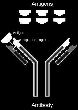
2 What is Antigen [Ag]? What is Antibody [Ab]? Immunoassay: is a test that uses the highly specific and selective antigen-antibody reactions forming antibody and antigen complexes [immuno-complexes] as a means of generating measurable results.
3
4 epitope antigen antibody
5 Enzyme Primary antibody antibody specified to specific antigen Secondary antibody antibody specified to Primary antibody
6 Also called protein immunoblot. Is a widely used immunoassay technique. To identify proteins specific proteins [antigens] in a sample of tissue homogenate or extract, based on their ability [the antigens] to bind to antibodies resulting in colour indicate the presence of this specific protein. Application?
7
8 To understand how proteins (antigens) can be analysed using antibodies raised against these proteins by Immunoblotting technique. To understand the steps in the development of Western and antigen-antibody interaction and detection. Electroblotting the pre-stained maker.
9 The mixture of proteins is separated based on molecular weight. These results are then electro-transferred to solid support producing a band for each protein. The transferred protein is detected by incubating the gel with specific primary antibody to the protein of interest, secondary antibody labelled with an enzyme which target the primary antibody, and substrate which in the end you will get coloured product. The colour indicates the presence of the protein of interest. The thickness of the band corresponds to the amount of protein present. Thus, the molecular weight and amount of the desired protein can be characterized from a complex mixture of proteins by western blotting.
10 Western blot performing steps The technique uses three elements to accomplish this task 1. Separating the sample mixture by size using SDS-PAGE. 2. Transfer to a solid support (electro-blotting), transfer the proteins bands from the gel to the membrane. 3. Marking target protein using a proper primary and secondary antibody to visualize.
11 1 st Phase: SDS-PAGE

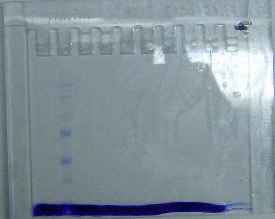
12 A protein sample is subjected to polyacrylamide gel electrophoresis. Protein sample SDS-PAGE To confirm the separation of the sample use: 1- Replica of the gel and stain it as usual. 2- Prestained marker. 3- Ponceau S.
 for use as size standards in protein electrophoresis (SDS-PAGE) and Western")
13 Prestained marker Figure: Protein Ladder is a mixture of nine (9) blue-, orange- and green-stained proteins (10 to 250kDa) for use as size standards in protein electrophoresis (SDS-PAGE) and Western blotting.
14 Prestained marker
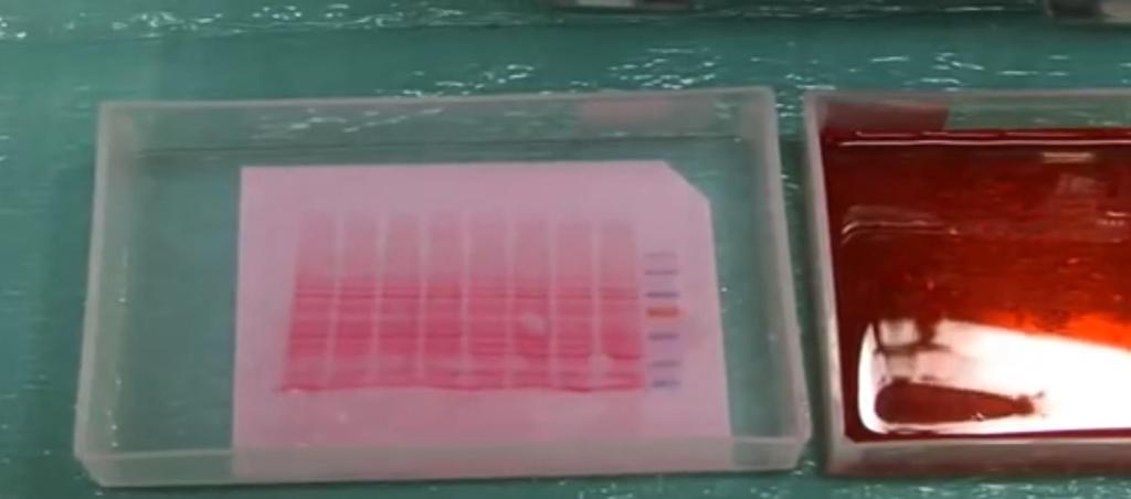
15 Ponceau S
16 2 nd Phase: Electroblotting

17 After that the gel is placed over a sheet of nitrocellulose, the protein in the gel is electrophoretically transferred to the nitrocellulose membrane. transfer step [Electroblotting] Membrane [with transferred proteins] Methods.

18 Transfer sandwich Note: The filter papers, gel and nitrocellulose membrane will soaked in transfer buffer.
19 3 rd Phase: Marking target protein to visualize
20 The nitrocellulose is then soaked in blocking buffer. Membrane [with transferred proteins] Blocking buffer: To block the nonspecific binding of proteins. The nitrocellulose is then incubated with the specific primary antibody for the protein of interest. Membrane [with transferred proteins] Specific primary antibody [specific for antigen of interest]
21 The nitrocellulose is then washed and incubated with a second antibody, which is specific for the first antibody [primary antibody]. Membrane [with transferred proteins +primary antibody] Secondary antibody [Specific for primary antibody]
22 Detection of specific protein using Western bolt [S] Substrate [E] [P] Colored product secondary antibody [Specific for primary antibody] Antigen of interest Primary antibody antibody specified to specific antigen

23 Thus the molecular weight and amount of the desired protein can be characterized from a complex mixture of proteins by western blotting.
24 Performing western blot: Ponceau S Staining: