Product datasheet. ARG30119 Pro-B Cell Marker Antibody panel (CD19, CD34, CD38, CD40, CD45)(FACS)
|
|
|
- Stephen Bryan
- 5 years ago
- Views:
Transcription
] anti-cd38 antibody [SP149] anti-cd40 antibody [HI40a] (FITC) anti-cd45 antibody [MEM-28] Goat anti-mouse IgG antibody (HRP) Goat anti-rabbit IgG")
1 Product datasheet Package: 1 kit ARG30119 Pro-B Cell Marker Antibody panel (CD19, CD34, CD38, CD40, CD45)(FACS) Component Cat. No. Component Name ARG62820 anti-cd34 antibody [4H11(APG)] anti-cd38 antibody [SP149] anti-cd40 antibody [HI40a] (FITC) anti-cd45 antibody [MEM-28] Goat anti-mouse IgG antibody (HRP) Goat anti-rabbit IgG antibody (HRP) ARG52764 ARG62839 ARG62856 ARG65350 ARG65351 Host clonality Reactivity Application Package Mouse mab FACS, WB, IHC-P, ICC/IF50 μg Rabbit mab FACS, IHC-P 50 μl Mouse mab FACS 50 tests Mouse mab Goat pab Ms FACS, ICC/IF, IHC-P, IP, 50 μg WB ELISA, IHC, WB 50 μl Goat pab Rb ELISA, IHC-P, WB 50 μl Summary Product Description B cell development occurs in the bone marrow and peripheral lymphoid tissues such as spleen. In the bone marrow, pro B Cell, pre B Cell and Immature B Cell develop. During this differentiation, several stages of rearrangement occur at immunoglobulin locus, resulting in the generation and surface expression of different types of receptors, such as pre-b-cell receptor. Different markers are also expressed at the surface of these cells throughout the development of B cell. Pro B Cells express CD19, CD34, CD38, CD40 and B220 on the surface of the cells. Arigo has these marker antibodies in the application of Flow Cytometry for the convenience of identifying Pro-B Cells in B cell populations. Cambier et al. Nature Rev Immunology 7: (2007) Alberts et al. Molecular Biology of the Cell: 1367 Parham. The Immune System Properties Note For laboratory research only, not for drug, diagnostic or other use. Bioinformation Resrarch Area Cancer antibody; Cell Biology and Cellular Response antibody; Controls and Markers antibody; Developmental Biology antibody; Immune System antibody; Metabolism antibody; Neuroscience antibody; Signaling Transduction antibody 1/6


2 Images ARG52708 anti-cd19 antibody [SP110] IHC-P image Immunohistochemistry: man Tonsil stained with CD19 antibody [SP110] (ARG52708) Immunohistochemistry: man Tonsil stained with CD38 antibody Immunohistochemistry: man Spleen stained with CD38 antibody Immunohistochemistry: man Thymus stained with CD38 antibody 2/6


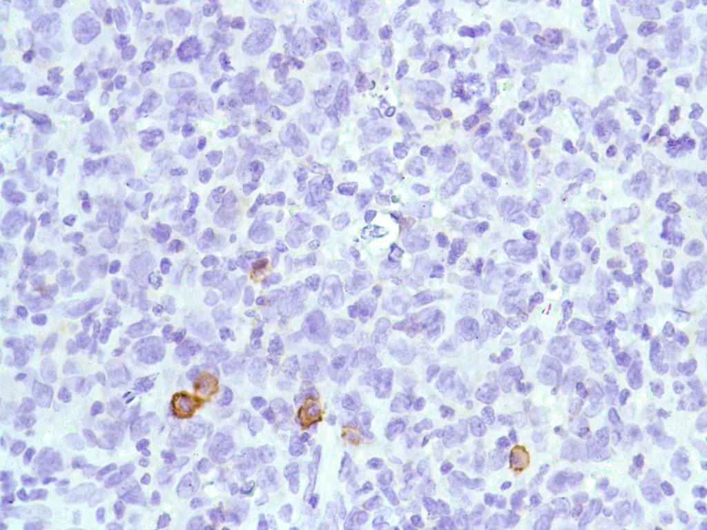

3 Immunohistochemistry: man Ovary stained with CD38 antibody Immunohistochemistry: man Prostate stained with CD38 antibody Immunohistochemistry: man B Cell Lymphoma stained with CD38 antibody Immunohistochemistry: man Prostate Adenocarcinoma stained with CD38 antibody 3/6


4 Immunohistochemistry: man Stomach Adenocarcinoma stained with CD38 antibody Immunohistochemistry: man Stomach stained with CD38 antibody Monoclonal antibody clone HI40a Flow Cytometry analysis image Flow Cytometry: man peripheral blood cells stained with antibody clone HI40a. Cells in the lymphocyte gate were used for analysis. Monoclonal antibody clone MEM-28 Flow Cytometry analysis image Flow Cytometry: man peripheral blood cells stained with antibody clone MEM-28. 4/6
 Cell nuclei")

![cells stained with ARG52708 anti-cd19 antibody [SP110] (green) or control](/docs-images/95/126259627/images/5-3.jpg "rabbit IgG (blue).")
5 Monoclonal antibody clone MEM-28 ICC/IF image Immunofluorescence: man peripheral blood mononuclear cell stained with clone MEM-28 (green) Cell nuclei was stained with DAPI (blue). ARG52708 anti-cd19 antibody [SP110] Flow Cytometry image Flow Cytometry: RAMOS cells stained with ARG52708 anti-cd19 antibody [SP110] (green) or control rabbit IgG (blue). Immunohistochemistry: man HK Lymphoma stained with CD38 antibody Immunohistochemistry: man Colon stained with CD38 antibody 5/6


6 Immunohistochemistry: man Colon Adenocarcinoma stained with CD38 antibody Immunohistochemistry: man Bladder stained with CD38 antibody Powered by TCPDF ( 6/6