Biotechnology 7 (4): , 2008
|
|
|
- Florence Elliott
- 5 years ago
- Views:
Transcription


1

2

3
4
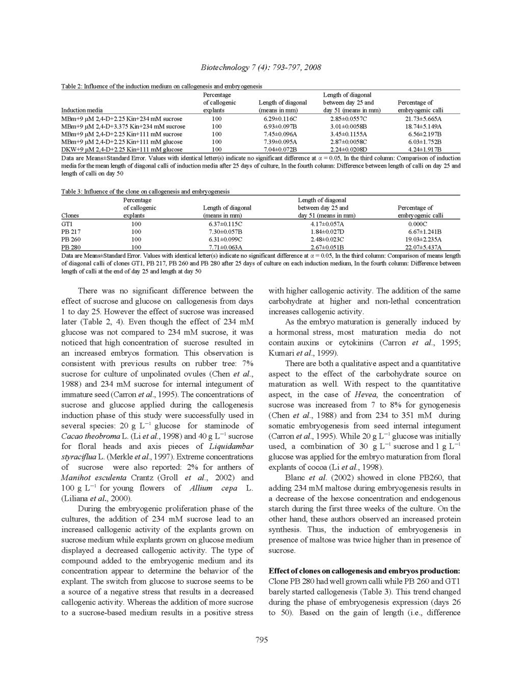
, the media containing sucrose induced the best growth of calli (Table 2).")
5 Biotechnology 7 (4): , 2008 b c Fig. 1: Embryogenesis callus of clone PB 280. About a dozen embryos at different stages: (a) globular, (b) torpedo and (c) cotyledonary. Bar represents 0.8 m between length at the end of day 25 and length at day 50), the media containing sucrose induced the best growth of calli (Table 2). Clone GT1 which began slowly had increased its callogenesis (Table 3). After 25 days of culture, embryos of clones PB 217, PB 260 and PB 280 were observed at the globular stage particularly on medium MBm supplemented with 9 µm 2, 4-D/3.375 µm Kin and sucrose. However the embryos at different stages of embryogenesis organisation (Fig. 1) from globular, heart to torpedo stages were observed during day 75 to day 100 on embryo development medium. At that phase, the majority of clone PB 217 embryos had senesced. This was especially observed for embryos from medium MBm supplemented with 111 mm glucose. The number of embryos (Fig. 1) for each embryogenic callus ranged from one to about ten. The most embryogenic clones were PB 260 and PB 280 whereas clone GT1 was not embryogenic in our experimental conditions (Table 2). These data showed that there is an effect of clone type both on callogenesis and embryogenesis. The fact that clone GT1 was not embryogenic despite a high callogenesis after day 25 could be due to a high accumulation of starch at the expense of a protein synthesis. The embryogenic rate of internal integument explants of clone GT1 was 7% in the first phases of culture. However the embryos did not grow (Carron et al., 1995). Embryos of clone PB 217 exhibited similar behaviour in MB medium supplemented with 111 mm glucose. One might argue that our experimental conditions did not allow a Table 4: Percentage of embryogenesis for each clone on each medium Percentage of embryogenic Callogenesis induction media calli PB 280/MBm+9 µm 2,4-D Kin+234 mm sucrose A PB 280/MBm+9 µm 2,4-D+2.25 Kin+234 mm sucrose A PB 260/MBm+9 µm 2,4-D+2.25 Kin+234 mm sucrose AB PB 260/MBm+9 µm 2,4-D Kin+234 mm sucrose B PB 260/MBm+9 µm 2,4-D+2.25 Kin+111 mm sucrose BC PB 217/MBm+9 µm 2,4-D+2.25 Kin+111 mm glucose BCD PB 260/DKW+9 µm 2,4-D+2.25 Kin+111 mm glucose CD PB 217/MBm+9 µm 2,4-D+2.25 Kin+234 mm sucrose 8.889CD PB 260/MBm+9 µm 2,4-D+2.25 Kin+111 mm glucose 6.667CD PB 280/DKW+9 µm 2,4-D+2.25 Kin+111 mm glucose 6.667CD PB 280/MBm+9 µm 2.4-D+2.25 Kin+111 mm sucrose 6.667CD PB 217/MBm+9 µm 2.4-D Kin+234 mm sucrose 6.667CD PB 217/MBm+9 µm 2,4-D+2.25 Kin+111 mm sucrose 5.333CD PB 280/MBm+9 µm 2,4-D+2.25 Kin+111 mm glucose 3.667CD PB 217/DKW+9 µm 2,4-D+2.25 Kin+111 mm glucose 3.333CD GT1/DKW+9 µm 2,4-D+2.25 Kin+111 mm glucose 0.000D GT1/MBm+9 µm 2,4-D Kin+234 mm sucrose 0.000D GT1/MBm+9 µm 2,4-D+2.25 Kin+111 mm sucrose 0.000D GT1/MBm+9 µm 2,4-D+2.25 Kin+234 mm sucrose 0.000D GT1/MBm+9 µm 2,4-D+2.25 Kin+111 mm glucose 0.000D Values with identical letter(s) indicate no significant difference at " = 0.05 the embryos at the globular stage to reach the necessary development stage before their transfer on maturation media. The best combinations clone/media were PB 280 or PB 260 with media supplemented with sucrose at 234 mm (Table 4). The frequency of embryogenesis with these combinations ranged from 19 to 59%. Chen et al. (1988) obtained about 35% of embryogenesis frequency. A low level of cytokinin in the dedifferentiation medium is sometimes responsible for the development of embryoids directly on the ovules, implying that they did not develop from callus. However, these embryoids grew abnormal after transfer to the differentiation medium and could only reach the torpedo stage (Chen et al., 1988). Embryos used in this study were not induced directly. The ploidy levels of the embryos were not determined in the present study. Usually karyotyping was carried out with young leaves or root tip cells from regenerated plantlets (Kumari et al., 1999; Chen et al., 1988). Previous cytological observation in Hevea indicated that regenerated plantlets were mixoploids (Chen et al., 1988). Several studies showed that gynogenic plants weren t absolutely haploids. In Spathiphyllum wallisii (Family Araceae), flow cytometry revealed the diploid nature of the plants (Eeckhault et al., 2001). In Onion, gynogenic plantlets were haploids (82%). The others were primarily diploid (15%). A few plants were mixoploid or tetraploid (Alan et al., 2004). CONCLUSION For the very first time, we have carried out gynogenesis in clones GT1, PB 217, PB 260 and PB
6