Introduction to histology and its methods of study
|
|
|
- Joanna Pearson
- 5 years ago
- Views:
Transcription
1 Introduction to histology and its methods of study Li shulei Department of Histology & Embryology
2 1 What is histology Definition Cell: smallest units functions in the human body Tissue System Histology is a science that studies Several the normal organs with related microstructure cells functions are similar of the in morphology human body or and the relationship between Motor related in function Cells and the system extracellular body s s structure matrix and Nervous functions. system Epithelial Circulatory tissue system Connective Respiratory tissue system Muscular Digestive tissue system Nervous Urinary tissue system Reproductive system Endocrine system Organ: different kinds of tissues
3 2 Why to study histology Anatomy: macrostructure Biochemistry: chemical compounds and processes Pathology: the relation between disease and the structures and functions of the body Although most medical students are not going to become histologists, a thorough knowledge of histology is fundamental for you as future doctors.
4 3 How to research on histology Preparation of tissue for microscopic examination Paraffin section Frozen section Microscopy Problems in the interpretation of tissue sections

5 3 How to research on histology Knife Section Block MICROTOME - a fancy meatslicer - holds the wax block, & cuts off thin slices, as the block is slowly advanced mechanically Light beam Glass slide Light microscope
6 Paraffin section Obtaining the specimen Fixation Dehydration Clearing Embedding Sectioning Staining

7 Obtaining the specimen fresh as possible and small pieces
8 Remove the water & replace with wax-solvent Imbed the oriented specimen in molten wax 70 % ethanol 80 % ethanol Dehydrating series Fresh tissue 95 % ethanol 100 % ethanol Clearing xylene Embedding label 4% formaldehyde fixative paraffin wax

9 Tissue processor Automatic tissues processor moves the tissues around through the various agents on a preset time scale.


10 Tissue embedding Tissues are infiltrated in molten wax to replace the xyline. The molten wax drop into a plastic box; then Put the tissues into the box. The molten wax solidify into a block with the tissue inside.
11 After it is solid, hold the wax block & cut slices Knife Section 1-10μm Block MICROTOME - a fancy meatslicer - holds the wax block, & cuts off thin slices, as the block is slowly advanced mechanically Water-bath Glass slide Mount the thin slices (sections) on slides Lift out floating section on the slide

12 Sectioning with microtome Rotation of the drive wheel moves the tissue-block holder up and down. Each turn of the drive wheel advances the specimen holder a controlled distance. After each forward move, the tissue block passes over the knife edge, which cuts the sections.


13 Sectioning with microtome Rotation of the drive wheel moves the tissue-block holder up and down. Each turn of the drive wheel advances the specimen holder a controlled distance. After each forward move, the tissue block passes over the knife edge, which cuts the sections.

14 Picking sections up from water bath sections are floated on a warm water bath that helps remove wrinkles.


15 Paraffin section Unstained section on glass slide Tray of unstained slides in drying oven Sections are picked up on a glass slide and placed in a warm oven to help the section adhere to the slide.
16 Staining Deparaffinized: running through xylene to alcohol to water Dye: acidic or basic compounds; electrostatic linkages with tissues Hematoxylin & Eosin (H H & E) E staining Hematoxylin: : stains cell nucleus and other acidic structure blue Eosin: stains the cytoplasm and collagen pink Basophilia: : affinity for basic dyes Acidophilia: : affinity for acid dyes Neutrophilia


 and the")
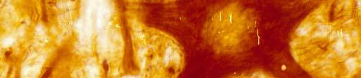
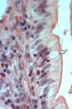
17 Light Microscope It is a cross-section of kidney medullar which is made up of lots of tubules. The wall of them is epithelial cells. The cell nucleus is basophilic (blue) and the cytoplasm is acidophilic (pink). HE staining.

18 Silver staining Gold staining


19 Frozen section Snap frozen in a cold liquid or cold environment Frozen sections are performed with a cryostat. cryostat Cutting a frozen section


20 Frozen section It is necessary to get a rapid diagnosis of a pathologic process. It is also effective in the histochemical study of very sensitive enzymes or small molecules.
21 Microscopy Light microscopy Conventional light microscopy Phase-contrast microscopy Polarizing microscopy Fluorescence microscopy Confocal microscopy Electron microscopy Transmission electron microscopy (TEM) Scanning electron microscopy (SEM)
22 Conventional light microscopy Mechanical parts Optical parts Condenser collects and focuses light to illuminate the object Objective enlarges and projects the image of the object in the direction of the eyepieces. Eyepieces magnify this image and project it onto the viewer s s retina
 Objective (40X) = 400X Light source Base Schematic diagram of")
23 Eyepiece /Ocular Objective lenses LIGHT MICROSCOPE Stage Condenser Slide Body Max MAGNIFICATION Eyepiece (10X) Objective (40X) = 400X Light source Base Schematic diagram of light microscope
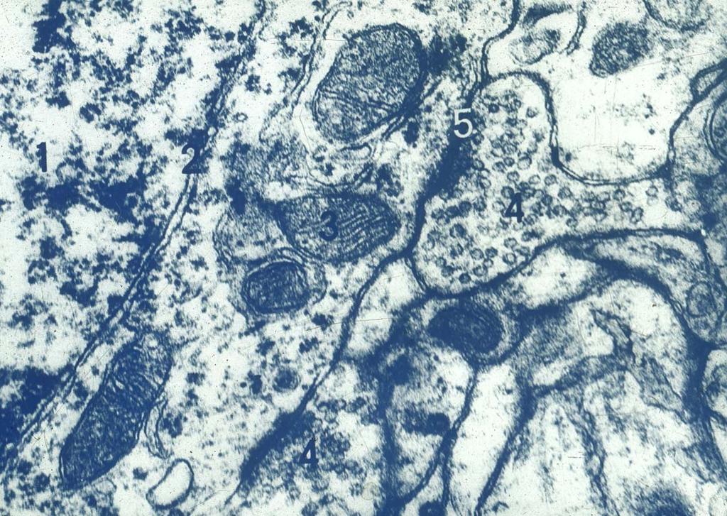
24 Phase-contrast microscopy & differential interference microscopy Phase-contrast microscopy light changes speed when passing through cellular and extracellular structures with different refractive indices. Differential interference microscopy produces an three-dimesional image Two types of microscopy are used to observed living cells.


25 A B Cultured neural crest cells seen with different optical techniques. A: Conventional light microscopy. B: Phase contrast microscopy. C: Nomarski differential interference microscopy. C

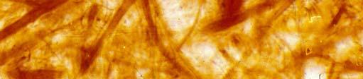
26 Under polarized light microscopy, collagen fibers appear brilliant or yellow.
27 Fluorescence microscopy Fluorescence: substances irradiated by certain light emit light with a longer wavelength. Fluorescence microscopy tissue sections are irradiated with ultraviolet (UV) light and the emission is in the visible portion of the spectrum. The fluorescent substances appear brilliant or colored on a dark background.
 emits yellow light, and the RNA-rich cytoplasm appears reddish")
28 Cytoplasm DNA Photomicrograph of kidney cells stained with acridine orange. DNA (within the nuclei) emits yellow light, and the RNA-rich cytoplasm appears reddish or orange.
29 Confocal microscopy A laser source Different layers of the specimen are seen in different focus simultaneously. Merged image of a three-dimension Clearer image


30 Different layers of the specimen are seen in different focus simultaneously. A merged image of a threedimensional object could be got.

31 a 3-D image of cultured cells

32 The image of specimen is clearer than in common fluorescence microscope.
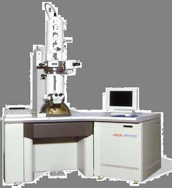
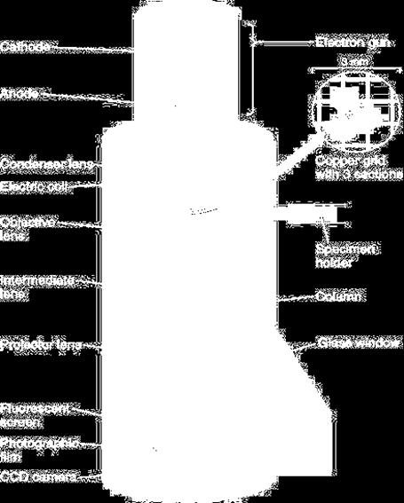
33 Transmission electron microscope high resolution (0.1nm)
34 electron lucent TEM micrograph of hepatocyte electron dense
35 Scanning electron microscopy pseudo-three three-dimensional views of the surfaces A very thin metal coating The electron beam interacts with this metal coating and produces reflected or emitted electrons.

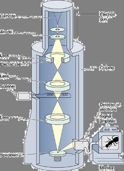
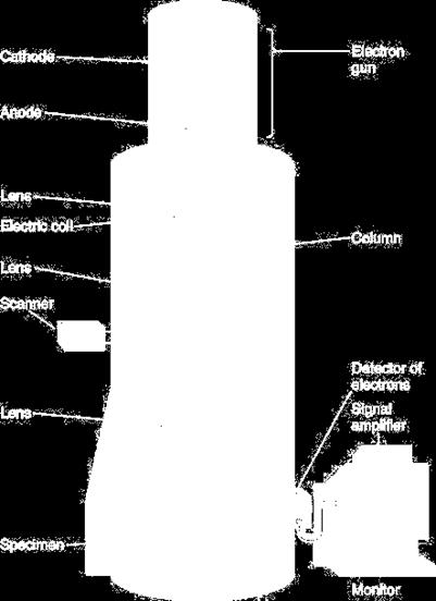
36 Schematic view of a transmission and scanning electron microscope

37 SEM micrograph of the epithelium of stomach
38 Problems in the interpretation of tissue sections Distortions & artifacts caused by tissue processing shrinkage Artificial spaces Wrinkles of the section precipitate of stain artifact Totality of the tissue Two dimensions & three dimensions

39 Shrinkage caused by tissue processing artificial spaces between the colloid and the follicular wall in the section of thyroid gland. Shrinkage of cells in hyaline cartilage


40 Lipid droplets infat cells are lost during tissue preparation.


41 Artifacts caused by tissue processing Mucous granules goblet cell goblet cell Mucous granules containing glycoprotein in the cytoplasm of goblet cells are lost during tissue preparation.


42 Totality of the tissue nucleus Nissl bodies Sliver staining H&E staining Neurofibrils
43 M


44 A B C How different 3-dimensional structures may appear when thin-sectioned. A: Different sections through a hollow ball and a hollow tube. B: A section through a single coiled tube may appear as sections of many separate tubes. C: Sections through a solid ball (above) and sections through a solid cylinder (below).
45 Important questions Hematoxylin & Eosin (H & E) staining basophilic acidophilic